13.2 sliding filament model
structure of actin vs myosin
myosin filament has a globular head region that connects with binding sites on the actin filament. Two regulatory proteins, tropomyosin and troponin, modulate this binding.
actin vs myosin
actin | myosin |
Thin filaments (8nm dia) | Thick filaments (16nm) |
Has Myosin Binding sites. | Contain myosin heads that can bind to actin. |
Individual molecules are helical (2 of them) | Individual molecules have a common shaft and a protruding globular head.  |
2 Regulatory proteins called Tropomyosin + Troponin are associated w it. | heads can make cross bridges + contain ATP binding sites and an enzyme called ATPase. |
Tropomyosin normally blocks myosin binding site. | |
Troponin can bind to Calcium ions. |
Myosin heads have:
A hinge enabling movement.
One site for binding to actin.
Another site for ATP binding, providing energy.
Actin filaments have:
Sites for myosin head attachment, known as actin-myosin binding sites.
Tropomyosin and troponin proteins attached, playing a regulatory role.
troponin and tropomyosin.
Tropomyosin molecules coil around actin
Troponin complex attached to each tropomyosin molecule made of 3 polypeptides
One binds to actin
One binds to tropomyosin
One to calcium ions.
Tropomyosin normally blocks myosin binding site.
Calcium ions bind to troponin resulting in a change in shape. This causes the tropomyosin to move which then exposes the myosin binding site on actin.
changes in sarcomere during muscle contraction
muscle contraction involves actin filaments being pulled closer together between the myosin, slide over each other + towards M-line in centre of sarcomere → shortens sarcomere → muscle shortens

Key changes to the sarcomere during muscle contraction:
I band and H-zone in sarcomeres shorten due to increased overlap of actin and myosin filaments.
A bands remain constant in length.
simultaneous contraction of many sarcomeres in myofibril = shortening + contraction of entire muscle.
sliding filament theory
widely accepted model: explains muscle contraction at molecular level. details how muscles contract, shorten + generate force, when actin + myosin filaments slide past each other
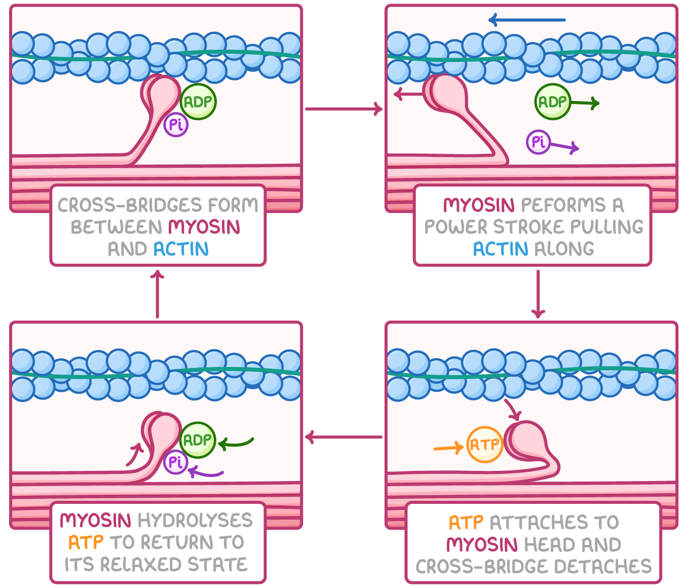
main steps in sliding filament theory:
Calcium ions (Ca2+) bind to troponin, altering its shape.
This change moves tropomyosin away from actin's binding sites, making them available for myosin.
Myosin heads attach to these exposed actin filaments, forming actin-myosin cross-bridges.
The myosin heads execute a power stroke, pulling the actin filament along and releasing ADP.
An ATP molecule binds to the myosin head, leading to its detachment from actin.
Ca2+ activates myosin's ATPase activity, breaking down ATP to ADP and phosphate, releasing energy.
This energy resets the myosin head to its original position.
The myosin head reattaches to a new actin site further along filament.
Energy sources for muscle contraction
Muscle contraction cycles require considerable energy, provided in form of ATP.
this ATP can be generated through different pathways:
Aerobic respiration - suitable for prolonged, low-intensity exercise.
Anaerobic respiration - used during short, high-intensity exercise.
The ATP-creatine phosphate system - Creatine phosphate is quick source of energy in muscles + broken down for immediate ATP replenishment w out oxygen. used for short bursts of vigorous exercise.