CIE AS Biology 4.1: Structure of Membranes
The cell membrane separates the internal cell environment from the external environment. Materials needed for cell growth are brought into a cell while toxic substances are kept out of the cell. The exchange of materials is strictly controlled by the membrane.
- the membrane is partially permeable
- the exchange of materials occurs by diffusion, osmosis, and active transport
The and are two of the many membranes in a cell. Membranes are important for compartmentalization in a cell.
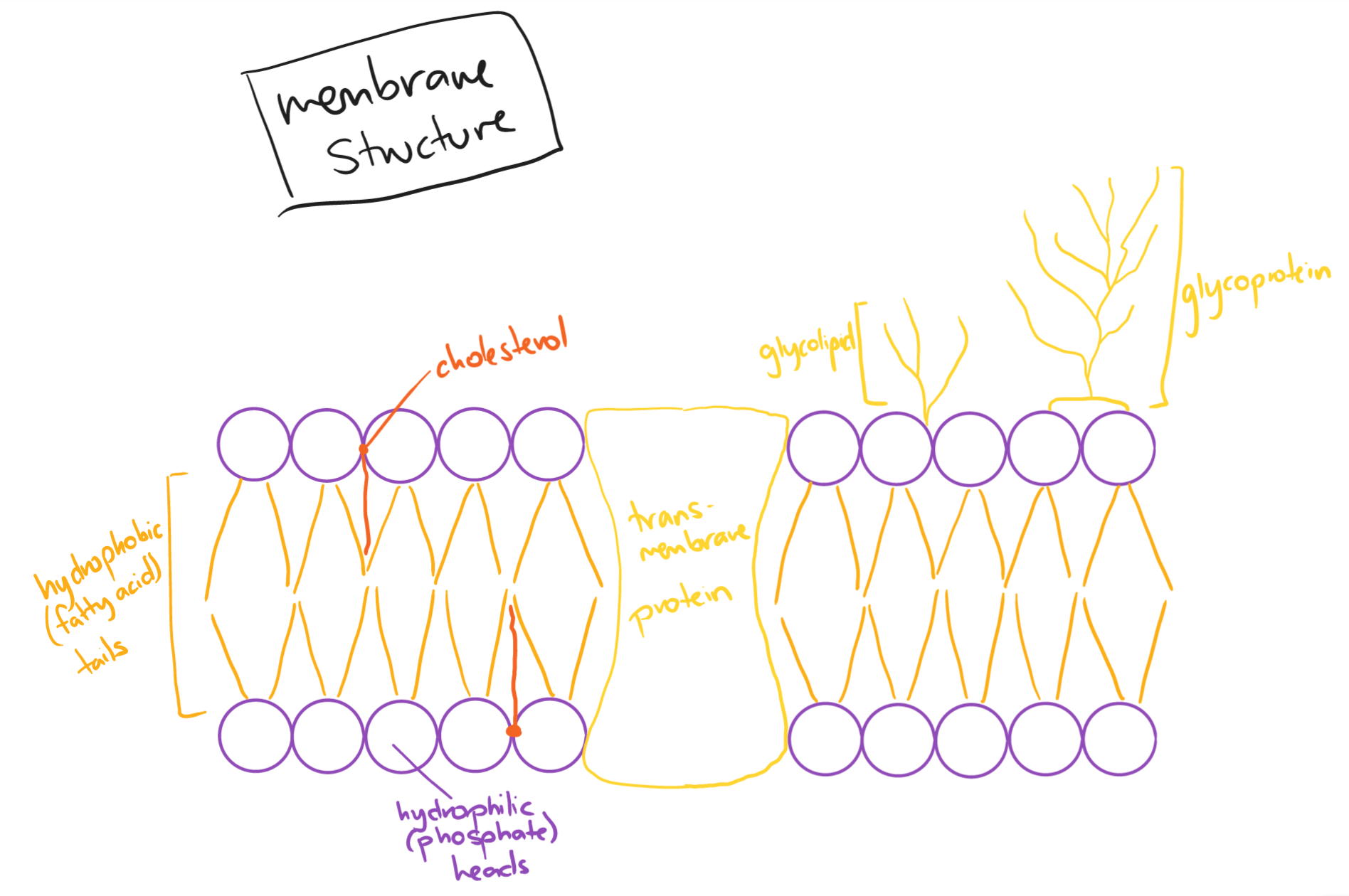
Components of the Membrane
The cell surface membrane contains phospholipids that form a bilayer, proteins such as glycolipids and glycoproteins, and cholesterol.
Phospholipid: contains a hydrophilic phosphate head and two hydrophobic fatty acid tails
Proteins:
- intrinsic membrane proteins: contained in the phospholipid bilayer; go right through the layer (transmembrane)
- : provide a hydrophilic pathway to allow ions and polar molecules to pass through the membrane (carrier and channel proteins)
- integral membrane proteins: permanently attached to the membrane
- extrinsic membrane proteins: found on one side of the membrane, often on the surface
- act as receptors/antigens
- peripheral membrane proteins: temporarily attached to the membrane
- glycolipids/glycoproteins: containing a short carbohydrate chain
- they are responsible for cell stability, cell signalling, endocytosis, and cell-to-cell recognition
Cholesterol: a steroid that maintains the flexibility and permeability of the cell membrane
 The fluid mosaic model of the membrane was first outlined in 1972 by S.J Singer and Garth L. Nicholson.
- The fluid mosaic model describes cell membranes as ‘fluid‘ due to:
- the movement of the phospholipids and proteins via diffusion
- the movement of the phospholipids within their own layers
- the different types of proteins are interspersed and move within the layer, though some may be fixed
- The fluid mosaic model describes cell membranes as a ‘mosaic‘ as their scattered pattern produced by the proteins within the bilayer forms a ‘mosaic-like‘ image when viewed from above
The fluid mosaic model is used to explain the passive and active movement between cells and their surroundings, cell-to-cell interactions, and cell signalling.