Protein Structure II: Hierarchical Organization and Structural Determination and Dynamics
Properties of the Peptide Bond and Polypeptide Chain
Foundational Chemistry: Proteins are linear polymers composed of amino acids. The formation of a polypeptide occurs through the condensation of amino acids to create peptide bonds.
Hydrogen Bonding: A single peptide bond contains both a carbonyl group () and an amino group (), allowing it to function as both a donor and an acceptor of hydrogen bonds.
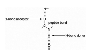
Electronic Character and Planarity: The peptide bond () possesses partial double-bond character due to resonance.
The C-N bond length is shorter than a standard single bond but longer than a typical double bond.
There is a partial negative charge () on the Oxygen atom and a partial positive charge () on the Nitrogen atom.
Because of this double-bond character, rotation around the bond is restricted, making the peptide bond planar.
Conformations: Due to the planarity, the peptide bond exists in two distinct conformations:
Trans: Side chains (SC) are on opposite sides of the peptide bond. This is the much-preferred conformation due to minimized steric hindrance.
Cis: Side chains (SC) are on the same side. This is rare and usually only occurs when specific structural constraints force the configuration.
Torsion Angles and the Ramachandran Plot
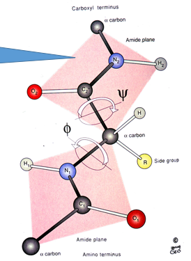
Rotational Degrees of Freedom: While the peptide bond itself is rigid, the polypeptide chain folds through rotation around the other two bonds in the backbone:
The (phi) bond: Between the Nitrogen and the -carbon ().
The (psi) bond: Between the -carbon and the carbonyl Carbon ().
Torsion Angles: These rotations create the and torsion angles, which define the path of the polypeptide backbone.
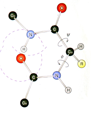
Steric Constraints: Not all angle values are possible. For example, setting both and results in a severe steric clash between atoms in the backbone and side chains.
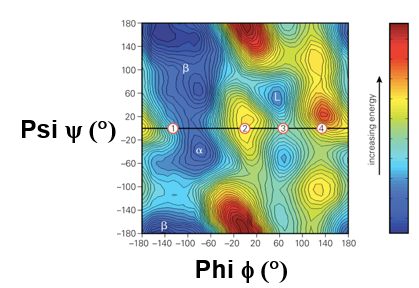
The Ramachandran Plot: A visualization of the allowed and disallowed regions for and angles.
Energy and Sterics: The plot reflects energy levels derived from steric constraints.
Clustering: Low-energy regions typically cluster into specific areas that correspond to common secondary structures (like -helices and -sheets).
Residue Specificity:
Glycine () is very permissive (it lacks a -carbon, allowing a wider range of angles).
Proline () is highly restricted due to its cyclic side chain.
Amino acids with bulky side chains are generally more restricted than smaller ones.
Historical Context of Protein Structure
The First Structures: Myoglobin and Hemoglobin were the first protein structures ever solved.
Recognition: John Kendrew and Max Perutz of the Laboratory of Molecular Biology (LMB) in Cambridge were awarded the Nobel Prize in Chemistry in 1962 for this work.
Scientific Observations: Upon solving the resolution model of sperm-whale myoglobin (published in Nature, 1958), John Kendrew remarked on the protein's extreme complexity and total lack of symmetry, which defied existing theories of protein structure that predicted more geometric regularity.
Hierarchical Organization of Protein Structure
Primary Structure: The covalent structure, consisting of the specific amino acid sequence.
Secondary Structure: Local non-covalent structures formed by backbone hydrogen bonds (motifs like the -helix and -sheet).
Tertiary Structure: The overall three-dimensional conformation of a single polypeptide chain.
Quaternary Structure: The arrangement and interactions resulting from multiple polypeptide chains (subunits).
Secondary Structure Motifs: The -Helix
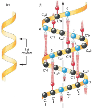
Geometry: The backbone follows a right-handed path.
Hydrogen Bonding: Stabilized by a regular H-bond pattern where the Carbonyl Oxygen of residue (; acceptor) bonds to the Amino Hydrogen of residue (; donor).
Specific Dimensions:
residues per turn.
Pitch (height of one full turn): .
Rise per residue: .
Side Chain Orientation: Side chains point outwards from the helical axis.
Amino Acid Propensities:
Favorable for a-helix formation: Alanine (), Leucine (), Methionine (), Arginine (), and Lysine ().
a-helix breakers: Glycine () and Proline ().
Glycine: entropic penalty for constraining angles, and it lacks a , meaning no backbone shielding
Proline: Disrupts the helical H-bond pattern (it lacks an amide hydrogen for donating) and introduces a physical kink in the helix due to its ring structure.
Amphipathicity: Because of the residues/turn, some side chains end up on nearly the same side (e.g., residues vs. ). This often creates an amphipathic helix with one hydrophobic face and one hydrophilic face.
Specialized Alpha Helical Structures
Coiled-Coils: Formed when alpha helices wrap around each other into a left-handed supercoil.
Heptad Repeat: Based on the residue repeat, helices with hydrophobic residues at positions and (also and ) can align to form a stable dimer.
Leucine Zippers: A common type of coiled-coil where Leucine is found at critical hydrophobic interface positions (favourable).
Function: Used for both homodimerization and heterodimerization of proteins.
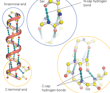
Helix Capping: Termini of helices break the regular H-bond pattern and require "caps" for stability.
N-cap: Often involves Serine (), Threonine (), Aspartic Acid (), or Asparagine () side chains hydrogen-bonding with the backbone amino group at the N-terminus.
C-cap: Often involves a Glycine () residue that allows for a main chain inversion to maximize backbone hydrogen bonding at the C-terminus.
Secondary Structure Motifs: The -Sheet
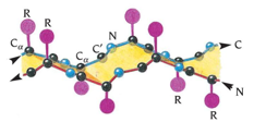
Antiparallel Beta Sheets:
Adjacent strands run in opposite directions ( vs. ).
Nearly linear, well aligned inter-strand H-bonds
Peptide dipoles are complementarily aligned.
Side chains alternate projecting to opposite faces of the sheet.
Strands are often connected by short turns.
Parallel Beta Sheets:
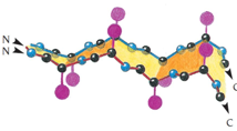
Adjacent strands run in the same direction ().
Inter-strand H-bonds are skewed or offset, making them slightly weaker than those in antiparallel sheets.
Peptide dipoles are aligned in parallel.
Side chains alternate projecting to opposite faces.
Strands are usually linked by longer loops or intervening helical segments.
Mixed Beta Sheets: Contain both parallel and antiparallel pairings within the same sheet. These often occur due to complex chain connectivity (e.g. long loops), which make a purely parallel or antiparallel layout impossible
help reconcile packing needs favorable loop placement even if not maximally favorable by backbone H-bonding alone
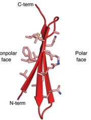
Amphipathicity and Surface: Similar to a-helices, individual strands can be amphipathic with opposing hydrophilic and hydrophobic sides. This allows -sheets to outline the protein surfaces, with one side facing the solvent and the other the interior.
Turns and Loops
Function I: Connectivity: They connect different elements of secondary structure. Minimal connections of amino acids are designated as "turns."
Function II: Functional Contribution: Longer loops lack strict structural constraints, making them highly versatile for:
Binding ligands.
Facilitating protein-protein interactions.
Allostery (mediating conformational switches).
Tertiary Structure: Motifs and Domains
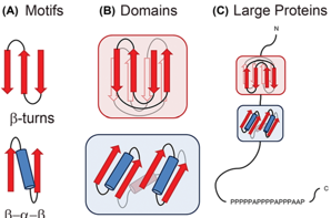
Motifs: Small organizations of secondary structure elements (usually elements), such as the Helix-loop-helix. Alpha and Beta sheets can organize locally to form small motifs.
They are like "phrases" in a sentence.
Do not fold stably on their own
They typically require the surrounding protein structure to remain stable.
the same motif can be found in many different domains
Domains: Discrete, independent units of tertiary structure, combination of multiple motifs.
They form and maintain their structure independently
Structure is closely linked to function.
Proteins can be single-domain or multi-domain.
The same domain can be found in many different proteins.
Proteins sharing the same domain are often functionally related.
Thermodynamics of Protein Folding
Contributors to Folding:
Attractive Interactions: Decrease in enthalpy () for the protein (e.g., H-bonds, Van der Waals).
Hydrophobic Effect: Increase in entropy () for the solvent. This is often the dominant driving force for folding.
Loss of Flexibility: Decrease in entropy () for the protein as it enters a structured state. This hinders folding.
Balance: 1 +2 both contribute but 2 is often much stronger, 3 hinders but loosens against the sum of 1+” (for folded proteins)
Membrane Proteins
Environment Adaptation: some proteins have evolved to populate the interior of lipid bilayer.
Residue Orientation: these proteins have adapted their structural properties, hydrophobic residues face the membrane interior shielding the polar backbone
Structural Trends: Most membrane proteins are -helical; -sheet membrane proteins are relatively rare.
Techniques for Determining Protein Structure
X-ray Crystallography:
Requires generating protein crystals (array of proteins in a periodic lattice), which is often difficult.
X-ray beams diffract off the crystal, creating a diffraction pattern.
Patterns are used to compute an electron-density map.
Limitation: The "phase problem"—some information required to build the structure is missing from the raw diffraction data.
Nuclear Magnetic Resonance (NMR):
Uses spin-active nuclei () align in a strong pulsed magnetic field ().Their relaxation emits a signal with characteristic spectra depending on their microchemical environment.
multidimensional NMR ,aps resonances to atoms and yields restraints such as distance and couplings
combination of restraints in computational refinement to produce a conformer ensemble consistent with data
great for solution structures and dynamics but size limited
Strength/Limit: Excellent for dynamics and proteins in solution, but limited by protein size.
Cryogenic Electron Microscopy (Cryo-EM):
Proteins are vitrified (frozen in glass-like ice) and imaged with electron beams.
Millions of noisy 2D projections in random orientations are computationally reconstructed into a 3D electron-density map.
The "Resolution Revolution": Previously only for large molecules ("blobology"), Cryo-EM now reaches resolutions comparable to X-ray crystallography.
Protein Dynamics
Proteins are not static; they move across various scales:
Femtoseconds (): Bond vibrations.
Picoseconds (): Rotamer flipping (side chain movement).
Nanoseconds (): Loop movements.
Micro- to Milliseconds ( to ): Large-scale collective motions and conformational changes.
Increasing the time and length scale of these motions generally corresponds to crossing a higher energy barrier.