Confocal microscopy
Confocal Microscopy: Overview and Advantages
Confocal microscopy offers several advantages over conventional widefield optical microscopy:
Depth of Field Control: Allows precise focusing on specific planes within thicker specimens.
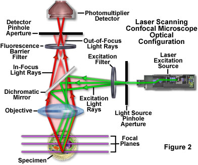
Image Clarity: Reduces background fluorescence, thus minimizing interference from out-of-focus light, resulting in sharper images.
Optical Sections: Capable of generating serial optical sections, aiding in the study of thick specimens.
The technique employs spatial filtering to eliminate out-of-focus light, enhancing the resolution of images. Recent advancements have increased its popularity due to the ability to generate high-quality images from specimens prepared for fluorescence microscopy, significantly benefiting cell biology applications involving both fixed and live samples. Confocal microscopy has become one of the most significant advances in optical microscopy due to these capabilities.
Comparison to Conventional Microscopy
In widefield optical epi-fluorescence microscopy, secondary fluorescence from the specimen obscures details, particularly in thicker specimens leading to a loss in fine detail.
Confocal microscopy improves upon this by excluding out-of-focus fluorescent signals, although it offers only marginal improvements in axial and lateral resolution compared to widefield methods but still less than transmission electron microscopy.
Historical Development
Marvin Minsky invented the concept of confocal microscopy in the mid-1950s for neural imaging but faced technological limitations related to light sources and data processing until advances in the late 20th century accelerated its development.
Subsequent advancements, including those by M. David Egger and G. Fred Brakenhoff, led to practical instruments capable of imaging living specimens more effectively.
Principles of Operation
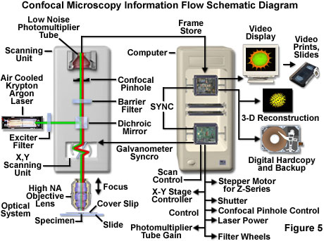
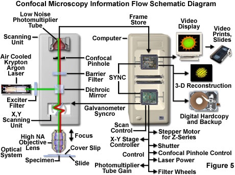
Confocal microscopy utilizes a laser excitation source, coupled with scanning and optical filtering techniques, to capture images at defined focal planes:
Light Path: Laser light passes through a pinhole aligned with the focal plane, allowing only in-focus light to reach the detector, significantly reducing background noise.
Scanning Mechanism: Using galvanometer mirrors, the laser beam scans across the specimen in a raster pattern.
Image Acquisition and Processing
Images are reconstructed point by point from the fluorescence emitted by the specimen, allowing detailed visualization of complex structures in living and fixed samples.
The system supports various configurations that enhance the capability for multidimensional imaging.
Advantages and Disadvantages
Advantages:
Thin optical sections enhance image clarity and detail.
Non-invasive imaging is possible for both living and fixed specimens.
Advanced software allows for complex modeling and analysis of multi-dimensional data.
Disadvantages:
Limited excitation wavelengths from lasers compared to widefield systems.
Potential cellular damage from high laser intensity.
Higher costs can limit availability in smaller labs, though cost-sharing models exist.
Conclusion
Confocal microscopy has fundamentally changed the field of microscopy, providing unparalleled capabilities for imaging cellular structures and functions, instrumental in both research and applied biology.