Western Blot Data Analysis
Explanation of Data/Results
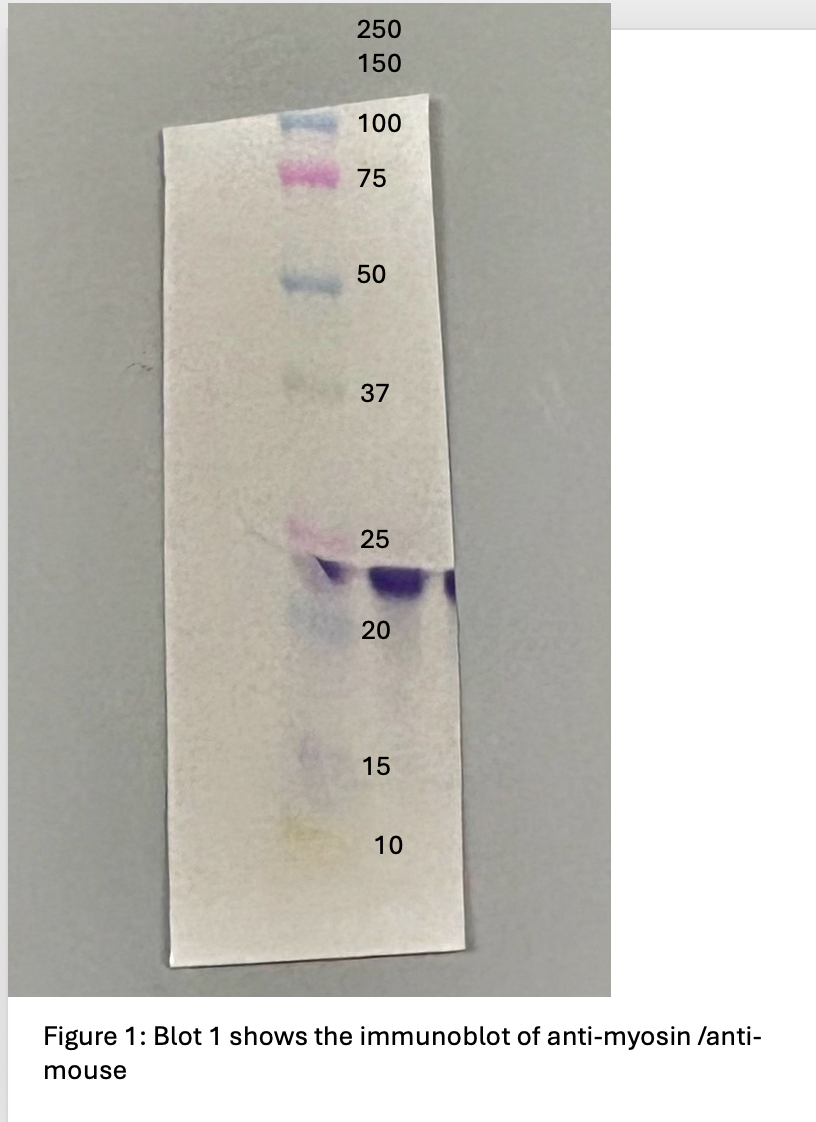
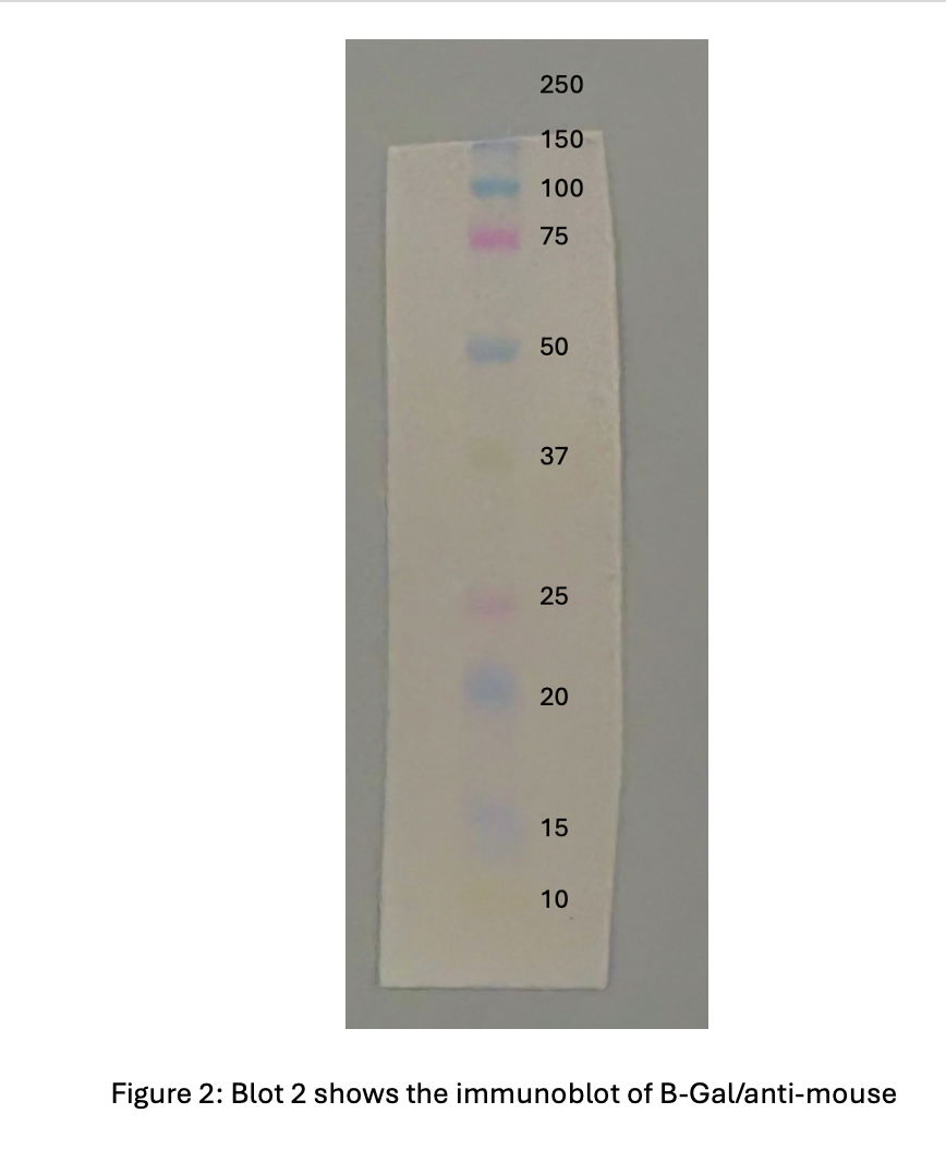
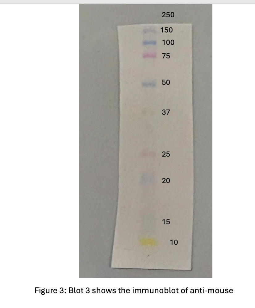
After preforming the SDS-PAGE and the Western Blot the unknown identity revealed to be myosin which was theorized from the previous SDS-PAGE experiment. The antibodies chosen for this experiment were anti-myosin , B-gal, and anti-mouse. Anti-myosin and B-gal were chosen as primary antibodies because these were the two main suspects for the identity of the protein. The secondary antibody was chosen as anti-mouse and used for all three blots. Only the third blot contained just the secondary antibody was used as a negative control. Based of the results shown in figure 1., blot 1 is the only blot that showed a stain for the binding of myosin supporting the hypothesis that if myosin is the correct identity then the blot containing anti-myosin antibodies will reveal a stain.
Myosin itself once dissembled from the SDS-PAGE, will seperate into its heavy and light chain. The light chain itself is 15-20kDa, once observing figure 1. blot 1, the appearance of the antibody stain is in the 20-25kDa area which is really close and confirms the identity of the protein.
The reason why HPR produces a visible color in the myosin western blot is because HRP enzyme which is attached to the secondary antibody that is going to oxidize 4-chloro-1-napthol and release the purple stain. That is why it is important for the primary antibody to bind first so it can find the protein and then the secondary will bind the primary antibody and release the color. This explains why there is no results in the figure 3. because the blot itself has no primary antibody to bind and therefore the secondary antibody cannot bind anywhere to release the stain. Figure 2. is explained by the reason that it was not the correct primary antibody therefore not being able to bind anywhere and again leading to the secondary antibody’s HRP to activate.
Discussion:
The goal of this experiment was to confirm the identity from the pervious SDS-PAGE experiment by preforming a western blot and adding antibodies the correct antibodies. With this the suspected idenitity of unknown protein 3 was myosin and based on the data and results, I support my hypothesis.
A positive control that I would include if this experiment were to be repeated is using the correct antibody which in this case is anti-myosin but from a different source to give me the same results and ensure that the antibodies are accurately detecting. A negative control would be include a primary antibody but not include the secondary antibody so there leads to no result in visible band shown.
If this experiment were to be repeated I would ensure to place the nitro-cellular membrane fully on the gel because as seen in figure 1-3. the top part of the band was cut off. Luckily for this experiment there was no need for the full molecular weight bands to be shown but for future experiments it can be important.
Based on the data /results, the HPR from the secondary anitbody was able to bind to the primary anitbody and release a purple band revleaing the idenitty of unknown protein 3 to be myosin. First SDS-PAGE was used to run the protein in a gel, and then the gel bands were transfered onto a nitrocellulus membrane in order to allow for anitbodies to be incubated onto the membrane. Once incubated the anitbodies either revelead or didnt reveal a puprle band indicating wether or not the unknown protein idenity matched the hypothesis.
In our next experiment the location of the protein within a cell will be determined. This will be done through cellular localization and fluorescence. The myosin protein will be identified in the cell whether it be the nucleus, the cell membrane, or the cytoplasm. where ever the cell glos is where the protein is located in. Localization of a specific protein uses localization microscopy. Flourscence happens when the molcules are ecvited by photoson therefore it is when a transfer of energy happens .