Msc. Anatomy Blueline
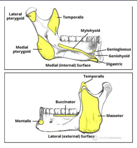
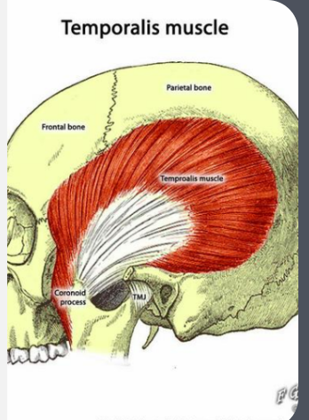
Temporalis
Actions:
Elevate Mandible
Protrude Mandible
Retrude Mandible- Posterior Fibers!
Lateral Excursion- 2° only!
Attachments:
Temporal fossa
Condyle of mandible (medial aspect)-Coronoid Process
Innervation- V3
EMBRYONICALLY: Originates from the 1st pharyngeal arch
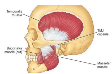
Masseter
Actions
Elevate
Protrusion
Attachments
Origin:
Superficial part- Maxillary process of Zygomatic bone
Deep part- Zygomatic arch of the Temporal Bone
Insertion
Condyle of Mandible
Innervation: V3
Embryonically: Originates from 1stpharyngeal arch
Medial Pterygoid
Actions
Elevate
Protrusion
Lateral Excursion- 1°
Attachments
Origin:
Superficial head, maxillary tuberosity &pyramidal process of palatine bone.
Deep head originates from the lateral pterygoid plate of the sphenoid bone
Insertion
Angle of mandible (ramus); medial aspect
INNERVATION: V3
EMBRYONICALLY: Originates from the 1stpharyngeal arch
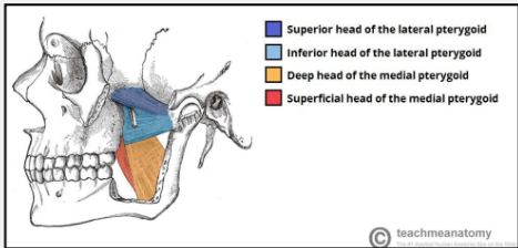
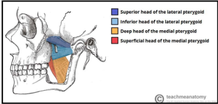
Lateral Pterygoid
Actions
Depress mandible
protrudes
Lateral Excursion-1°
INNERVATION: V3
EMBRYONCIALLY: Originates from 1st pharyngealarch
ATTACHMENTS:
Origins and insertions
Superior Head:
The upper/superior head originates on the infratemporal surface and infratemporal crest of the greater wing of the sphenoid bone and inserts onto the articular disc and fibrous capsule of the temporomandibular joint.
Inferior Head:
The lower/inferior head originates on the lateral surface of the lateral pterygoid plate and inserts onto the neck of condyloid process of the mandible
Digastric
Actions
Depress mandible at the beginning of the action, assisted by the myohyoid and geniohyoid.
Attachments
Posterior Belly: Arises from the mastoid notch, which is medial to the mastoid process of the temporal bone. The muscle becomes a tendon, becomes a tendinous pulley and passes through the hyoid bone.
Anterior Belly: The anterior belly arises from a depression on the inner side of the lower border of the mandible called the Digastric fossa of Mandible
the two bellies end in an intermediate tendon which perforates the Stylohyoideus muscle, and is held in connection with the side of the body and the greater cornu of the hyoid bone by a fibrous loop, which is sometimes lined by a mucous sheath
The two bellies of the digastric muscle have different embryological origins, and are supplied by different cranial nerves.
Posterior Belly: Originates from the 2nd pharyngeal arch (CN VII)
Anterior Belly: Originates from the 1st pharyngeal arch (V3 via mylohyoid nerve)
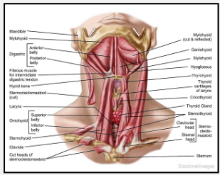
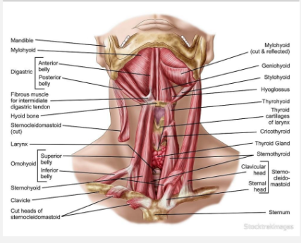
Mylohyoid
Actions
Elevates the Hyoid bone and the tongue
If other muscles are involved in keeping the hyoid bone fixed, then it depresses the mandible.
Attachments
origin: Mylohoid line of mandible
Insertion: Body of the Hyoid bone.
INNERVATION: Mylohyoid nerve from the inferior alveolar
EMBRYONICALLY: 1st pharyngeal arch
Summary
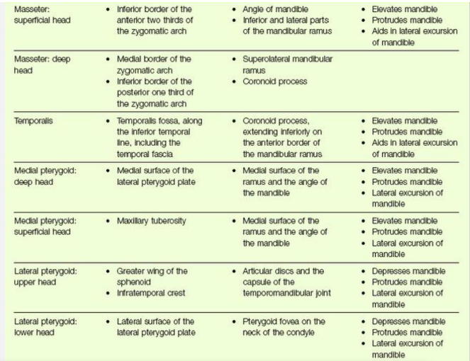
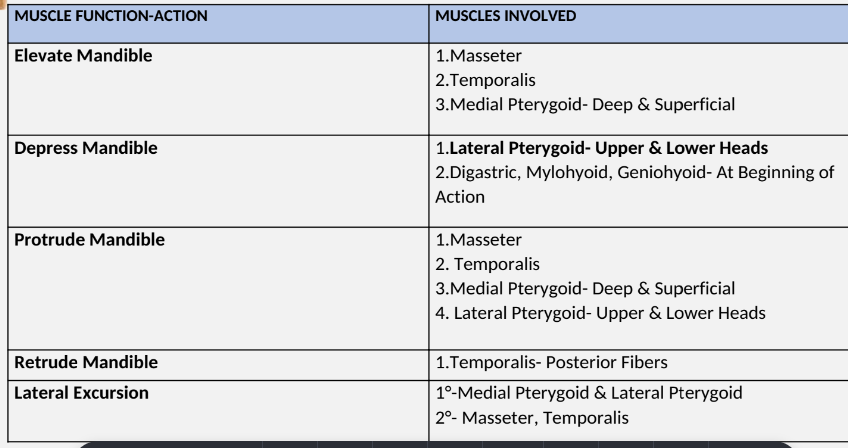
Summary Muscle Action
Cervical Muscles
More than 20 pairs of cervical muscles work synchronously to stabilize and control head movement.”
“ Cervical muscles are anatomically distinct from the masticatory muscles, however these groups of muscles have a bidirectional influence on each other and are co-activated during functional tasks.”
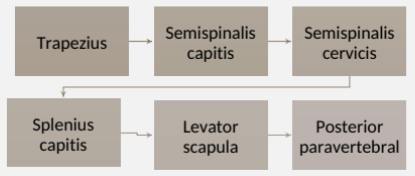
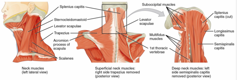
Posterior Cervical Muscles
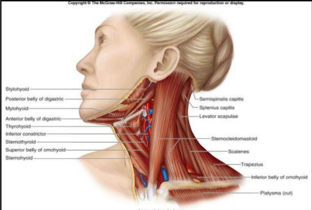
Anterolateral cervical muscles
Sternocleidomastoid (SCM)
Plastysma
Scalene
Anterior prevertebral
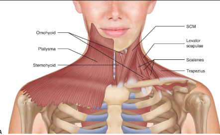
Cervical and Masticatory Muscle interactions
The masticatory muscles and the cervical muscles act in concert, influencing each other in the processes of chewing, swallowing and speaking. They should be viewed as a complex when evaluating a patient.
Myofascial pain and dysfunction can occur in any of these muscles, influencing patient perception of pain and resolution of pain.
In the area of orofacial pain (OFP), particularly in TMD, muscle disorders are the most common presentation, representing over 50% of diagnoses.
Referred Cervicogenic Pain
It is important to attempt to generate referred pain from the splenius capitis (located in the depression just posterior to the sternocleidomastoid muscle along the base of the skull) and trapezius muscles because referred cervicogenic pain is relatively prevalent among TMD patients.
Palpating the neck muscles just below the occipital protuberance most commonly generates referred pain to the forehead, periorbital, vertex, temple, occipital, postauricular, and ear.
Patients may have mild to moderate temporalis and/or masseter muscle palpation tenderness, and then when their suboccipital muscles are palpated, their eyes “light up” with the temporalis or masseter muscle pain complaint.
Take Aways
Knowing the anatomy of the muscles of mastication is important.
Understand what each of the muscles do during function. (mscs. Mastication)
Know and understand the key cervical muscles as they relate to the bi-directional influence they have with the muscles of mastication.
Key cervical muscles that are evaluated
SCM
Trapezius
Splenius Capitus