Digestive system
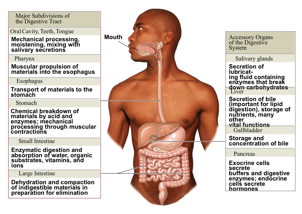
Digestive (GI) tract
Mouth
Pharynx
Esophagus
Stomach
Small/large intestines
Rectum
Anus
Accessory Organs:
Teeth
Tongue
Salivary glands
Gall bladder
Liver
Pancreas
Digestive system functions:
Ingestion
Mechanical processing
Digestion (duh)
Secretion
Absorption
Excretion
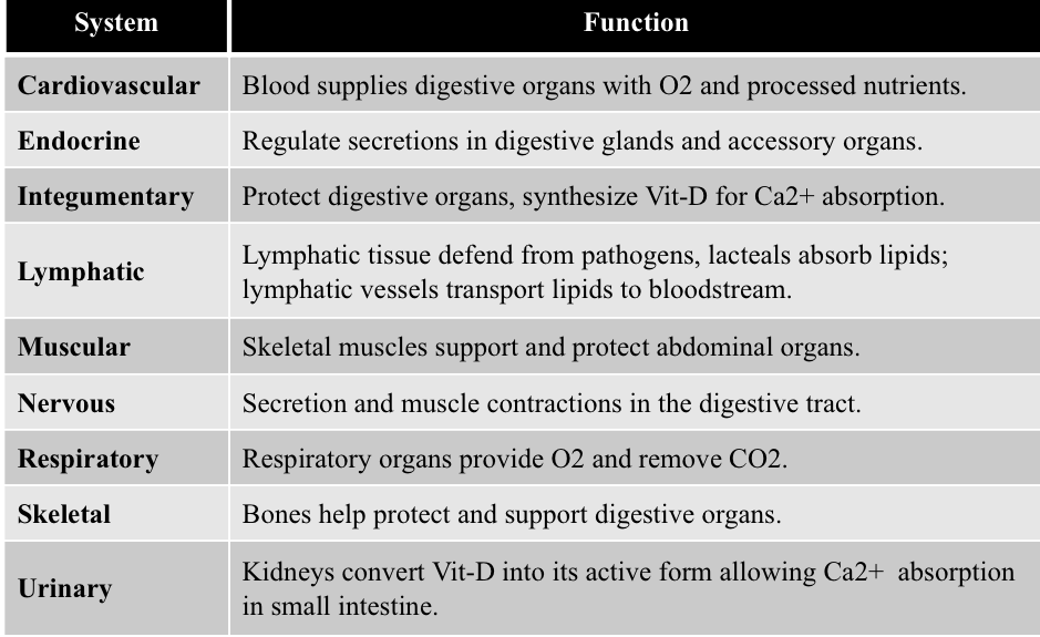
Digestive and other systems:
Histology of the GI tract:
Mucosa
Consists of an inner lumen
Consists of the outer epithelium and the inner lamia propira
Stratified squamous in high-pressure areas (oral cavity, pharynx, esophagus, rectum)
and simple columnar in the rest, with secretory cells.
The muscularis mucosae moves mucosal folds.
Folded to increase surface area.
4 types of epithelium:
Cutaneous
Synovial
Mucos
Subcutaneous
The Submucosa:
Dense irregular connective tissue
Binds mucosa to muscularis externa
Innervated (continuous blood vessels and lymphatics)
Submucosal plexus (a neural network) on the outer border
Assists with the secretion of enzymes into the system
Muscularis Externa:
Inner circular and external longitudinal smooth muscle layers
San, which ends at the myenteric plexus
Contains:
Parasympathetic ganglia increase activity
Sympathetic ganglia; decreases activity
Helps with churning and mixing
The Serosa:
Covers the muscularis externa, along most of the GI tract, forming the peritoneal cavity
Consists of
Mesenteries
specialized peritoneum stabilizes some of the GI tract
Innervated (contains blood vessels, nerves, and lymphatics
Adventitia
outer layer of the oral cavity, pharynx, esophagus, and rectum. (Collagen-rich)
Oral (buccal) cavity:
Lined with oral mucosa
Senses material before swallowing
Mechanically process
Lubricates with saliva and mucus
Begins enzymatic digestion of carbs and lipids
Lateral walls: cheeks and continue to the lips
Roof: palate (made of the maxilla bone)
Floor: tongue
Perform cheOropharynx: starts at the base of the tongue
The tongue:
Manipulates food within the oral cavity (compresses/ distorts it)
Senses taste, touch, and temp
Assists in chewing
Lingual tonsils: paired lymphoid nodules at the base of the tongue
Salivary glands:
~1-1.5 L saliva/day
99.4% water+ mucins, ions, buffers, metabolites, enzymes.
Salivary amylase breaks down carbohydrates
parotid salivary gland:25%
Sublingual salivary glands:5% ( produces the least amount of saliva), merocrine secretion
Submandibular gland: 70% (produces the majority of saliva), apocrine secretion
Teeth:wing or mastication
Made of dentin
Neck: area between crown and root
Crown: covered in enamel (crystalline form of calcium phosphate) requires Ca2+, phosphates, and vitamin D3
Pulp cavity: receives blood vessels and nerves through the root canal and the root of the tooth
The periodontal ligament with cementum binds the root of teeth to the bone
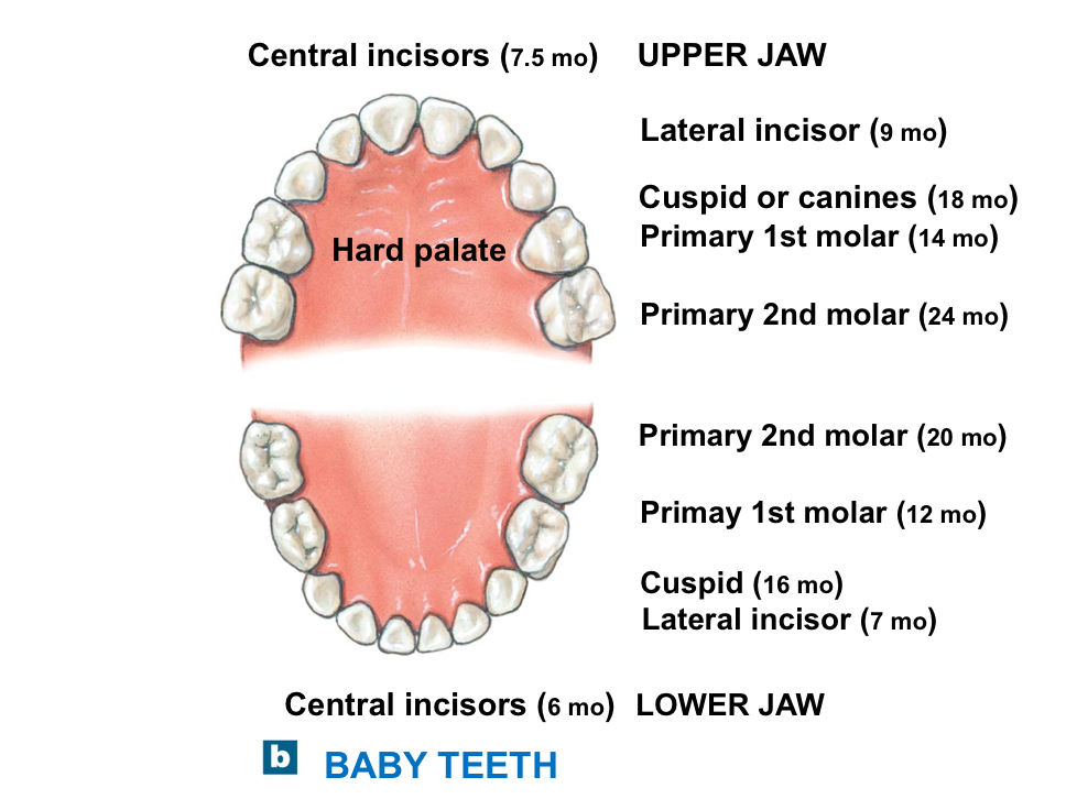
There are 20 baby teeth.
The bottom jaw’s incisors erupt first
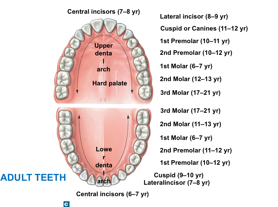
There are 32 adult teeth
The third molar erupts last
Pharynx
Food passes through the oropharynx and laryngopharynx to the esophagus
Mucosa is made up of stratified squamous epithelium
Laminate propila contains mucous glands and tonsils
Esophagus
A muscular tube of ~25 cm (10 in) long and 2 cm (0.75 in) in diameter
The passageway of food from the pharynx to the stomach
Posterior to the trachea.
Enters abdominal cavity through the esophageal hiatus in the diaphragm.
Lined with stratified squamous epithelium.
Sphincters are present at both ends, made of a circular pattern of smooth muscle cells.
Deglutition
Initiated voluntarily
Proceeds automatically
The Stomach:
Functions:
Temporary storage of ingested food.
Mechanical breakdown of ingested food
Chemical digestion by acids and enzymes
Production of intrinsic factor, a compound necessary for the absorption of Vit B12.
Chyme: Ingested material mixed with glandular secretions, ns producing a viscous, highly acidic soup. (Moves faster when the meal contains fewer promises and more carbs, caffeine, and alcohol)
Stomach Anatomy:
Cardia: connects with the esophagus
Fungus: bulge of the superior stomach
Body: a large area between the fungus and the pylorus
Pylorus: distal, connects the stomach to the small intestine. Regulates the flow of chyme to the small intestine
The Gastric Wall:
Lined with simple columnar epithelium with numerous mucous cells.
Mucous cells secrete alkaline mucus that protects the epithelium.
Gastric Pits: Shallow depressions that open to the surface
Neck cells undergo active mitosis, replacing mucosa cells.
Gastric glands:
Parietal cells
Intrinsic factor for VitB12 absorption. HCl lowers the pH of gastric juices to 1.5-2
Kills microorganisms and activates enzymes.
Pepsinogen+ HCL= Pepsin
Chief cells
Kills microorganisms and activates enzymes.
G cells: Gastrin
Mucous cells: life spans ~ 3-7 days
Parietal and chief cells produce ~1.5L of gastric juice in a day
Digestion:
Pepsin initiates protein digestion into small peptides
Salivary amylase will digest carbohydrates until pH ~4.5. (initiated in the mouth
No nutrients are absorbed in the stomach because,
Mucosa covered by alkaline mucous.
Epithelial cells lack transport mechanisms.
Gastric lining is impermeable to water.
Digestion is incomplete, and nutrients are still complex.
Small intestines:
intestinal wall: 3 levels of increased surface area
Circular folds: (plicae circulares) transverse ridges
Villi: finger-like projections of mucosa covered with columnar epithelium
Microvilli: (brush border) modified apical surface of columnar cells
intestinal movement:
Weak peristaltic contractions: initiated by a rural network independent of the CNS
Gastroenteric reflex: initiated by distension of the stomach. Increased peristaltic contractions in the duodenum
Gastrointestinal reflex: triggered by gastrin, reflexes in the ileocecal valve
~5hrs from duodenum to end of ileum
Juices: mostly water from mucosa, rest by intestinal glands (~1.8 L/day)
Hormones:
Gastrin: stimulates the production of acid and enzymes
Secretin: increases buffer and bile secretions
CCK: increases enzyme production (pancreas) and ejection of bile (gall bladder)
GIP: Inhibits the gastric actI it’s and causes release of insulin. (Inhibits increased gas production and releases insulin)
The Pancreas:
Location: behind the stomach, extending from the duodenum towards the spleen ~80 g. Secretes ~1 L of pancreatic juices mostly (Na2HCO2)
Major tissues and function:
Pancreatic islets: endocrine type secretes insulin, glucagon
Pancreatic acinar cells: exocrine type, produce digestive enzymes, juices, and buffers
Histology:
Pancreatic acini: pouches of acinar cells
Pancreatic ducts: formed by the convergence of many smaller ducts
Islets ~1% contains:
Alpha cells that secrete glucagon
Beta cells that secrete insulin
Pancreatic secretions: CCK stimulates the production of 4 classes of enzymes:
Carbohydrase: digests sugars and starches (ex. amylase)
Lipase: breaks down lipases
Nucleases: break down nucleic acids
Proteases: breakdown of proteins, ~ 70% ( ex. trypsin, chymotrypsin)
High acid chyme triggers secretin release, which increases bicarbonate levels to buffer chyme.
Liver:
Lobes are divided into 100,000 lobules (functional unit)
Liver cells:
Hepatocytes: major type, arranged within lobules in the form of plates like spokes of a wheel, secrete bile, synthesize plasma proteins, serve as a transporter
Kupffer cells: phagocytic type provides immunity/ protection
Bile: secreted by hepatocytes within bile canaliculi - common bile duct- duodenum
Additional bile is returned to the gall bladder
Bile is a mixture of water, inconsistent, bilirubin from destroyed RBS, cholesterol, and bile salt.s
Bile salts are synthesized from cholesterol in the liver and are required for normal digestion and absorption of fats
Blood circulation:
Blood arrives via the hepatic artery and hepatic portal vein
At the sinusoids, hepatocytes absorb nutrients, toxins, and secrete plasma proteins
High blood glucose triggers the synthesis of glycogen
Low blood glucose triggers glycogen breakdown
Blood leaves sinusoids into the central veins, the hepatic vein, and then the IVC
Functions:
Metabolic: extract nutrients/ toxins from the blood
monitors and adjusts organic nutrients
The large intestine:
Horse-shoe shaped, frames around the small intestine
Ileocecal valve to anus
consists of the cecum, colon, and rectum
Functions:
reabsorption of water, compacting chyme into feces
absorption of vitamins freed by bacterial action
storage of feces prior to defecation
Cecum:
AN expanded pouch
Chyme enters through the ileocecal valve
compaction begins here
Appendix: found on the posteromedial surface
contains lymphoid nodules for immune function
Inflammation is called appendicitis (removal is called appendectomy)
The Colon:
Large Diameter, no villi, abundant mucous ccells. The external surface had
Haustra: Elongated pouches
Taenia coli: 3-longitudinal bands of smooth muscle
Segments:
Ascending colon
Descending colon
transverse colon
transverse colon
sigmoid colon
The rectum:
An expandable organ for storing feces
Anal canal is the last portion
anus consists of-
internal anal sphincter- made of smooth muscularis externa, under ANS control
external anal sphincter: made of skeletal muscle under voluntary control
The large intestine (functions cont.)
Main function: absorption/conversion
Water (1300ml of total 1500ml a day)
bile salts
Vit K (fat-soluble) for the production of clotting factor
Vitamin B5 (water-soluble)synthesis of neurotransmitters and steroid hormones
Vitamin H (Biotin) essential for glucose metabolism
conversion bilirubinnn to an absorbable form that is eliminated via urine
Bacterial breakdown of peptides into ammonia, nitrogen compounds, and hydrogen sulfide
Ammonia and other toxins are absorbed. The liver converts them into harmless forms and eliminates them through the kidneys
Indigestible carbs serve as a source of nutrients for bacteria that form gas
All non-absorbable forms remain in feces, giving a brown color
Defecation: Involves two feedback loops,
Short loop:
Stretch receptors in the rectal walls stimulate local peristalsis
moves feces towards the anus
Long loop:
Parasympathetic reflexes trigger the involuntary relaxation of the internal sphincter
conscious relaxation of the external sphincter to eliminate feces
Aging Effects:
Chief cells of a baby’s stomach produce renmate (a coagulation factor). It gives more time to help digest milk's nutrients.
Increase in ulcer, dehydration
Increase in cancer rate (colon, stomach)
Decline in osmoreceptor sensitivity leads to dehydration
Bone loss leads to tooth loss, cavities, and gingivitis (osteopenia, not osteoarthritis)
loss of taste and olfactory sensations