Digestive System: Anatomy, Physiology, and Digestive Processes (Chapter 25)
Introduction
Overview: most nutrients cannot be used in their ingested form and must be broken down into smaller components before the body can utilize them.
Digestive system as an disassembly line: break down nutrients into usable forms and absorb them for distribution to tissues.
Gastroenterology: the study of the digestive tract, its disorders, diagnosis, and treatment.
25.1 General Anatomy and Digestive Processes
Digestive Function 1: Five stages of digestion
1. Ingestion: selective intake of food.
2. Digestion: mechanical and chemical breakdown into usable forms of chemical monomers.
3. Absorption: digestive system moves nutrients from GI tract into cells of the body, uptake of nutrient molecules into epithelial cells and then into blood/lymph.
4.Compaction: absorption of water and consolidation of indigestible residue into feces.
5. Defecation: elimination of feces.
Digestive Function 2: Mechanical digestion
Physical breakdown of food into smaller particles.
Cutting and grinding by teeth; churning by stomach and small intestine.
Exposure of more surface area to digestive enzymes.
Digestive Function 3: Chemical digestion
Series of hydrolysis reactions that convert macromolecules into monomers.
Enzymes produced by salivary glands, stomach, pancreas, and small intestine.
Results:
Polysaccharides → monosaccharides
Proteins → amino acids
Fats → monoglycerides and fatty acids
Nucleic acids → nucleotides
Some nutrients (e.g., vitamins, amino acids, minerals, cholesterol, water) are absorbed directly without digestion.
General anatomy: Digestive tract vs accessory organs
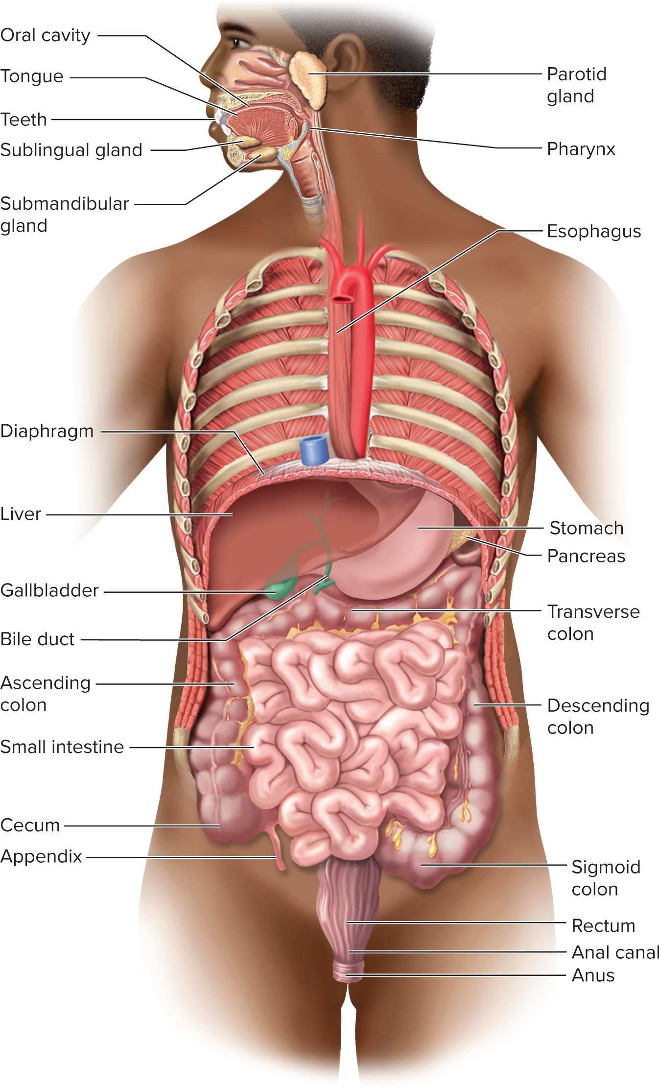
Digestive tract (alimentary canal): ~30 ft long muscular tube from mouth to anus.
mouth, pharynx, esophagus, stomach, small intestine, and large intestine
Gastrointestinal (GI) tract is the stomach and intestines
Accessory organs: teeth, tongue, salivary glands, liver, gallbladder, pancreas.
Digestive tract is open to environment at both ends
Most material in it has not entered the body tissues
– Considered to be external to the body until it is absorbed by the epithelial cells of the alimentary canalOn a strict sense, defecated food residue was never in the body
Most of digestive tract follows a basic structural plan with the digestive tract wall consisting of layers:
Layers of the digestive tract (from lumen outward)
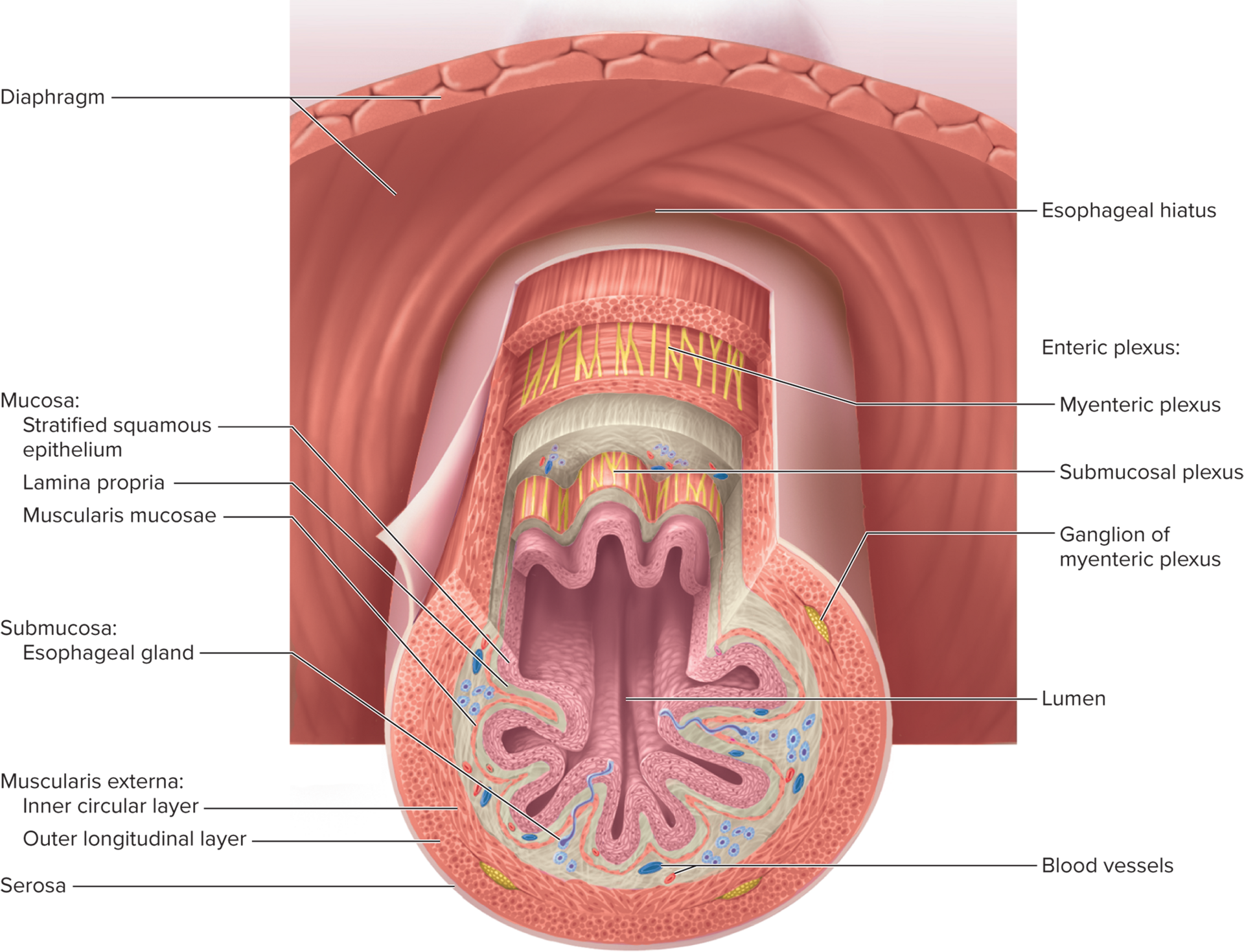
Mucosa: epithelium, lamina propria, muscularis mucosae.
Submucosa: loose connective tissue with vessels, nerves, and sometimes glands (MALT extends here).
Muscularis externa: inner circular layer and outer longitudinal layer; sphincters formed where the circular layer thickens.
Serosa: areolar tissue + mesothelium; or adventitia where serosa is absent.
Mucosa details
Epithelium: simple columnar in most of tract; stratified squamous in mouth, esophagus, and lower anal canal.
Lamina propria: loose connective tissue.
Muscularis mucosae: thin smooth muscle, tensing mucosa to create ridges and grooves to enhance contact with food. Improves efficiency of digestion and nutrient absorption
MALT: mucosa-associated lymphatic tissue with abundant lymphocytes and nodules.
Submucosa details
Thick layer of loose connective tissue.
Contains blood vessels, lymphatics, nerve plexus, and sometimes mucus-secreting glands that dump lubricating mucus into the lumen
MALT can extend into submucosa in some GI parts.
Muscularis externa details
Usually two muscle layers: inner circular (thickens to form valves) and outer longitudinal.
Sphincters: thickened circular layer at certain regions that regulate the passage of material through the tract
Outer longitudinal layer: Responsible for the motility that propels food and residue
through the tractMotility: propulsion of material through the tract.
Serosa and peritoneum relationship
Serosa: outer serous membrane with areolar tissue and mesothelium.
Adventitia: fibrous connective tissue binding pharynx, parts of esophagus, and rectum to surrounding tissue.
Enteric nervous system (ENS)
Nervous network in esophagus, stomach, and intestines regulating motility, secretion, and blood flow; >100 million neurons.
Can function independently of CNS but is modulated by CNS.
Composed of two major networks:
Submucosal (Meissner) plexus: controls glandular mucosal secretions and movements of muscularis mucosae.
Myenteric (Auerbach) plexus: parasympathetic ganglia and nerve
fibers between the two layers of the muscularis externacontrols peristalsis and contractions of muscularis externa.
Relationship to peritoneum and mesenteries
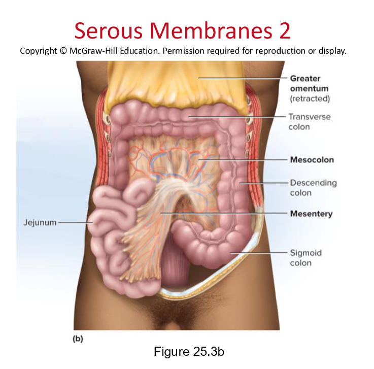
Mesenteries: connective tissue sheets suspending stomach/intestines from the abdominal wall; allow mobility and provide vessels and nerves.
Functions: support organs, allows strenuous contractions with free movement prevent twisting, holds abdominal viscera in proper relationship to each other, provide pathways for vessels and nerves, contain lymph nodes.
Parietal peritoneum—a serous membrane that lines the wall of the abdominal cavity
Peritoneal subdivisions and attachments:
Visceral vs parietal peritoneum; peritoneal cavity; lesser omentum; greater omentum; mesentery of small intestine.
Forms dorsal mesentery: a translucent two-layered membrane extending to the digestive tract
The two layers of the mesentery separate and pass around opposite sides of the organ forming the serosa
– Come together on the far side of the organ and continue as another sheet of tissue, called the anterior (ventral) mesentery
• May hang freely in the abdominal cavity
• May attach to the anterior abdominal wall or other organs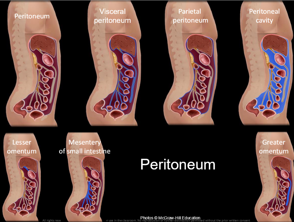
Peritoneum structures and relations
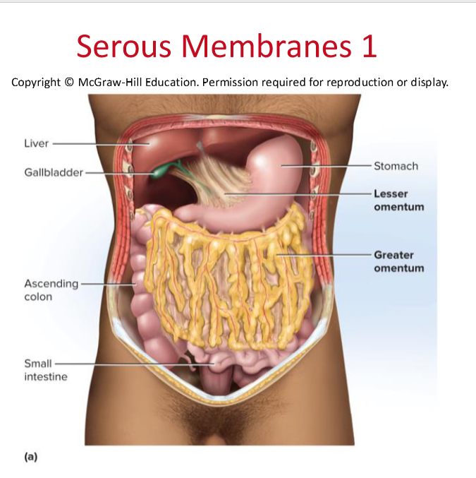
Lesser omentum: from lesser curvature to liver.
Greater omentum: hangs from greater curvature over intestines; forms deep pouch; encases spleen and colon surfaces via mesocolon.
Mesocolon: anchors colon to abdominal wall.
Intraperitoneal vs retroperitoneal organs: intraperitoneal (enclosed by mesentery on both sides) such as stomach, liver, parts of intestine; retroperitoneal (against posterior wall) such as duodenum, pancreas, parts of large intestine.
Regulation of the Digestive Tract
Motility and secretion of the digestive tract are controlled by neural, hormonal, and paracrine mechanisms
Neural control
Short (myenteric) reflexes: stretch or chemical stimulation acts through myenteric plexus
Stimulates paristaltic contractions of swallowing
Long (vagovagal) reflexes: parasympathetic stimulation of digestive motility and secretion
Hormones
such as gastrin and secretin released into the blood regulate various aspects of digestion, including enzyme secretion and gastric acid production.
Paracrine secretions like histamine and prostaglandins
Chemical messengers that diffuse through the tissue fluids to stimulate nearby target cells
Sphincters of the digestive tract
orbicularis oris: The first sphincter of the digestive system, surrounds the entrance to the oral cavity.
Upper esophageal: separates the pharynx from the esophagus and regulates the passage of food into the esophagus.
lower esophageal: At the terminal end of the esophagus, which subconsciously controls the entrance of food into the stomach.
pyloric: regulates the movement of food from the stomach to the small intestine.
ileal papilla: The junction of the small and large intestines is marked by a pronounced muscular swelling, which regulates movement into the large intestine.
internal anal sphincter: A smooth muscle structure that is controlled involuntarily, facilitating the regulation of fecal passage from the rectum to the anal canal.
External anal sphincter: A striated muscle structure that is under voluntary control, allowing for the conscious regulation of fecal expulsion from the anal canal.
25.2 The Mouth Through Esophagus
The Mouth (buccal cavity)
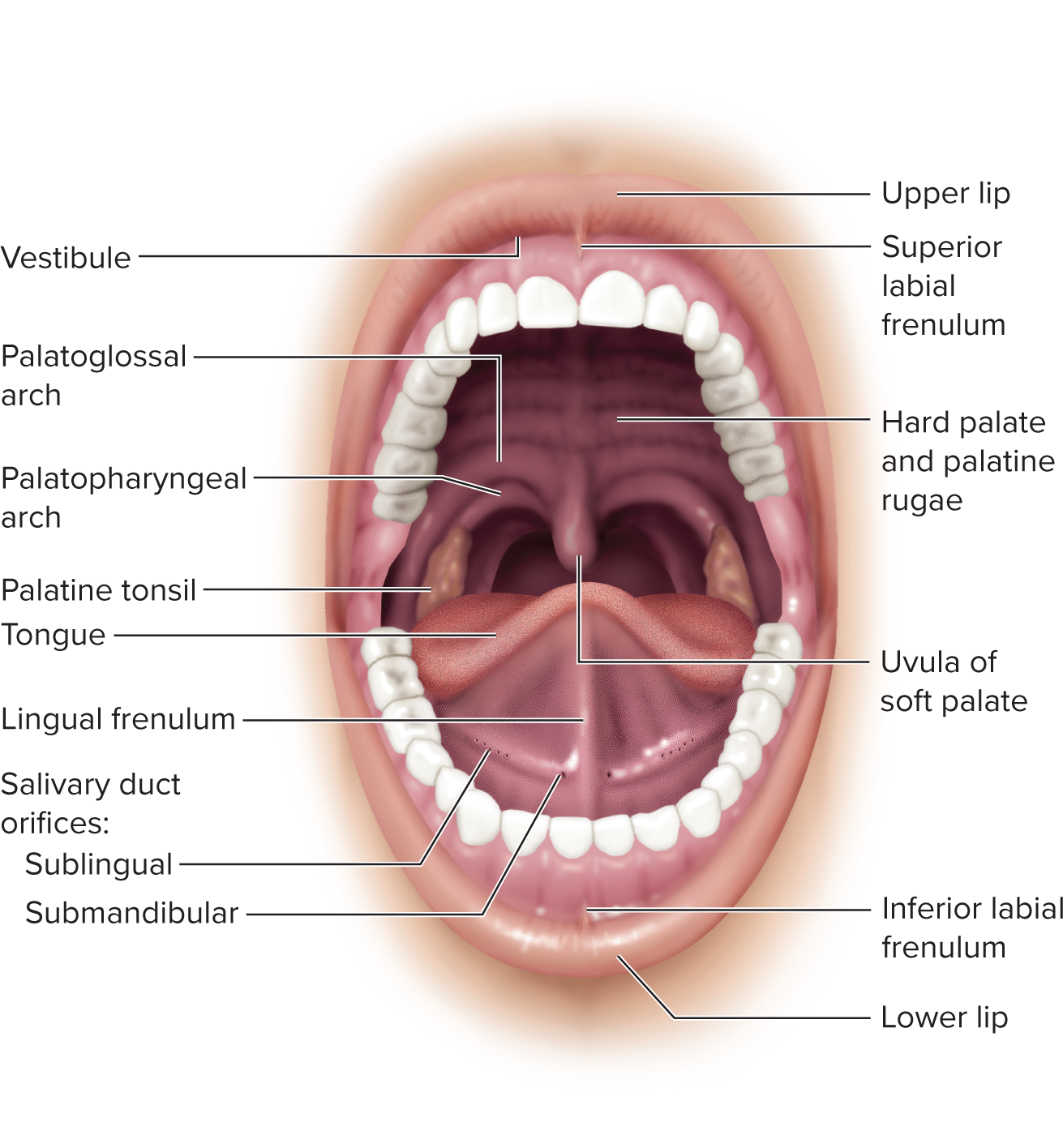
Functions: ingestion, taste, sensory responses to food, chewing and chemical digestion, swallowing, speech, respiration.
Boundaries: cheeks, lips, palate, tongue.
Oral fissure and Fauces: anterior opening between lips; posterior opening to the throat.
Epithelium: stratified squamous; keratinized in gums and hard palate; nonkeratinized in floor of mouth, soft palate, insides of cheeks and lips.
cheeks and lips: retain food and push between teeth, essential for speech, sucking, blowing, subcutaneou fat, buccinator muscle of cheek and orbicularis oris of the lips
orbicularis oris: is the first sphincter of the digestive system, surrounds entrance to the oral cavity
labial frenulum: median fold that attaches each lip to the gum between anterior incisors
vestibule: space between cheek or lips and teeth
Lips: cutaneous are: colored, has hair follicles and sebaceous glands
red vermillion area: hairless region where lips meet, tall dermal papilla allows blood vessls and nerves to come closer to epidermal surface, more senstive
labia mucosa: inner surface of lips facing gums and teeth
The Tongue
Muscular, manipulates food, senses taste/texture; cleans teeth post-meal.
Surface: nonkeratinized stratified squamous epithelium.
Lingual papillae: sites of most taste buds; vallate papillae mark boundary between body/root; lingual frenulum attaches body to floor of mouth.
Intrinsic vs extrinsic muscles: intrinsic for fine movements; extrinsic for stronger food manipulation (genioglossus, hyoglossus, palatoglossus, styloglossus).
Lingual glands: serous and mucous glands amid the extrinsic muscles secrete a portion of the saliva
Lingual tonsils: contained in the root
The Palate and Palatine Structures
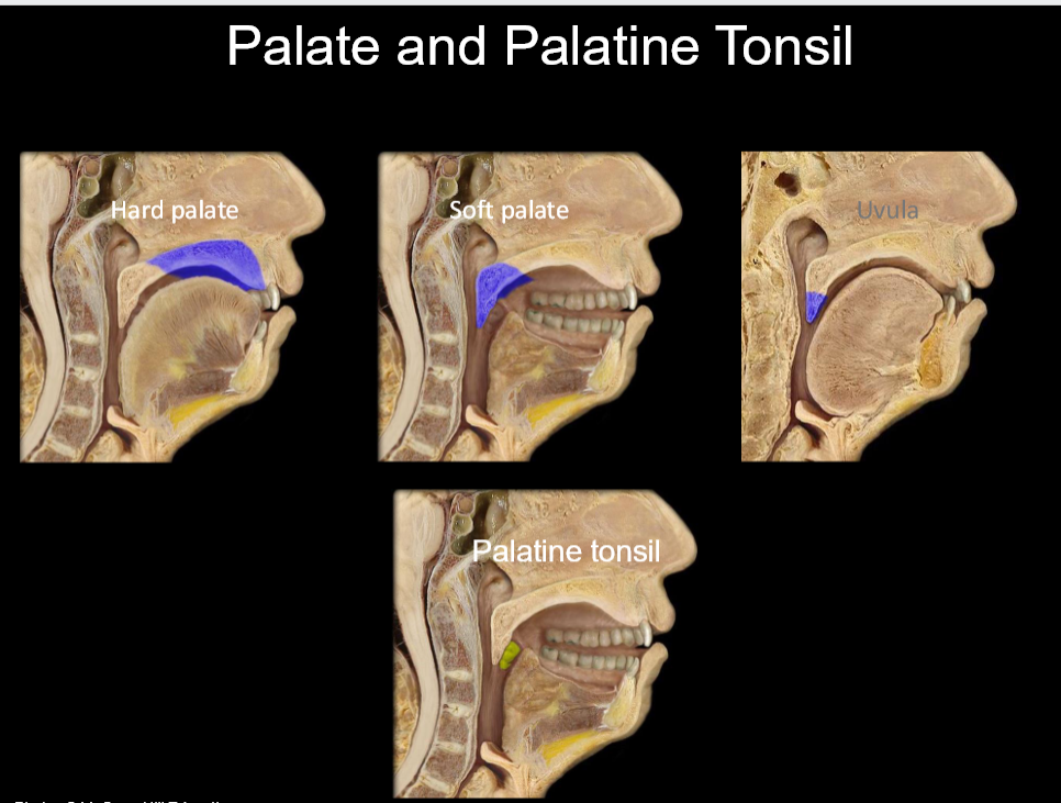
Palate—separates oral cavity from nasal cavity
– Makes it possible to breathe while chewing foodHard bony palate: anterior bony part with palatine processes; palatine rugae help tongue manipulation. supported by palatine processes of maxillae and palatine bones
Soft palate: posterior with spongy texture, flexible; composed of skeletal muscle and glandular tissue, uvula (retain food in mouth until ready); arches (palatoglossal and palatopharyngeal); palatine tonsils between arches.
The Teeth (Dentition)
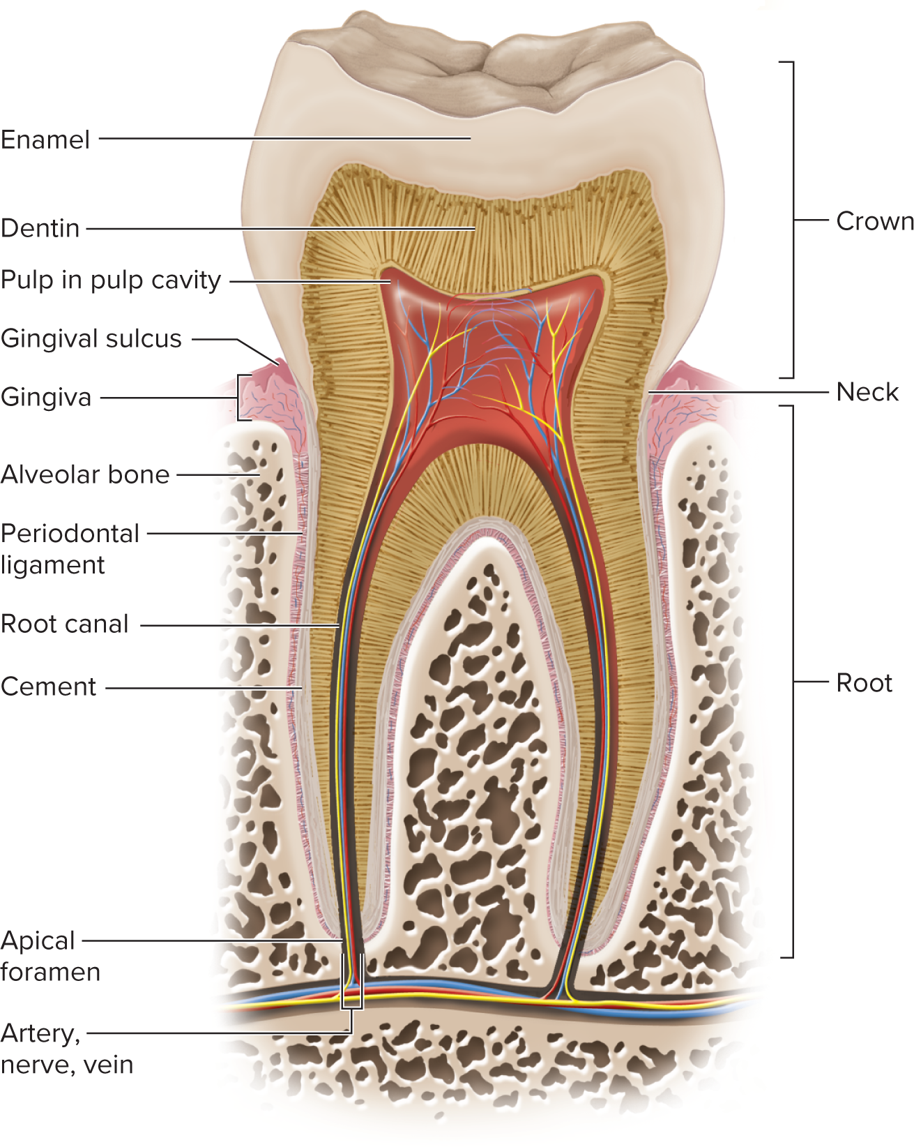
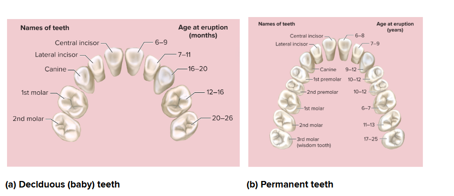
32 permanent teeth; 20 deciduous teeth, 16 in mandible, 16 in maxilla
Tooth regions: crown, root, neck; gingival sulcus; enamel covers crown/neck; dentin forms most tooth; cementum covers root; periodontal ligament anchors tooth in alveolus.
2 incisors—chisel-like cutting teeth used to bite off a piece of
food, 1 canine—pointed and act to puncture and shred food, 2 premolars—broad surface for crushing, shredding, and
grinding, 3 molars per side per jaw—even broader surface for crushing, shredding, and
grinding (total numbers vary by dentition stage).Alveolus (tooth socket in bone) and gomphosis joint (between tooth and bone; gingiva (gum covers alveolar bone).
Periodontal ligament—modified periosteum whose collagen fibers penetrate into the bone on one side and into the tooth on the other
Regions of a tooth:
Crown: portion above the gum, Root: the portion below the gum, embedded in
alveolar bone, Neck: the point where crown, root, and gum meet, Gingival sulcus: space between the tooth and theDentin: hard tissue that makes up the majority of the tooth beneath the enamel
Enamel: the hardest substance in the body, covering the crown of the tooth
Cementum—covers root (Cementum and dentin are living tissue and can regenerate)
Root canal—space in a root leading to pulp cavity in the crown (nerves and blood vessels) Apical foramen: pore at basal end of each root canal.
Pulp: the innermost part of the tooth containing blood vessels and nerves.
gum
Growth timeline: 20 deciduous teeth erupt 6–30 months; replaced by 32 permanent teeth between ages ~6 and ~25; 3rd molars (wisdom teeth) erupt 17–25 years and may be impacted.
The human mouth is home to more than 700 species of microorganisms, especially bacteria
Mastication (Chewing)
Function: breaks food into smaller pieces to be swallowed, mechanical digestion, increased surface area for enzymes.
Muscles involved: masseter, temporalis, medial and lateral pterygoids; cheek muscles (buccinator) and orbicularis oris aid in manipulating food.
Saliva and Salivary Glands
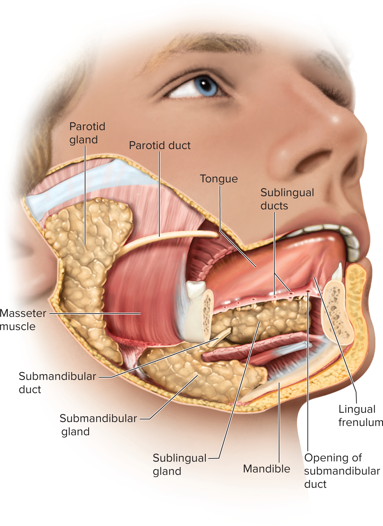
Functions: dampens mouth, begins starch/fat digestion, cleans teeth, inhibits bacteria, dissolves molecules for taste, binds food into bolus for swallowing.
Saliva composition: hypotonic solution of 97% to 99.5% water and the following solutes:
Salivary amylase: enzyme that begins starch digestion in the
mouthLingual lipase: enzyme that is activated by stomach acid and
digests fat after food is swallowedMucus: binds and lubricates a mass of food and aids in
swallowingLysozyme: enzyme that kills bacteria
Immunoglobulin A (IgA): an antibody that inhibits bacterial
growthElectrolytes: Na+, K+, Cl−, phosphate, and bicarbonate
pH: 6.8 to 7.0
Intrinsic glands (lingual in tongue produce lingual lipase, labial inside of lips, palatine roof of mouth, buccal inside cheek): constantly secrete saliva at a constant rate.
Extrinsic glands 3 pairs connected to oral cavity by ducts:
parotid: beneath the skin anterior to the earlobe
submandibular: halfway along the body of the mandible, empties at side of lingual frenulum near lower central incisors
sublingual: located in the floor of the mouth, multiple that empty posterior to papilla of submandibular duct
ducts channel saliva into mouth; mumps is inflammation of the parotid gland.
Histology of Salivary Glands
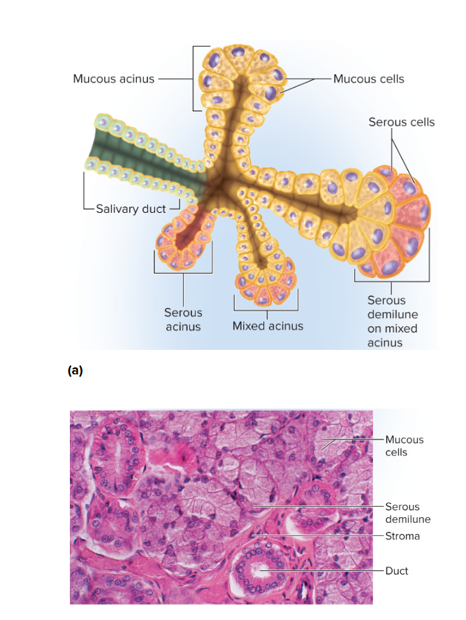
Compound tubuloacinar glands
– Branched ducts ending in aciniMucous cells secrete mucus
Serous cells secrete thin fluid rich in enzymes and electrolytes
Mixed acinus has both mucous and serous cells
Salivary physiology
Extrinsic salivary glands secrete about 1 to 1.5 L of saliva per day
Cells of acini filter water and electrolytes from blood and add amylase, mucin, and lysozyme
Salivation is regulated by salivatory nuclei in the medulla/pons; stimuli include presence of food, taste, smell, thoughts
parasympathetic stimulation yields copious enzyme-rich saliva
sympathetic yields less saliva with more mucus.
Bolus formation: saliva binds food into a soft, slippery mass.
The Pharynx and Swallowing
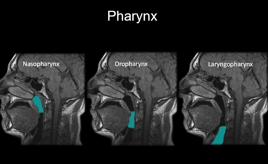
Pharynx: muscular funnel connecting oral/nasal cavities to esophagus and larynx; longitudinal muscles and pharyngeal constrictors propel food downward.
Swallowing center: nuclei in medulla oblongata coordinating swallowing; involves nerves V, VII, IX, XII.
Phases of swallowing:
Oral phase (voluntary): tongue forms bolus and pushes into oropharynx; epiglottis occludes airway.
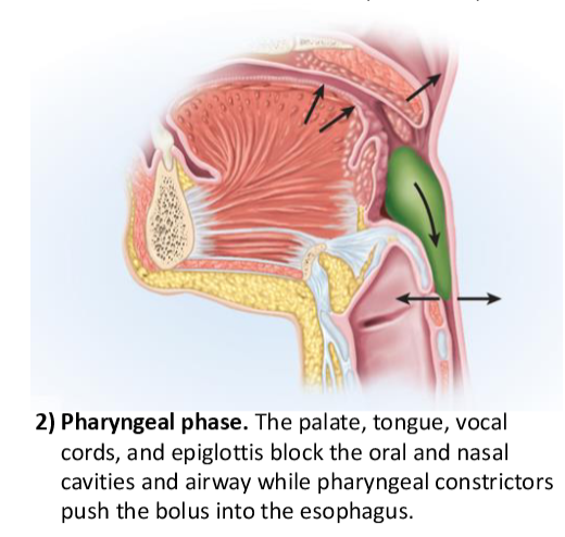
Pharyngeal phase (involuntary): constrictors push bolus downward; palate, tongue, vocal cords, and epiglottis block nasopharynx/mouth; airway protected.
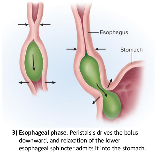
Esophageal phase (involuntary): peristalsis propels bolus to stomach; lower esophageal sphincter relaxes to admit bolus.
The Esophagus
Upper esophageal sphincter: separates the pharynx from the esophagus and regulate the passage of food into the esophagus
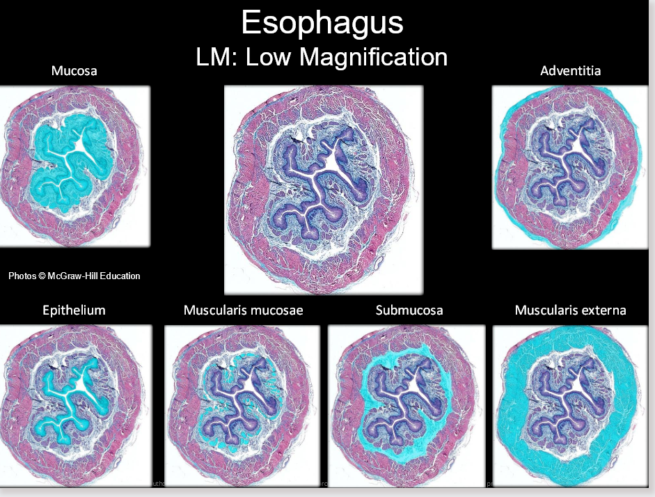
pharynx: muscular tube that connects the nasal cavity and mouth to the esophagus, playing a crucial role in swallowing and breathing.
Esophagus: straight muscular tube, 25–30 cm long; begins at C6, passes through diaphragm, ends at cardial orifice.
Extends from pharynx to cardiac orifice of stomach passing through esophageal hiatus in diaphragm
Lower esophageal sphincter: a ring of muscle at the junction of the esophagus and stomach, preventing the backflow of stomach contents into the esophagus.
Epithelium: nonkeratinized stratified squamous; submucosal glands secrete mucus.
Muscular layers: upper third predominantly skeletal muscle, middle mixed, lower third smooth muscle.
Adventitia rather than serosa.
Heartburn: reflux of stomach contents into esophagus.
Swallowing (deglutition)—a complex action involving over 22 muscles in the mouth, pharynx, and esophagus
Swallowing center: pair of nuclei in medulla oblongata that coordinates swallowing
Communicates with muscles of the pharynx and esophagus by way of trigeminal, facial, glossopharyngeal, and hypoglossal nerves
occurs in three phases: oral, pharyngeal, and esophageal
Oral phase: under voluntary control, food is manipulated within the mouth and pushed into the oropharynx by the tongue. epiglottis tips posteriorly and food bolus slides around it and into laryngopharynx
involuntary, prevents from reentering mouth or nasal cavity while breathing is suspended
25.3 The Stomach
a muscular sac in upper left abdominal cavity immediately inferior to the diaphragm
Primarily functions as a food storage organ
Internal volume of about 50 mL when empty
1.0 to 1.5 L after a typical meal
Up to 4 L when extremely full – can extend nearly as far as
the pelvis
Mechanically breaks up food, liquefies it, and begins chemical digestion of protein and fat
Chyme: soupy or pasty mixture of semi-digested food in
the stomach
Most digestion occurs after the chyme passes on to the small intestine
Gross anatomy
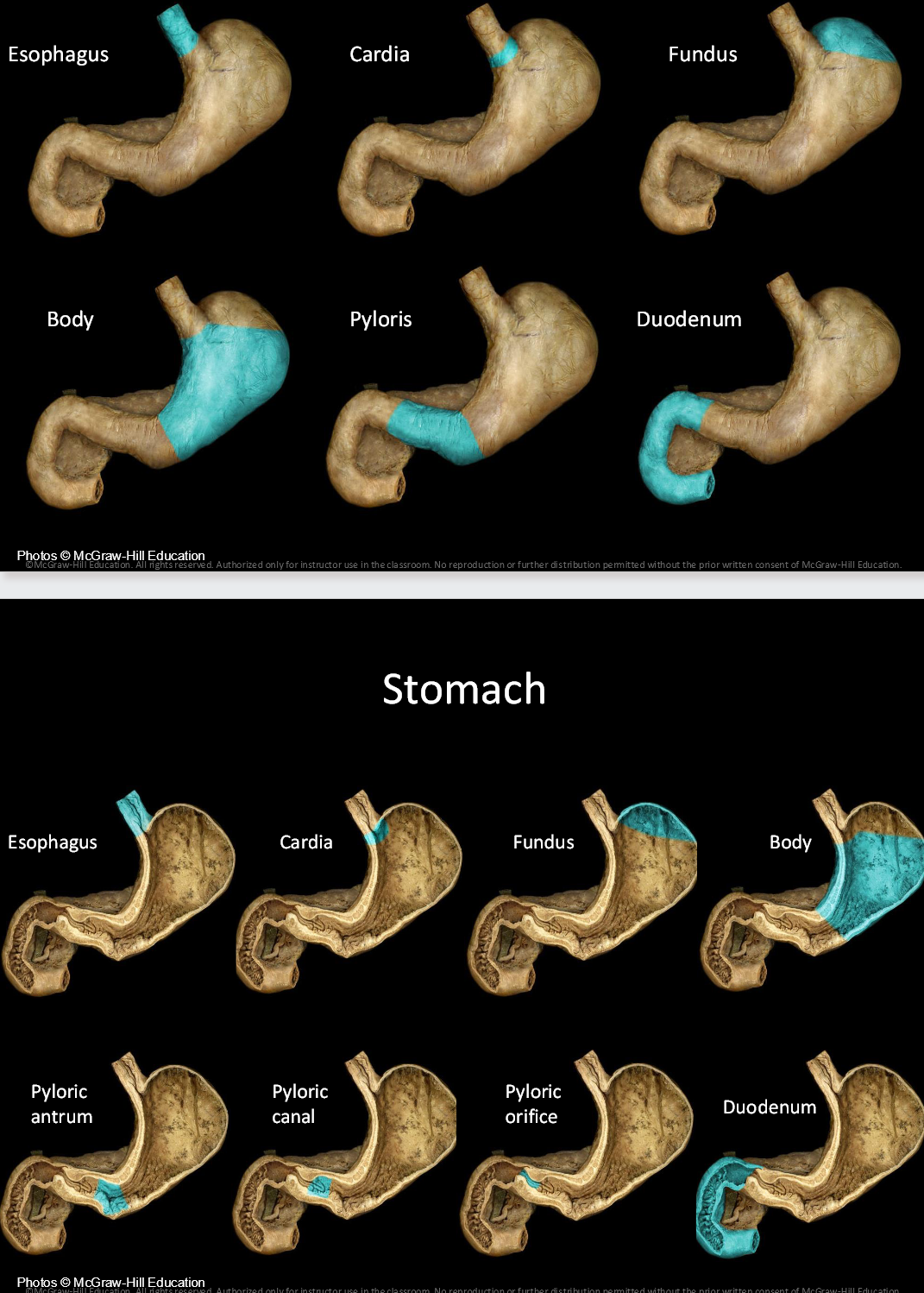
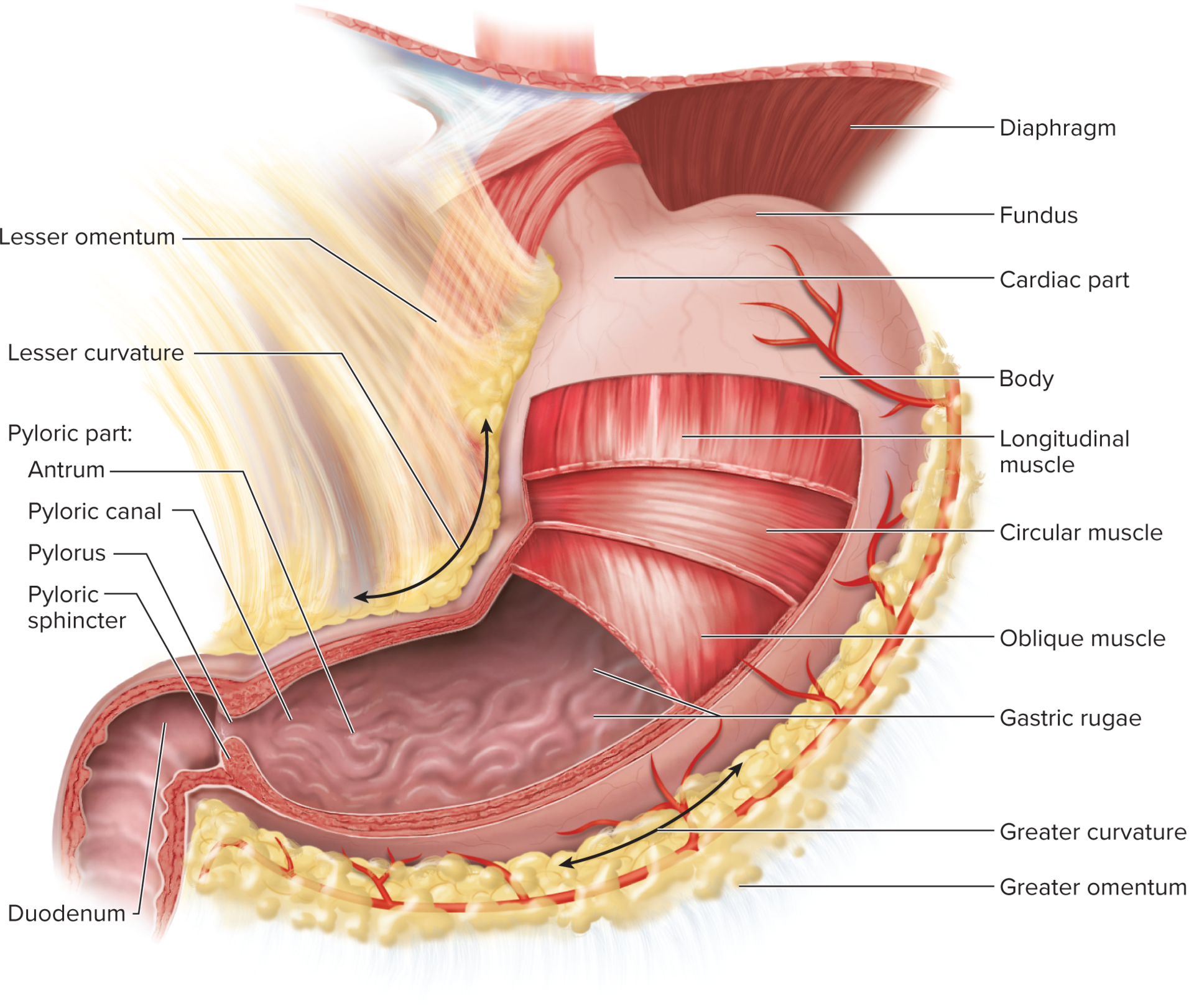
Stomach is a J-shaped sac in the upper left abdomen, inferior to the diaphragm.
4 Regions: cardial part (cardia), fundus, body (corpus), pyloric part (antrum and canal), pylorus leading to the duodenum.
Greater curvature (~40 cm) and lesser curvature (~10 cm); omenta attach to curvatures (greater and lesser).
pyloric region: This region plays a crucial role in regulating the passage of food from the stomach into the small intestine, where it becomes more digestible.
The pyloric sphincter acts as a valve, controlling the movement of chyme into the duodenum while preventing backflow into the stomach.
Stomach wall and muscle layers
Muscularis externa has three layers: inner oblique, middle circular, outer longitudinal.
Mucosa forms gastric pits and glands; surface epithelium is simple columnar; mucus secreting cells protect mucosa.
Gastric glands and cell types
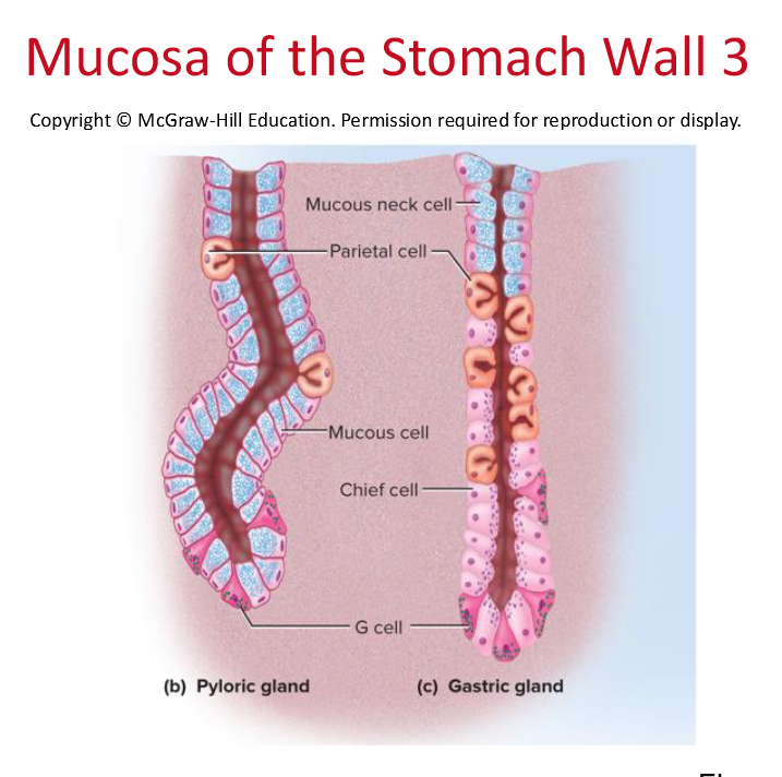
cardiac glands (cardial part): mucous cells.
Pyloric glands (pyloric part): mucous cells and endocrine cells.
Body/fundus gastric glands: mucous neck cells, regenerative (stem) cells, parietal cells, chief cells, enteroendocrine cells.
Parietal cells: secrete hydrochloric acid (HCl), intrinsic factor, ghrelin.
Chief cells: secrete pepsinogen and gastric lipase.
Mucous cells: mucus secretion; regenerative cells replenish epithelium.
Enteroendocrine cells: secrete hormones/paracrine signals that regulate digestion.
Secretions and their functions
Gastric juice: ~ ; mainly water, HCl, and pepsin.
mainly a mixture of water, hydrochloric acid, and pepsin
HCL (hydrochloric acid) creates an acidic environment that activates pepsinogen to pepsin, aiding in protein digestion and providing a barrier against pathogens.
Activates pepsin and lingual lipase; liquefies food; dissolves ferric ions to ferrous; contributes to nonspecific defense by destroying pathogens.
Pepsin (pepsinogen precursor): protease that begins protein digestion; activated by HCl; autocataylsis enhances activation.
Gastric lipase: minor role in fat digestion.
Intrinsic factor: essential for vitamin B12 absorption in the small intestine; stomach’s indispensable function; B12 involved in hemoglobin synthesis.
Hydrochloric acid secretion mechanism (gastric lumen)
first need chloride from circulatory system, and carbon dioxide to combine, forming bicarbonate in the parietal cells. This process creates a proton gradient that drives the secretion of gastric acid into the stomach.
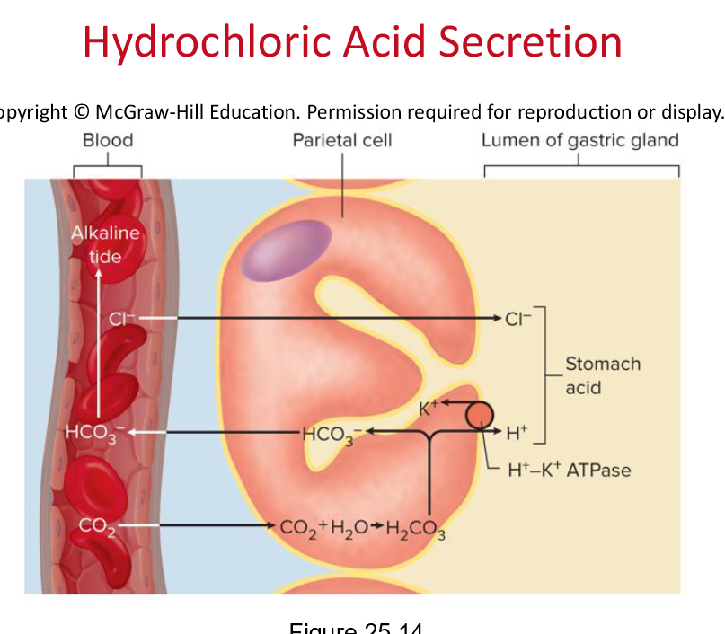
CO2 + H2O ⇌ H2CO3 ⇌ H+ + HCO3- via carbonic anhydrase.
H+ pumped into lumen by H+/K+ ATPase (proton pump); HCO3- exchanged for Cl- via chloride shift; Cl- joins with H+ to form HCl in lumen.
Alkaline tide: during digestion, bicarbonate concentration in blood rises.
Other gastric secretions
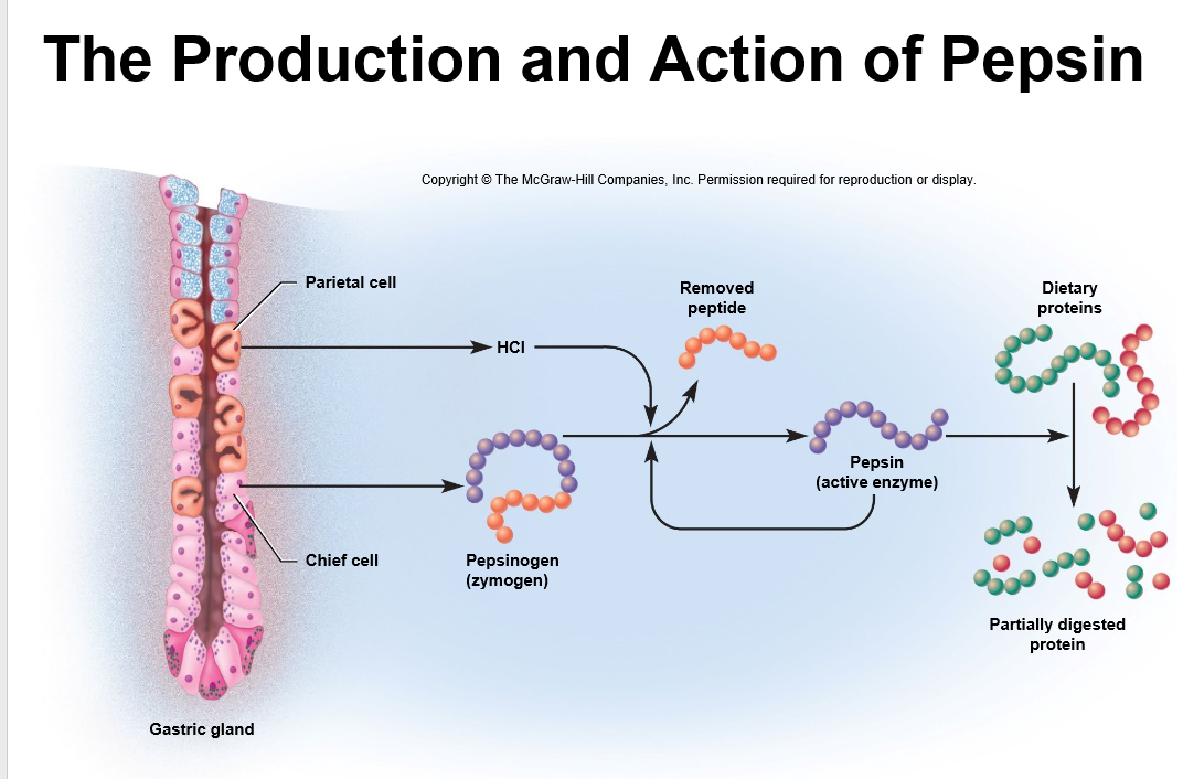
Pepsinogen activated to pepsin by HCl; autocatalytic conversion amplifies activation.
is used for the first stage of protein catabolism, breaking down complex proteins into smaller peptides, facilitating further digestion in the small intestine.
Pepsin: An enzyme produced in the stomach that plays a vital role in protein digestion by cleaving peptide bonds, allowing for the subsequent absorption of amino acids in the intestine.
Gastric lipase hydrolyzes triglycerides, releasing fatty acids and monoglycerides.
Intrinsic factor: a glycoprotein secreted by parietal cells essential for B12 absorption in ileum.
vitamin B12 is needed to synthesize hemoglobin
Secretion of intrinsic factor is the only indispensable function of the stomach
Gastric and pyloric glands have a variety of cells that produce a variety of chemical messengers
Most are hormones that enter blood and stimulate distant cells
Gastric protection and ulcers
Protective mechanisms: mucous coat, tight junctions, rapid epithelial turnover (3–6 days).
Breakdown of protection can lead to gastritis/peptic ulcers; often linked to Helicobacter pylori infection; antibiotics and supportive therapy used.
Regulation of gastric function
Nervous and endocrine control coordinate secretion and motility; three overlapping phases:
Cephalic phase: brain control prior to food intake; ~40% of acid secretion.
Gastric phase: stomach itself controls secretion; stretch and chemical stimuli increase secretion; about two-thirds of gastric secretion.
Short and long neural reflexes; histamine and gastrin also stimulate acid/enzyme secretion.
Intestinal phase: duodenum modulates gastric activity via hormones and reflexes; secretin/CCK/GIP; gastrin decreases; pyloric sphincter tightens to limit chyme entry.
Key chemical messengers:
Acetylcholine (ACh): stimulates parietal and chief cells; secretions during cephalic and gastric phases.
Histamine: paracrine from enterochromaffin-like cells stimulates acid secretion.
Gastrin: hormone from G cells in pyloric glands stimulates acid secretion.
Secretin and cholecystokinin (CCK): released by duodenum in response to chyme; regulate pancreatic/biliary secretion; influence gastric motility/secretion.
Gastric phases in detail
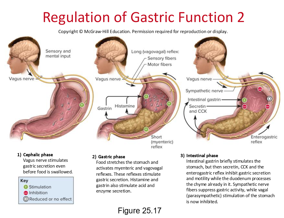
Cephalic phase: sight, smell, taste, or thought of food triggers hypothalamus → medulla → vagal stimulation → enteric nervous system → gastric secretion (≈ 40% of acid).
Gastric phase: distension and peptides in the stomach activate short (myenteric) and long (vagovagal) reflexes; histamine and gastrin amplify secretion; pH rises (pepsinogen activation) prior to food digestion.
Intestinal phase: chyme in duodenum triggers secretin/CCK and enterogastric reflex; gastric secretion/ motility initially rise, then inhibited; gastrin secretion declines; pyloric sphincter contracts to control chyme entry; GIP stimulates insulin release in preparation for nutrient absorption.
Important quantitative notes
Gastric juice production:
Pancreatic juice production:
Bile production: ~
Typical gastric capacity: up to about
Bolus volume entering duodenum per flow: ≈ per wave of chyme
Typical gastric emptying time: about for a meal; longer for high-fat meals (up to ~6 h)
Protection and pathology
Protective features prevent mucosal erosion by acid/pepsin; imbalance can lead to peptic ulcers.
Vomiting—forceful ejection of stomach and intestinal contents (chyme) from the mouth
Emetic center in the medulla oblongata integrates multiple muscle actions
Vomiting induced by:
Overstretching of the stomach or duodenum
Chemical irritants such as alcohol and bacterial toxins
Visceral trauma
Intense pain or psychological and sensory stimuli
Retching—thoracic expansion and abdominal contraction creates a pressure difference that dilates the esophagus
Vomiting—occurs when abdominal contractions and rising thoracic pressure force the upper esophageal sphincter to open
25.4 The Liver, Gallbladder, and Pancreas
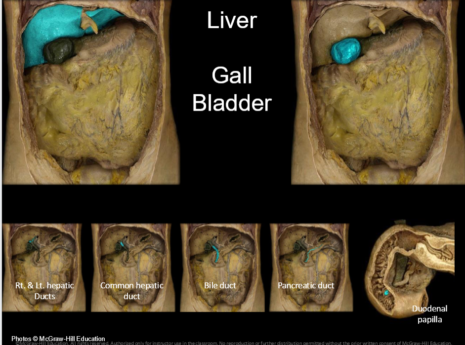
The Liver
Largest gland; located under the diaphragm; reddish-brown.
Functions include bile production, processing of nutrients, detoxification, synthesis of plasma proteins and hormones (e.g., albumin, angiotensinogen).
Gross anatomy: four lobes (right, left, quadrate, caudate); falciform ligament; round ligament (ligamentum teres).
Vascular supply: hepatic artery proper and hepatic portal vein; hepatic triads (branch of portal vein, hepatic artery, bile ductule).
Blood drainage: hepatic veins to inferior vena cava; bile flows through bile ducts to gallbladder or duodenum.
Gallbladder and bile ducts
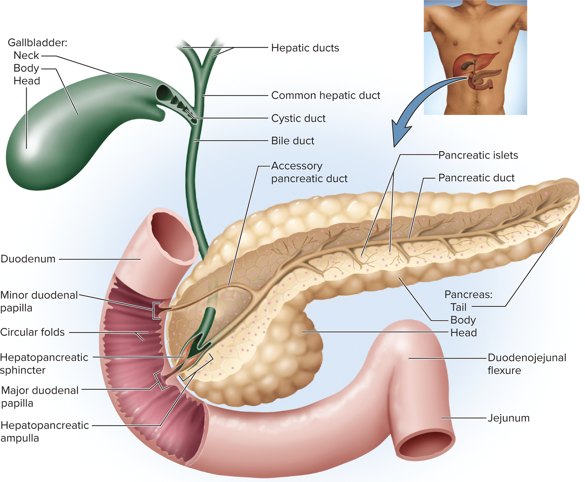
Gallbladder stores/concentrates bile; lined by mucosa; duct system includes cystic duct and common hepatic duct; bile flows to the duodenum via bile duct via hepatopancreatic ampulla (major duodenal papilla) controlled by hepatopancreatic sphincter (sphincter of Oddi).
Bile composition: bilirubin (pigment from heme), bile acids (bile salts) that emulsify fats, cholesterol, phospholipids; urobilinogen/stercobilin give urine/fecal color; gallstones can form from cholesterol/calcium bilirubinate crystals.
Enterohepatic circulation: ~80% of bile acids reabsorbed in the ileum and returned to liver; 20% excreted in feces; critical for cholesterol homeostasis.
The Pancreas
Both endocrine (islets of Langerhans) and exocrine (acini) functions.
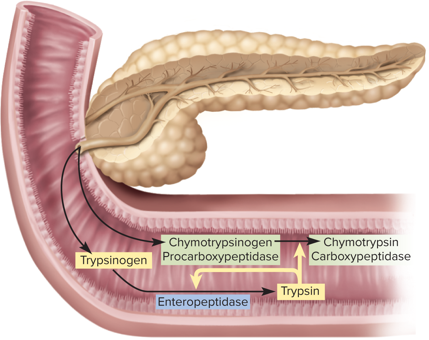
Exocrine pancreas secretes ~ of pancreatic juice daily; pancreatic juice includes enzymes (trypsinogen, chymotrypsinogen, carboxypeptidase, pancreatic amylase, pancreatic lipase) and bicarbonate-rich fluid.
Pancreatic duct runs the length of the gland; joins the bile duct at the hepatopancreatic ampulla and releases into the duodenum via the major duodenal papilla.
Pancreatic zymogens: trypsinogen (activated to trypsin by enteropeptidase in the small intestine); trypsin activates other zymogens (trypsin, chymotrypsinogen → chymotrypsin; procarboxypeptidase → carboxypeptidase).
Other pancreatic enzymes: pancreatic amylase, pancreatic lipase, ribonuclease, and deoxyribonuclease.
Regulation of secretion
Three main stimuli for pancreatic juice and bile release: acetylcholine (ACh), cholecystokinin (CCK), and secretin.
ACh: stimulates acini to secrete enzymes during cephalic phase; enzymes remain in acini/ducts until chyme reaches the duodenum.
CCK: released by duodenal mucosa in response to fats; stimulates pancreatic enzyme secretion and strongly stimulates gallbladder contraction and relaxation of hepatopancreatic sphincter to release bile into the duodenum.
Secretin: released by the duodenum in response to acidic chyme; stimulates duct cells of liver/pancreas to secrete bicarbonate; raises pH for enzyme activity.
25.5 The Small Intestine
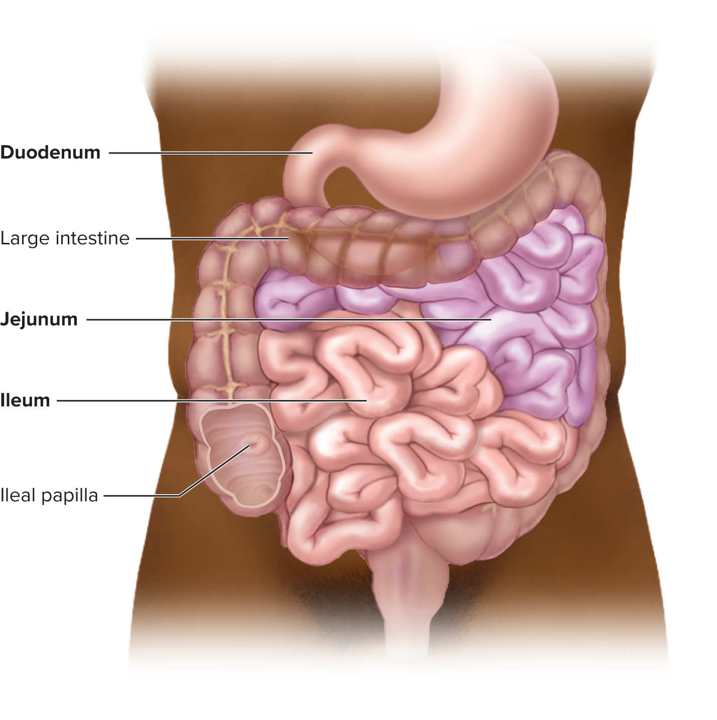
Gross anatomy
Primary site of chemical digestion and nutrient absorption; length ~ in a living person (up to ~8 m in a cadaver); diameter ~ .
Regions: duodenum (first ~25 cm), jejunum (roughly 40% of postduodenal length), ileum (last ~60%).
Duodenum receives chyme from stomach and secretions from liver/pancreas; major and minor duodenal papillae; duodenojejunal flexure marks transition to jejunum.
Jejunum: highly vascularized, thick walls; many circular folds (plicae circulares) and large villi; major site of digestion/absorption.
Ileum: thinner walls, fewer folds, Peyer patches (aggregated lymphoid nodules) prominent away from mesenteric attachment; ileocecal valve regulates flow into cecum; intraperitoneal with serosa.
Blood supply and lymphatics
Supplied by superior mesenteric artery (SMa) with 12–15 jejunal/ileal arteries.
Venous drainage to hepatic portal system via superior mesenteric vein.
Microscopic anatomy
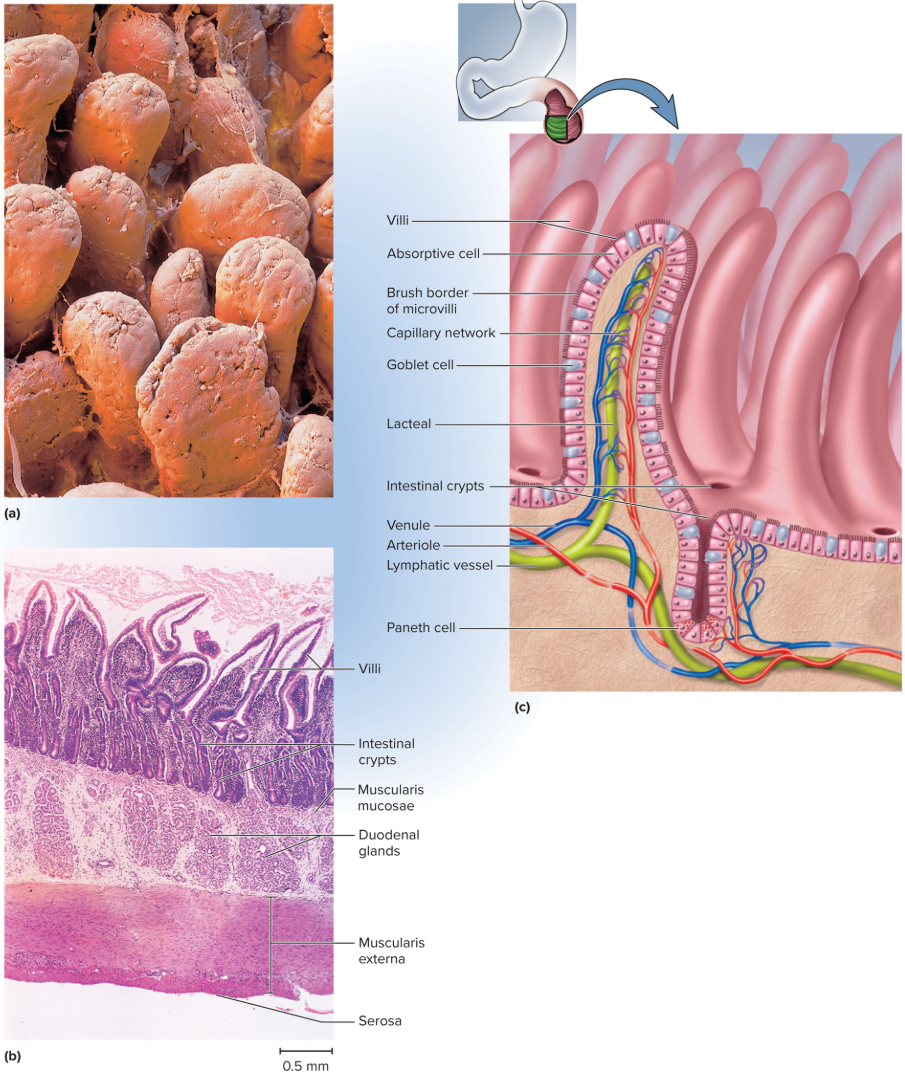
Lumen lined with simple columnar epithelium; mucosa/submucosa have circular folds, villi, microvilli, and brush border enzymes for contact digestion.
Villi: absorptive enterocytes and goblet cells; core lamina propria with capillaries and a lacteal (lymphatic) for lipid transport.
Microvilli: brush border enzymes (e.g., dextrinase, glucoamylase, maltase, sucrase, lactase) enabling final steps of digestion at the brush border; contact digestion requires chyme contact with mucosa.
Crypts of Lieberkühn: intestinal crypts with enterocytes, goblet cells; stem cells in lower halves replenish epithelium; Paneth cells secrete lysozyme, phospholipase, defensins.
Duodenal glands: mucus-rich bicarbonate secretions to neutralize stomach acid.
Intestinal surface area and absorption
Surface area amplification: circular folds (2–3×), villi (×10), microvilli (×20) → total amplification ~ × relative to a single flat surface.
Intestinal enzymes and absorption of macronutrients
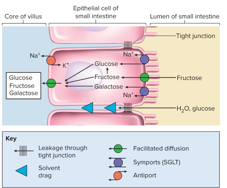
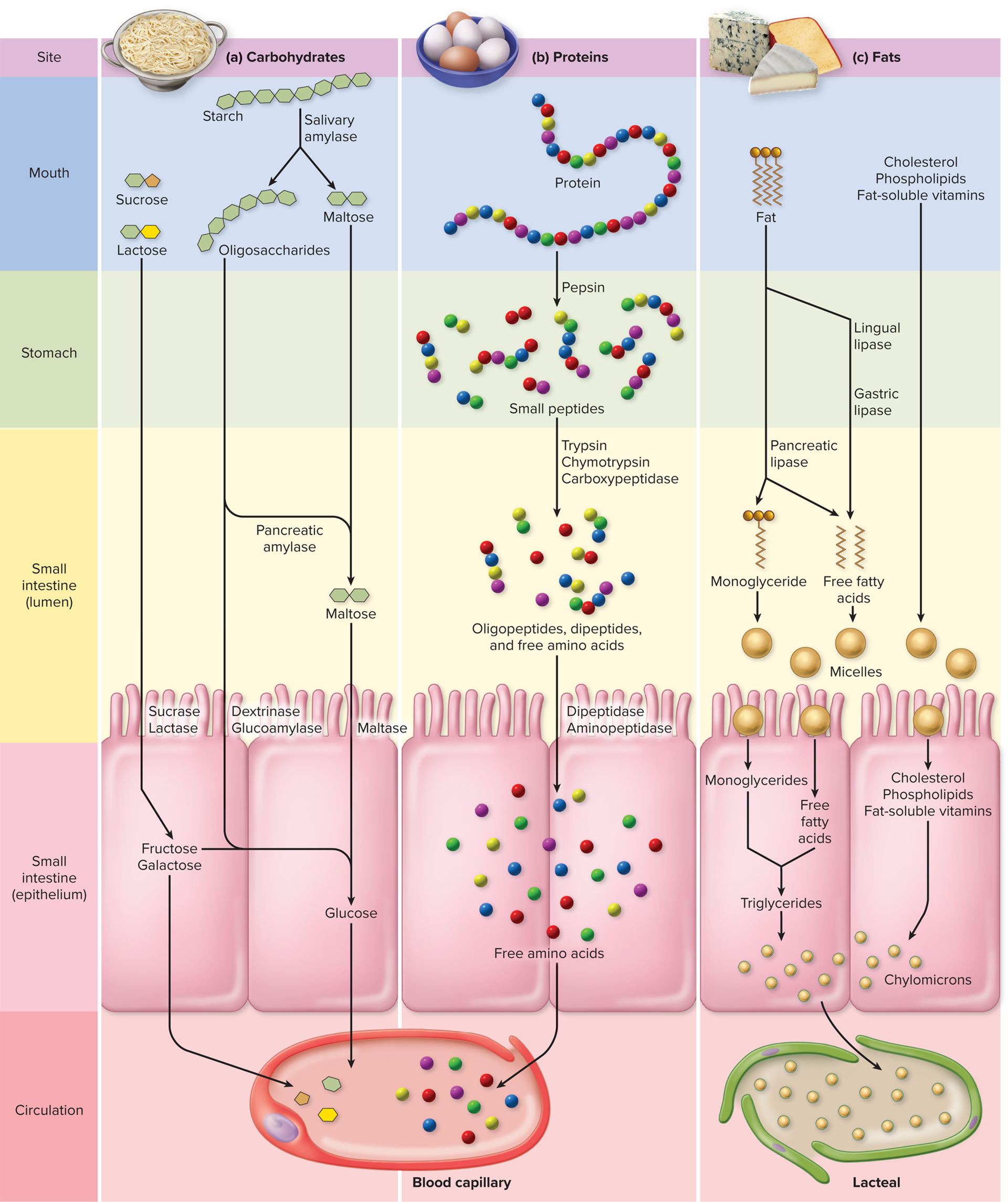
Carbohydrates: starch → oligosaccharides → disaccharides (maltose) → glucose; brush border enzymes complete digestion; monosaccharides absorbed via SGLT (glucose/galactose cotransport) and GLUT transporters; most absorbed sugars reach hepatic portal system.
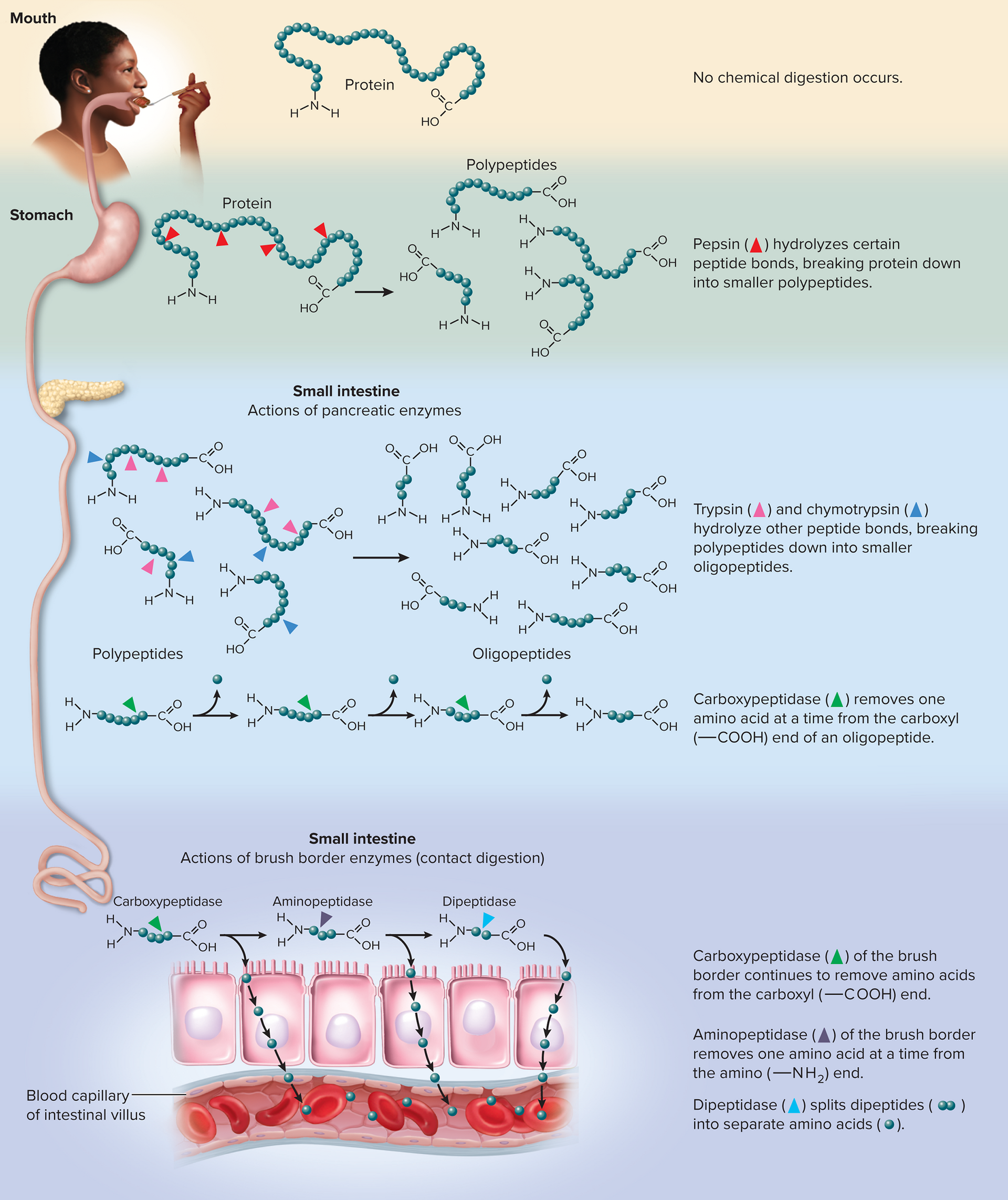
Proteins: pepsin acts in stomach; pancreatic proteases continue digestion in small intestine; brush border peptidases complete amino acid release; amino acids absorbed by Na+-dependent cotransport, then diffuse into blood.
Nucleic acids: pancreatic nucleases degrade DNA/RNA to nucleotides; brush border nucleotidases/phosphatases complete digestion.
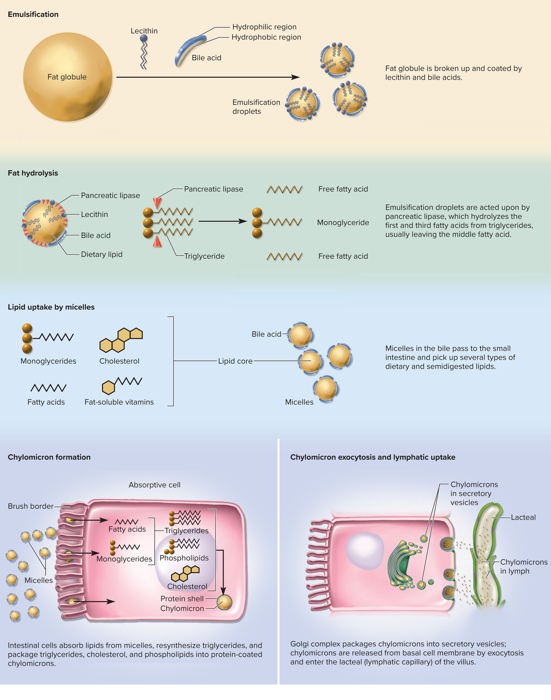
Lipids: emulsified by bile acids and lecithin; pancreatic lipase digests triglycerides to free fatty acids and monoglycerides; micelles ferry lipids to absorptive cells; chylomicrons formed in enterocytes and transported via lacteals in lymphatics.
Lipid digestion and absorption details
Lipases act on triglycerides; pancreatic lipase preferentially removes the first and third fatty acids, leaving a monoacylglycerol and two FFAs.
Micelles transport lipids to absorptive cells; lipids enter epithelial cells by diffusion and are resynthesized into triglycerides; packaged into chylomicrons for lymphatic transport.
Vitamins and minerals
Fat-soluble vitamins (A, D, E, K) absorbed with lipids; water-soluble vitamins absorbed by diffusion or specific transporters; Vitamin B12 requires intrinsic factor for ileal absorption.
Minerals: iron and calcium absorption regulated; iron absorption controlled by hepcidin; calcium absorption through transcellular and paracellular routes; parathyroid hormone and vitamin D regulate calcium homeostasis.
Water absorption and osmosis
The small intestine absorbs most of the water (part of 9 L/day intake). Water reabsorption follows osmotic gradients created by nutrient absorption; acidification/alkalinization of luminal contents influences water movement.
25.6 Chemical Digestion and Absorption
Carbohydrates
Starch digestion: starch → oligosaccharides → maltose → glucose.
Salivary amylase initiates digestion in the mouth; denatured by stomach acid; pancreatic amylase resumes in the small intestine; brush border enzymes complete hydrolysis to monosaccharides.
Lactose intolerance: deficiency in lactase leading to undigested lactose in colon; osmotic effects cause diarrhea; yogurt/cheese may be tolerated due to bacterial lactose metabolism.
Proteins
Digestion begins in the stomach with pepsin in acidic pH; continues in small intestine via pancreatic enzymes (trypsin, chymotrypsin, carboxypeptidase).
Brush border peptidases complete amino acid liberation; amino acids absorbed by cotransport and diffusion into bloodstream.
Lipids
Lipid digestion requires emulsification by bile acids and lecithin; pancreatic lipase acts on triglycerides;
Micelles transport lipids to absorptive cells and chylomicrons ferry lipids into lymphatic system.
Nucleic acids, vitamins, and minerals
Nucleases digest DNA/RNA to nucleotides; brush border enzymes finalize digestion.
Vitamins absorbed as described (A, D, E, K with fats; B and C water-soluble; B12 requires intrinsic factor).
Minerals absorbed along the small intestine with various mechanisms (Na+ cotransport with sugars/amino acids; calcium and iron absorption regulated by hormones).
25.7 The Large Intestine
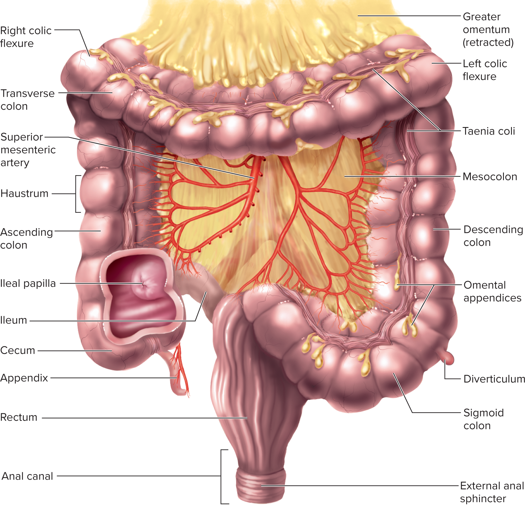
The large intestine, primarily responsible for water and electrolyte absorption, also plays a critical role in the fermentation of indigestible carbohydrates and the storage of fecal matter before excretion.
Gross anatomy and regions
Begins at the ileocecal valve as cecum; contains appendix.
Regions: ascending, right/hepatic flexure, transverse, left/splenic flexure, descending, sigmoid colon; rectum and anal canal at distal end.
Haustra and taenia coli produce segmental contractions and pouches; omental (epiploic) appendages are fatty protrusions along the colon.
Functional overview
Primary role: absorb water/electrolytes; form and store feces.
Large intestine hosts a large microbial population (gut microbiome) that digests cellulose and produces vitamins B and K.
Gas production (flatus) from bacterial activity; much gas is reabsorbed.
Absorption and motility
Transit through colon takes about 36–48 hours; most time in the transverse colon.
Haustral contractions occur roughly every 30 minutes to mix residue and enhance absorption.
Mass movements (1–3 times daily) propel feces toward the rectum; stimulus by gastrocolic and duodenocolic reflexes.
Defecation and neural control
Rectal stretch triggers intrinsic defecation reflex (myenteric) and parasympathetic defecation reflex.
External anal sphincter and puborectalis muscle must be voluntarily relaxed to defecate; abdominal Valsalva maneuver increases abdominal pressure to aid defecation.
Microbiota and health implications
Gut microbiome with ~800 bacterial species; aids in digestion, vitamin synthesis, and immune regulation.
Gas composition includes hydrogen sulfide, indole, skatole; some hydrogen gas may pose risk during certain surgical procedures if altered.
25.7 The Large Intestine (continued) — Key clinical and physiological notes
Microbes are essential for vitamin synthesis and metabolic activities; they can contribute to flatulence product and overall colonic health.
Disorders and clinical relevance: hemorrhoids, inflammatory conditions, and colorectal cancer risk relate to bowel habits, diet, and microbiota balance.
25.8 Regulation of Secretion and Digestion (brief mention)
Regulation of secretion involves nervous and endocrine systems; feedback from the small intestine modulates stomach activity via hormones and neural reflexes.
Hormones involved include gastrin, secretin, CCK, and GIP, which coordinate pancreatic/biliary secretions and GI tract motility.
Quick reference:
Gastric phases: cephalic ~40 ext{%} of acid secretion; gastric phase accounts for most secretion; intestinal phase provides negative feedback via secretin/CCK/GIP.
Milk and stomach protection: mucous coat, tight junctions, rapid renewal; turnover
Important clinical figures: cholelithiasis (gallstones); peptic ulcers; H. pylori infection; vomiting reflex mechanics; ulcerogenic potential of NSAIDs not detailed here but widely relevant.
Connections to foundational principles and real-world relevance
The digestive system integrates anatomy, physiology, microanatomy, and neuroendocrine signaling to convert dietary macromolecules into absorbable units and to regulate energy balance, hydration, and nutrient distribution.
The ENS exemplifies a semi-autonomous nervous system with reflexive capabilities, while CNS input modulates its activity—illustrating the brain-gut axis.
The liver, gallbladder, and pancreas coordinate with the small intestine to optimize digestion of fats, proteins, and carbohydrates, highlighting integration of exocrine secretions with digestive motility.
The peritoneal attachments (mesenteries, omenta) illustrate how anatomy supports organ mobility, protection, and vascular access, essential for understanding surgical approaches and abdominal pathophysiology.
Implications and practical considerations
Pathologies such as peptic ulcers, GERD, gallstones, pancreatitis, and malabsorption syndromes illustrate how disruptions in secretion, enzyme action, pH balance, or motility can impair digestion and nutrition.
The importance of microbiota in the large intestine highlights a broader view of health, immunity, and metabolic function.
Pharmacology frequently targets the GI tract (e.g., proton pump inhibitors, antacids, enzymes supplements), reflecting the central role of digestion in health and disease.