W6: Microbiology of the eye
6.1 Overview
Learning objectives:
Describe the basic anatomy and physiology of the eye
Describe the composition and diversity of the ocular microbiota
Describe the factors that influence the ocular microbiota
Describe how the ocular surface microbiota is altered in disease
6.3 Ocular Surface Microbiota
The ocular surface is a non-sterile, low-biomass anatomical site
The ocular surface structures is a mucosal site: it interacts with the external environment, providing significant exposure to particulate matter and infectious agents.
ocular surface harbours a low-biomass endogenous microbiota capable of performing competitive inhibition to protect against opportunistic infections.
Low biomass:
containing a low quantity of microbial cells or microbial genetic material.
Studies of the ocular surface estimate ~0.06 bacterial cells per conjunctival cell, compared to ~10 bacteria per gut epithelial cell
The ocular surface is one of the least colonised mucosal surfaces of the human body
competitive inhibition:
occurs where different microbial species compete for the same resources, such as nutrients or space, within a given environment.
The endogenous microbiota plays an important role in competitive inhibition against opportunistic infection by physical occupation of space, resource consumption, and the production of metabolites that may have antimicrobial effects on pathogens
Composition of the ocular microbiota
The composition of the ocular microbiota refers to the infectious agents that are present and their relative abundance.
Similar to other mucosal sites, the composition of the ocular surface microbiota may differ between individuals
however, there is emerging evidence of a core microbiome, consisting of infectious agents that are consistently found at the ocular surface of most healthy individuals.
The left eye and right eye harbour a mix of bacteria and fungi; composition of the ocular surface microbiota is not equal between the left and right eyes in some individuals
Some microbes are common across many/most individuals, whereas others are not
The core microbiota is present across most individuals:
At the genus level, the core ocular surface microbiota consists of two bacterial genera (Cutibacterium and Staphylococcus) and one fungal genus (Malassezia)
Cutibacterium spp. make up the largest portion of the microbial community
Major bacteria on the ocular surface
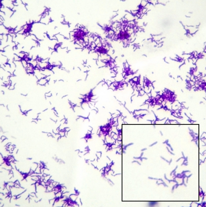
Cutibacterium spp.
Cutibacterium acnes is the most prevalent Cutibacterium species on the ocular surface, and is also a key component of the skin endogenous microbiota
Normally exist as commensals but can cause opportunistic ocular infections in susceptible hosts, such as chronic blepharitis or post-surgical endophthalmitis
Gram positive bacilli (rod-shaped)
Cutibacterium mastitidis on the ocular surface stimulates T cells in the conjunctiva to secrete immune molecules into the tear film, which provide protection against common ocular pathogens such as Pseudomonas aeruginosa and Candida albicans.
Staphylococcus spp.
Staphylococcus epidermidis is the most prevalent Staphylococcus species on the ocular surface, and is also a key component of the skin endogenous microbiota
Normally exist as commensals but can cause opportunistic ocular infections in susceptible hosts
Gram positive cocci (spherical-shaped)
S. epidermidis on the ocular surface stimulates epithelial cells to produce low-levels of antimicrobial peptides. This helps to maintain an 'immune vigilant' state at the ocular surface without triggering overt inflammation.
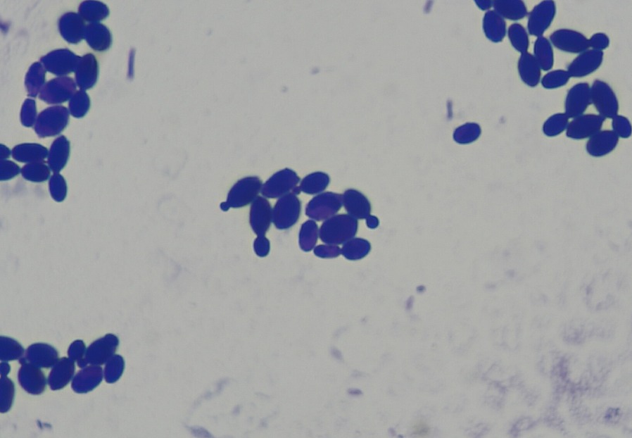
Malassezia spp.
Malassezia restricta is the most prevalent Malassezia species on the ocular surface
M. restricta is a lipophilic yeast typically associated with sebaceous-rich areas such as the lid margin but also the skin covering the scalp and face
M. restricta metabolises sebum-derived lipids, which may contribute to regulation and homeostasis of the ocular surface
Reproduces via budding
SUMMARY:
Notably, there is no consensus on the composition of the ocular surface microbiota. For example, some studies identified additional genera within the core microbial community and defined the core ocular surface microbiota as including the following genera:
Corynebacterium, Staphylococcus, Acinetobacter, Streptococcus, Pseudomonas, Cutibacterium and Bacillus
Diversity of the ocular surface microbiota
The diversity of the ocular microbiota refers to the variety of different microbes and how they are distributed.
The ocular surface microbiota typically has low diversity due to:
Antimicrobial molecules in tears
Mechanical clearance via blinking
Regional variation in the ocular surface microbiota
The ocular surface microbiota varies by region due to differences in glandular secretions, exposure to the environment, and tissue structure.
For example, the graphs below demonstrate that the composition of the ocular microbiota at the level of phylum differs between anatomical sites.
Methodology: how is the microbiota characterised:
Step 1: specimen collection
specimen from the body site of interest is collected.
for the eye this commonly involves collection of conjunctival swabs
a key challenge for ocular surface, where there is a low-biomass microbiota, is the potential from contamination from the environment or during the specimen collection process that may confound results.
Step 2: DNA extraction:
DNA is extracted from the collected specimen. The extracted material will contain DNA from host cells, as well as DNA from infectious agents that may be present.
Step 3: DNA sequencing
The extracted DNA is run through a machine, which generates the sequences of the DNA fragments that are present in the specimen.
This approach can be targeted to only sequence the DNA of a specific class of infectious agent. For example, 16S rRNA sequencing is performed to sequence the bacterial DNA sequences. Alternatively, all DNA within the specimen can be sequenced (metagenomics) but this is costly.
Step 4: Computational analysis
Once the DNA is sequenced, researchers use computer programs to match the sequences to known microbes in databases.
It is then possible to determine:
Which microbes are present
The relative abundance of each type
Note that the method of specimen collection and sequencing can greatly influence the results, therefore it can be challenging to compare microbiome studies.
6.4 Factors that influence ocular surface microbiota
As shown on the previous page, while a core ocular surface microbiota is thought to exist there are also additional members of the ocular surface microbial community that differ between individuals.
Host factors that influence ocular surface microbiota
Age: The ocular surface microbiota of infants and children exhibit increased diversity compared to adults. The composition shifts across lifespan.
sex: steroid-hormone dependent changes lead to changes in ocular surface microenvironment.
the female ocular surface microbiota has been reported to have a lower abundance of P.aeruginosa and S. epidermidis and higer abundance of E. coli compared to males.
Contact lens wear: Contact lens wear alters the diversity of bacterial genera within the ocular surface microbiome.
The effect of contact lens wear on these microbial communities depends on the type of contact lens, the duration of wear (e.g., daily wear versus extended wear), and the age group.
Topical ophthalmic therapy:
Topical antimicrobials can reduce the diversity of microbes within the ocular surface microbiota.
artificial tears lower the culture-positive rate of bacteria in the conjunctival sac, mainly because of physical wash-out.
Environmental and lifestyle factors that influence ocular surface microbiota
Geographical location: Differences in the abundance of community members of the ocular surface microbiota have been reported at species level based on climatic variations.
Humidity, UV exposure, and airborne particulates affect microbial community structure.
Eyeglasses and undertaking sporting activities: Associated with shifts in microbial diversity at the ocular surface, possibly due to airflow, sweat, and mechanical protection.
Alterations of the ocular surface microbiota in disease
disruptions in the ocular surface microbiota are closely linked to pathologies.
the ocular surface microbiota is altered in systemic conditions such as allergies and diabetes mellitus. These alterations are linked to increased susceptibility to ocular infection and inflammation.
Restoring balance to the ocular surface microbiota
Probiotic: live microorganisms; when consumed or applied in adequate amounts confer health benefits by restoring/maintaining microbial balance.
Prebiotic: Compounds that serve as a nutrition source for probiotics.
Topical application of probiotics and prebiotics may help reintroduce beneficial bacteria and suppress opportunistic pathogens, especially after topical antibiotic use or in conditions like dry eye, blepharitis, and allergic conjunctivitis.
Certain probiotic strains can enhance mucosal immunity by promoting cells to secrete antimicrobial molecues, supporting tear film stability and epithelial integrity.