Neurons and Neuronal communication: Synapses & synaptic transmission
Course Overview and Learning Objectives
Course Component: 4BBY1030 Cell Biology & Neuroscience 2024-2025.
Instructor: Dr. Philip R. Holland, Institute of Psychiatry, Psychology & Neuroscience (IoPPN), King's College London (KCL).
Primary Learning Outcomes:
- Describe the fundamental structure and physiological function of a synapse.
- Explain the mechanisms by which neurons communicate with each other across a synaptic junction.
The Scientific Concept and History of the Synapse
The Discovery of the Synaptic Cleft:
- Sir Charles Sherrington established the concept in the early 1900s.
- He discovered that pre-synaptic and post-synaptic neurons are not physically continuous but are separated by a physical gap known as the synaptic cleft.Chemical Transmission Discovery:
- Otto Loewi established in the 1920s that synaptic transmission across the cleft is mediated by chemical messengers.
- These messengers include chemical neurotransmitters and neuromodulators.
Structural Classification of Synapses
Anatomical Types Based on Connection Site:
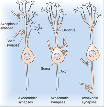
- Axodendritic Synapse: A connection between an axon terminal and a dendrite. (most common)
- Axospinous Synapse: A specific type of axodendritic connection where the axon connects to a dendritic spine.
- Shaft Synapse: A connection directly onto the dendritic shaft.
- Axosomatic Synapse: A connection between an axon terminal and the cell body (soma). synapse on cell body
- Axoaxonic Synapse: A connection between an axon terminal and the axon of another neuron. synapse at axonKey Neuronal Components Involved:
- Dendrite → Soma (Cell Body) → Axon
Mechanisms of Gap Junction Communication (Electrical Synapses)
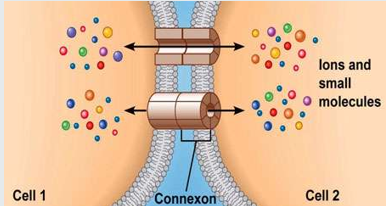
Role: communication between cells by having ion channels allowing ion exchange and movement
General Structure:
- Direct physical connection between Cell 1 and Cell 2.
- Molecules such as ions and small molecules pass through specialized channels called Connexons.
Synaptic communication:
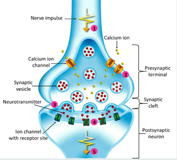
- Synaptic Cleft: The distance between neurons is approximately . Transformation of an electrical impulse into a neurotransmitter chemical in order to be reconverted back into an electrical impulse
- Axon Terminal: The presynaptic ending.
- Post-Synaptic Density (PSD): The protein-rich area on the post-synaptic membrane.
- Dendritic Spine: The receiving structure on the post-synaptic neuron.Step-by-Step Transmission Process:
1. Resting State: The synapse remains inactive until a signal arrives.
2. Action Potential (AP) Arrival: The nerve impulse (AP) reaches the presynaptic terminal.
3. Calcium Influx: Voltage-gated channels open in response to depolarization. ions enter the terminal.
4. Exocytosis: The entry of triggers synaptic vesicles to fuse with the membrane, releasing their neurotransmitter content into the synaptic cleft.
5. Diffusion: The neurotransmitter (NT) diffuses across the synaptic cleft.
6. Receptor Activation: NT binds to ion channels with specific receptor sites on the postsynaptic neuron and activates postsynaptic cell.
7. Postsynaptic Response: The opening of ion channels (e.g., ) leads to a change in membrane potential (graded potential).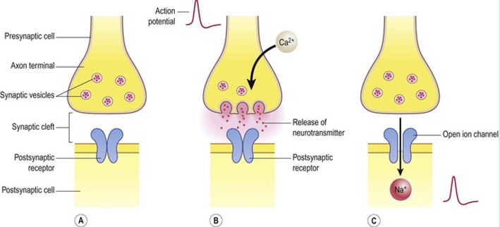
Graded Potentials and Summation
Definition of Graded Potentials:
- A wave of depolarization throughout the cytoplasm, potential will diminish quicklyTriggering an Action Potential:
- Occurs at the Axon Hillock.
- A neuron must be depolarized from its resting potential of to the threshold of to fire an AP.
- Peak AP voltage reaches approximately .
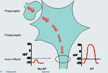
if only one synapse is firing impulses on a post synaptic nerve, it is less likely to reach the threshold to instigate an action potential however:
Types of Summation:
- Spatial Summation: multiple synapse firing at one nerve increases the chances of reaching the threshold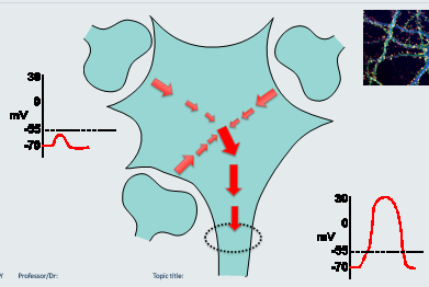
- Temporal Summation: one/two synapses firing at one nerve repeatedly in a short period increases chances of reaching threshold
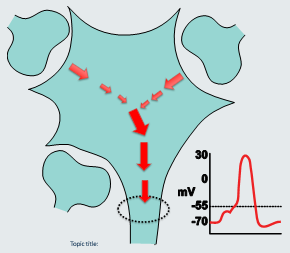
Post-Synaptic Potentials and Integration
Excitatory Postsynaptic Potential (EPSP):
Often mediated by the neurotransmitter Glutamate.
glutamate sent causes Na+ to entre post-synaptic neuron causing depolarization causing excitatory postsynaptic potential (inc chance of generating AP)
Involves the influx of ions.
Generates depolarization (making the neuron more likely to fire).
- Magnitude example: or .
Inhibitory Postsynaptic Potential (IPSP):
Often mediated by the neurotransmitter Gamma-aminobutyric acid (GABA)
GABA sent causes Cl- to enter post synaptic neuron causing hyperpolarization causing inhibitory post synaptic potential (dec change of generating AP)
Involves the influx of ions or efflux of ions.
Generates hyperpolarization (making the neuron less likely to fire).
Integration:
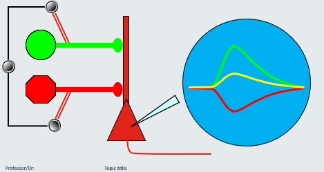
- EPSPs and IPSPs are sub-threshold events which determine whether a neuron will reach threshold to fire an action potential or not.
- They act to cancel each other out or add up to determine if the threshold for an action potential is reached.
Receptor Types and Kinetics
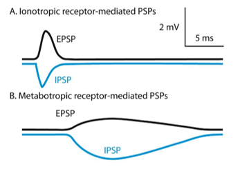
Ionotropic Receptors:
- These are ligand-activated ion channels.
- Kinetics: Fast-acting; they open and close quickly.
- Function: Directly generate post-synaptic potentials (EPSP via ; IPSP via or ).
- Typical Response Timing: Approx .Metabotropic Receptors:
- These are coupled to G-proteins.
- Kinetics: Slower acting compared to ionotropic receptors.
- Function: Generate longer-lasting and more varied cellular responses.
Frequency Coding and Neural Signaling
Mechanism:The strength of a stimulus is encoded by the frequency of action potentials, not their amplitude.
as intensity of a stimulus increases (e.g. greater mechanical pressure) the frequency rate of action potential increases (more action potentials are fired but to the same amount - all of nothing)
A graded potential in the trigger zone determines the frequency of firing.
Action potential peaks at while resting stays at .
Application (Example - Optogenetics):
- Blue Light: Can be used to open cation channels, leading to an influx of positive ions and depolarization.
- Yellow Light: Can be used to open channels, leading to an influx of negative ions and hyperpolarization.
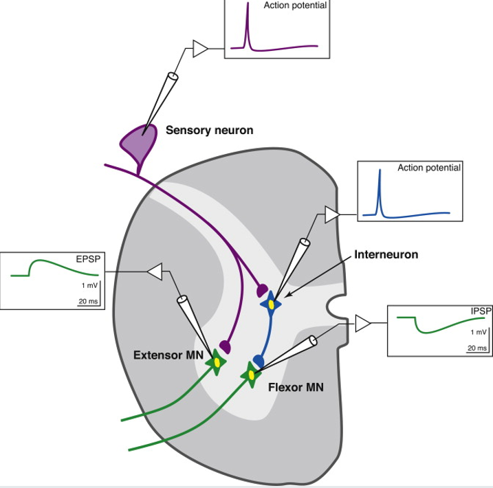
Network Communication: The Knee Jerk Reflex
Participating Anatomy:
- Sensory Neuron: Cell body located in the dorsal root ganglion. - located outside the CNS
- Motor Neurons: Extensor Motor Neuron (MN) and Flexor Motor Neuron (MN). located in the dorsal root ganglia in the spinal cord
- Interneuron: Connects the sensory signal to the inhibitory motor pathway. located in the spinal cord
- Muscles: Quadriceps (Extensor) and Hamstring (Flexor).
- Spinal Cord Structure: White matter and Gray matter.Reflex Action Potential Sequence:
tapping sends vibration to the patellar tendons in the quadriceps
detected by sensory neurons Sensory neuron fires an action potential.
This generates an EPSP (approx over ) in the Extensor Motor Neuron leading to contraction of the Quadriceps.
The sensory neuron also synapses with an inhibitory interneuron.
The interneuron fires an action potential, which generates an IPSP (approx over ) in the Flexor Motor Neuron, preventing the Hamstring from contracting.