Ch. 3 Biological Basis of Behavior
Overview
Neuroanatomy
Nervous System / Brain / Endocrine System
Genetics / Genetic Disorders
Neuroanatomy
The study of the parts and function of neurons.
Neurons are individual nerve cells that make up your central nervous system.
Every neuron is made up of discrete parts.
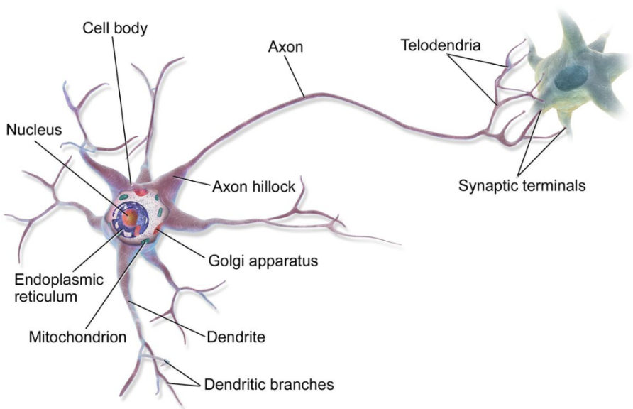
Neuroanatomy Terms
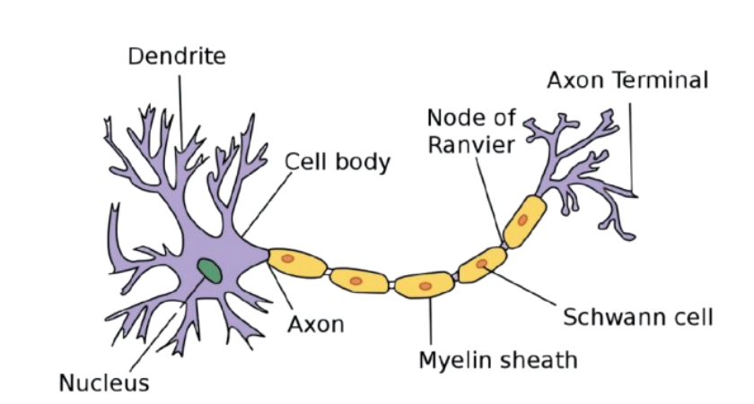
Dendrites - Rootlike parts of the cell that stretch out from the cell body. Grow to make synaptic connections with other neurons.
Cell Body - Contains the nucleus and other parts of the cell needed to keep it living (mitochondria, nucleus, Golgi apparatus, ribosomes, etc.)
Axon - Wire like structure ending in the terminal buttons that extends from the cell body.
Myelin Sheath - A fatty covering around the axon of some neuron.
Terminal Buttons - the branched end of the axon that contains neurotransmitters.
Neurotransmitters - Chemicals contained in the terminal buttons that enable neurons to communicate. Neurotransmitters fit into receptor sites on the dendrites of neurons like a key fits into a lock.
Nodes of Ranvier - The small gaps between the myelin sheath. These gaps are where neural transmission takes place on myelinated neurons.
Synapse - The gap between the terminal button and the dendrite.
How a Neuron Fires
Neural firing is an electrochemical process. Electricity travels within the cell from the dendrites to the terminal buttons.
Neurotransmitters travel between cells in the synapses of neurons. Electricity does not jump across the synapse; neurotransmitters travel across.
Action Potentials
Neuron has a slightly negative charge (-70mV).
Neurotransmitters from one neuron land on the dendrites of another neuron. The neurotransmitters fit into receptor sites.
If the threshold is reached, the neuron becomes permeable to positive ions and they rush down the neuron causing it to fire.
All-or-nothing principle - neurons either fire or don’t.
Neurotransmitters
Serotonin - involved in mood, sleep, pain, sensitivity, and arousal.
GABA - an inhibitory neurotransmitter.
Glutamate - an excitatory neurotransmitter.
Norepinephrine - involved in alertness/arousal.
Neurons (afferent, inter, efferent)
Afferent/sensory neurons - take information from our senses to the spinal cord.
Interneuron - found in the spinal cord, relays signals between (afferent) sensory neurons, and (efferent) motor neurons; involved in the process of sensory-motor integration.
Efferent neurons - conducting cells that carry information from the central nervous system (the brain and spinal cord) to muscles and organs throughout the body.
A subset of movements are controlled by direct transmission from afferent to efferent cells at the level of the spinal cord.
Reflexes: Quick and involuntary responses to environmental stimuli.
Central Nervous System (CNS)
The CNS is composed of the brain and spinal cord.
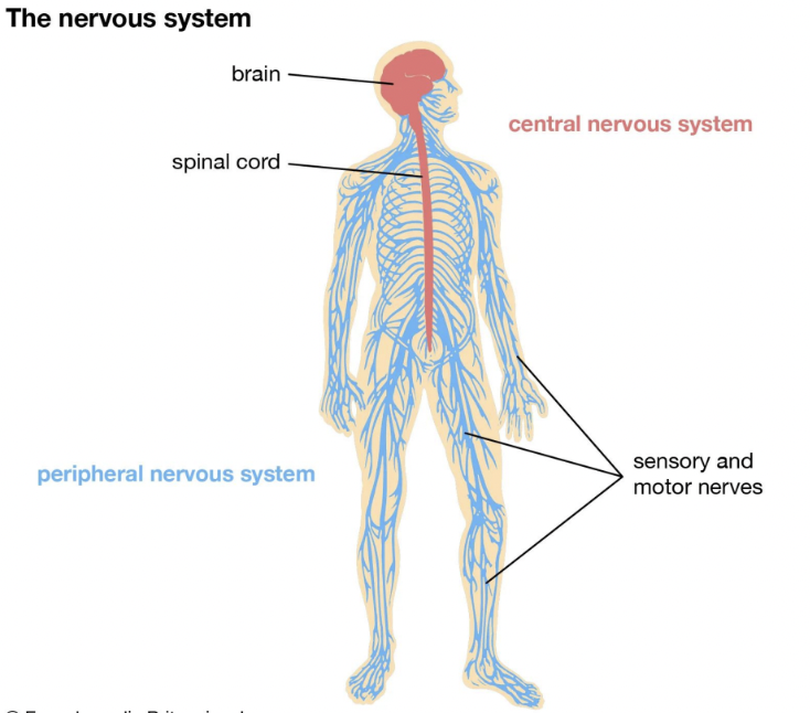
Peripheral Nervous System (PNS)
The PNS consists of all the other nerves in your body other than those in the brain and spinal cord.
Divided into two categories: somatic and autonomic.
Somatic Nervous System
The somatic nervous system controls our voluntary muscle movements. The motor cortex of the brain sends impulses to the somatic nervous system, which controls the muscles that allow us to move.
Autonomic Nervous System
The autonomic nervous system is responsible for control of the bodily functions not consciously directed, such as breathing, the heartbeat, and digestive processes.
The autonomic nervous system is divided into the sympathetic nervous system and the parasympathetic nervous system.
Sympathetic Nervous System
The sympathetic nervous system mobilizes our body to respond to stress. It is involuntary.
This part of our nervous system carries messages to organs, glands, and muscles. The sympathetic nervous system controls heart rate, blood pressures, respiration, and slows non-vital functions such as digestion and reproductive organs.
Also called the Fight or Flight System
Parasympathetic Nervous System
The parasympathetic nervous system is responsible for slowing down our body after a stress response.
The parasympathetic nervous system puts on the brakes to slow down the body’s autonomic nervous system.
Often referred to as the Rest and Digest System.
Studying the Brain
Ways we can study the brain:
Accidents, Lesions, Electroencephalogram, Computerized Axial Tomography, Magnetic Resonance Imaging, Positron Emissions Tomography, Functional Magnetic Resonance Imaging.
Accidents
Accidents give clues about brain function.
Famous case (1848) of a railroad worker, Phineas Gage who was hit by a piece of rebar in the front of his head. Having sustained damage to his frontal lobe, and having become highly emotional and impulsive after, doctors concluded that the frontal lobe was somehow regulating emotion.
Lesions
Lesion is the removal or destruction of the brain.
A famous example is doctors who lesioned mentally ill patients who had no other treatment options. Lesions give clues about function.
Researchers discovered that lesioning a part of the frontal lobe would make the patients calm and relieve some serious mental conditions.
Electroencephalogram (EEG)
EEG detects currents given off by the electrochemical reactions going on in the brain. Neurons use an electrochemical process to send signals.
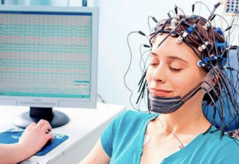
Researchers can examine what type of waves the brain produces during different stages of consciousness and use this information to generalize about brain function.
Widely used in sleep research.
Computerized Axial Tomography
CAT or CT scan is a sophisticated x-ray.
A CAT scan uses x-ray cameras that rotate around the brain and combine all pictures into a detailed three dimensional picture of the brain’s structure.
Magnetic Resonance Imaging (MRI)
The MRI is similar to a CAT scan. Determines the structure; gives a more detailed image than a CAT.
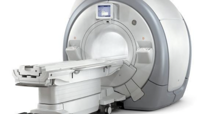
Uses magnetic fields to measure the density and location of brain material.
Does not expose patients to x-rays like a CAT scan.
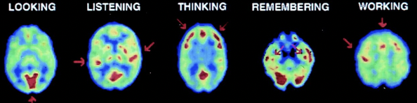
Positron Emissions Tomography (PET)
PET scans let researchers see what areas of the brain are most active during certain tasks. It will determine brain function.
In PET scans of the brain, a radioactive atom is applied to glucose (blood sugar).
If one area of the brain is using more glucose during some activity, it is a good indication that that area is associated with that mental state or that behavior.
Functional Magnetic Resonance Imaging (fMRI)
fMRI combines elements of MRI and PET scans.
An fMRI scan can show details of brain structure with information about blood flow to the brain, function.
Brain Terminology Overview
Hindbrain: Medulla, Pons, Cerebellum
Midbrain: Reticular Formation
Forebrain: Thalamus, Hypothalamus, Amygdala and Hippocampus, Cerebral Cortex.
Hindbrain
Consists of structures in the top part of the spinal cord.
Controls basic biological functions that keep us alive (respiration, heart rate, blood pressure).
Includes: Medulla, Pons, and Cerebellum
Medulla
Involved in control of blood pressure, heart rate, and breathing.
Also known as Medulla Oblongata and is located above the spinal cord.
Pons
Located just above the medulla and toward the front.
Connects the hindbrain with the midbrain and forebrain. Also involved in control of facial expressions.
Cerebellum
Located on the bottom rear.
Coordinates some habitual muscle movements such as tracking a target with our eyes.
Cerebellum means little brain, named this probably because it looks like a miniature brain.
Forebrain
Areas of interest in the forebrain:Thalamus, Hypothalamus , Amygdala, Hippocampus. These four areas make up the limbic system.
Thalamus
Located on top of the brain stem.
Responsible for receiving sensory signals coming from the spinal cord and sending them to the right areas of the forebrain.
Hypothalamus
Small structure below the thalamus.
Controls body temperature, sexual arousal, hunger, and thirst.
Also controls biological rhythms ( wake/sleep patterns).
Hippocampus
Looks like two arms surrounding the thalamus and is vital to our memory.
Memories are processed through the hippocampus and then sent to other locations in the cerebral cortex for permanent storage.

Research shows that memories or information must pass through this area first in order to be encoded.
Individuals with brain damage of the hippocampus are unable to retain new information.
If the hippocampus is damaged, old information may also be lost.
Amygdala
Very important to certain emotions like fear and aggression.
The thalamus, hypothalamus, amygdala, and hippocampus are grouped together and are called the limbic system.
Cerebral Cortex
Gray wrinkled stuff that is densely packed with neurons.
The surface of the cerebral cortex is wrinkled. The big wrinkles are called Fissures.
Fissures increase the availability of surface area. The more wrinkles, the more surface area contained within the skull.
The cerebral cortex is divided into two hemispheres: left and right.
The left hemisphere gets sensory messages and controls the motor function of the right half of the body. The right hemisphere gets sensory messages and controls the motor function of the left half of the body. This is called contralateral control.
Hemispheric specialization refers to the different and specific functions performed by the two hemispheres of the brain.
Split brain patients, patients whose corpus callosum has been cut to treat epilepsy, cannot orally report information only presented to the right hemisphere, since the spoken language centers of the brain are usually located in the left hemisphere.
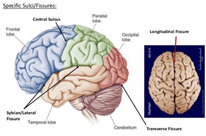
Cerebral Cortex: Frontal Lobe
The frontal lobe is a large area of the cerebral cortex located at the top front part of the brain.
Within the front lobe, and the very, is the prefrontal cortex, and is believed to be essential in directing thought processes.
The prefrontal cortex is said to act as the brain’s central executive. Researchers believe this part of the brain to be responsible for abstract thought and emotional control.
Broca’s Area - in the frontal lobe and is responsible for controlling the muscles involved in producing speech.
Damage to Broca’s area might leave one unable to make the muscle movements needed for speech.
Language Areas of the Cortex
Wernicke’s Area - located in the temporal lobe.
Wernicke’s area interprets both written and spoken speech. Damage to this area affects our ability to understand language.
Damage to Wernicke’s area may result in our speech lacking syntax and grammatical structure.
Parietal Lobe
Located behind the frontal lobe and at the top of the brain.
The parietal lobe contains the sensory cortex aka the somatosensory cortex.
The sensory cortex is a thin vertical strip that receives incoming touch sensations from the rest of our body.
Sensory and Motor Cortex
The motor cortex is the region of the cerebral cortex involved in the planning, control, and execution of voluntary movements.
The sensory cortex, like the motor cortex, is organized top down: the top of the sensory cortex receives sensations from the bottom of the body, progressing down the cortex to the bottom, which processes signals from our face and head.
Temporal Lobe
The temporal lobe processes sound sensed by our ears. Sound waves are processed by the ears, turned into neural impulses, and interpreted in our auditory cortices.
Occipital Lobe
At the very back of the brain, farthest from the eyes.
The major function of the occipital lobe is to interpret information from our eyes in our visual cortex.
Impulses from the retinas in our eyes are sent to the visual cortex to be processed.
Endocrine System
The endocrine system is a set of glands that secrete hormones that affect many different biological processes in the body.
Controlled by the hypothalamus. The hypothalamus produces separate hormones that stimulate or inhibit hormone production in the pituitary gland. The pituitary gland is known as the master gland.
Endocrine System: Adrenal Glands
The adrenal glands produce adrenaline (epinephrine) which signals the fight or flight response.
This causes the autonomic nervous system to increase heart rate, breathing, and blood pressure. It prepares the body for action.
Stressful situations cause the pituitary gland to release adrenocorticotropic hormone, which stimulates the adrenal gland.
The adrenal medulla secretes epinephrine (adrenaline) and norepinephrine (noradrenaline). These initiate the fight or flight response.
Endocrine System: Reproductive Glands
Women’s ovaries and men’s testes produce sex hormones. Women produce estrogen in their ovaries and men produce testosterone in their testes.
The different levels of these hormones explain some differences in behavior like aggression.
Genetics
Many traits, like body shape, introversion, and height result from the combination of nature (genetic code) and nurture (environment).
Every human cell contains 46 chromosomes in 23 pairs.
The genetic material that makes up chromosomes is called deoxyribonucleic acid (DNA).
Segments of DNA, called genes control the production of specific proteins that control some human traits.
A genotype comprises all of the possible combinations of genes. A phenotype is the observable trait
Genes can either be dominant or recessive.
A dominant trait is more likely to be expressed than a recessive trait.
Genetics: Twins
Identical twins develop from one fertilized egg called a zygote.
Research by Thomas Bouchard says that monozygotic twins given up at birth showed that IQ does depend slightly on the environment in which they are raised.
But IQ of separated twins are still highly correlated, so genetics still play a role in IQ.
Effective psychological environment - when people treat twins in similar ways bc of their similar physical traits.
Chromosomal Abnormalities
Sometimes chromosomes do not combine normally.
Turner’s syndrome is where the infant only has a single X chromosome.
Babies and children with Turner’s generally have different traits such as shortness, webbed necks, and differences in physical sexual development.
Klinefelter’s syndrome is when males have an extra X, resulting in XXY.
Sexual development is different and personality like extreme introversion occurs.
Down syndrome: babies born with down syndrome are born with an extra chromosome on the twenty first pair.
This results in a rounded face, shorter fingers and toes, slanted eyes set far apart, and intellectual difficulties.