RENAL SYSTEM
renal system 1 - lecture 15
OVERVIEW OF KIDNEY FUNCTION
in general animals have a tube-like structure ( composed of multiple cell types)
that’s involved in ion balance and fluid balance
vertebrates have multiple roles in homeostasis :
ion balance
osmotic balance
blood balance
pH of the blood
excretion of unwanted products
hormone production
strong link between the cardiovascular system + renal system
EVOLUTION OF RENAL SYSTEM
ancient aquatic animals use cellular transport, maintaining differences between ICF and sea water ( e.g. by ion pumps)
development of an OUTER EPITHELIAL LAYER was a major evolutionary step (ecf separated from the external environment)
through evolution increasingly complex mechanisms emerged for generating differences between the ecf and the sea water (external environment)
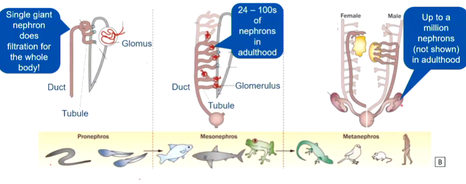
three renal structures are observed during vertebrate evolution and development 😀
DEVELOPMENT
( structures get progressively more complicated during development)
pronephros
(simplest kidney)
blunt endings to tubes + cilia presence
mesonephros
first bowman’s capsules appear
( c shaped cup) which increases SA
metanephros (kidney)
emergence of ureter
many smaller nephrons clustered in kidney
EVOLUTION
through evolution there is an increase in numerous nephrons ( more efficient filtration units)
endothermy is the ability of an organism to regulate its body temperature internally, maintaining a constant temperature regardless of external conditions.
with the shift to endothermy, the mass of kidney remains consistent even when we take into account the body size
the size essentially makes sense in all organisms.
an increase is seen in the filtration + reabsorption rates
nephrons are more densely packed in more complex organisms
birds and mammals show the greatest efficiency in filtration and reabsorption rates
renal cells initially evolved to transport nutrients but developed additional functions
EVOLUTION OF THE NEPRHON
the basic structure of the nephron is largely conserved between species
birds contain both reptilian and mammalian type of neurons
all mammals have a loop of Henle, but the length of the loop of Henle varies through species ( longer in species that are in harsh conditions)
the basic nephron structure in vertebrates =
filtering unit (glomerulus capsule)
proximal + distal tube
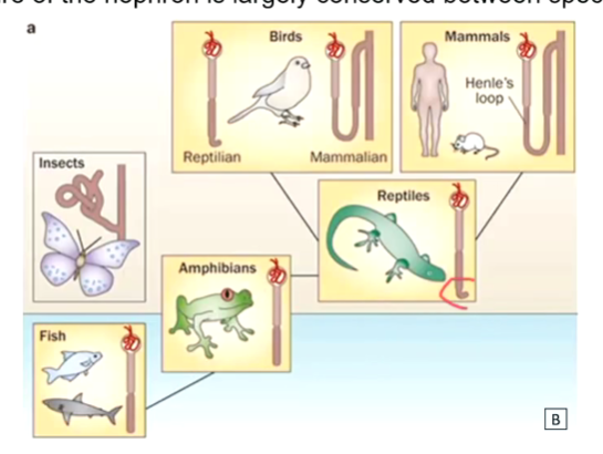
salt transport in different classes
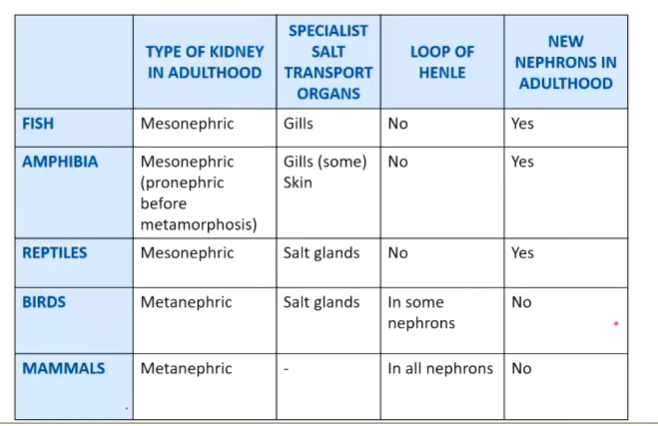
mammals - no additional salt transport organs
INTRODUCTION TO THE RENAL SYSTEM
kidneys are critical for maintaining mammalian extracellular fluid composition
the nephron is the key functional unit
nephrons have a complicated structure
regions are specialized for different functions
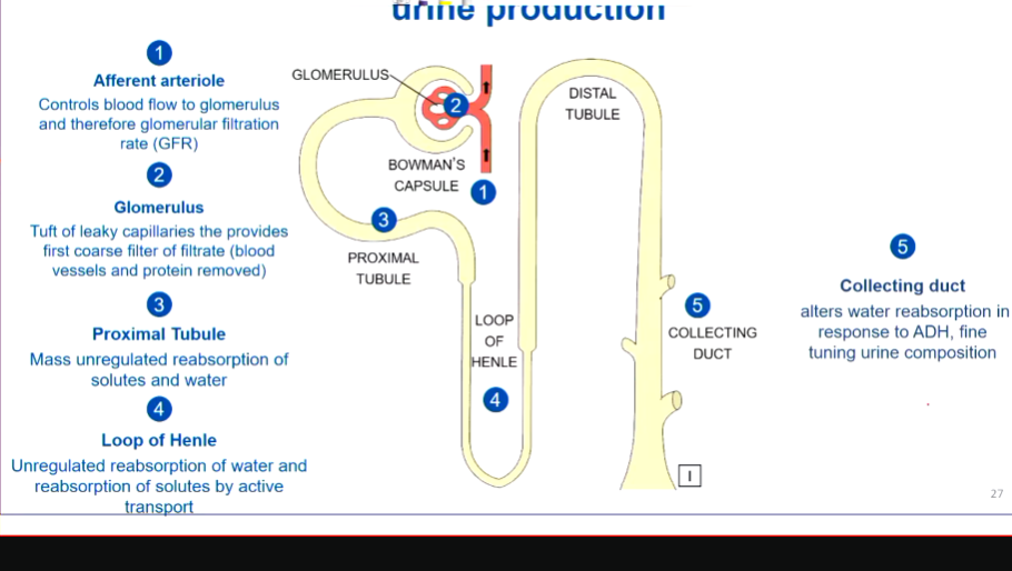
INTRODUCTION TO THE NEPRHON
lots of parts here
the filtration rate is determined by the afferent arteriole + efferent arteriole because of the pressure gradient they have
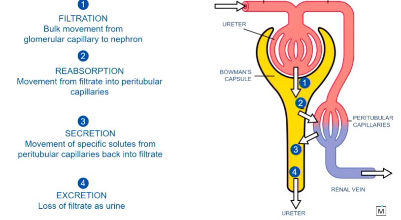
the space between the glomerulus and glomerulus capsule = mass filtration ( everything except cells, blood cells + proteins will get into your kidney)
not a fine process, think of it as a massive recycling point
the loop of Henle region all about creating gradients that help to conc urine
tweaking of filtration to get osmolarity happens at the end section
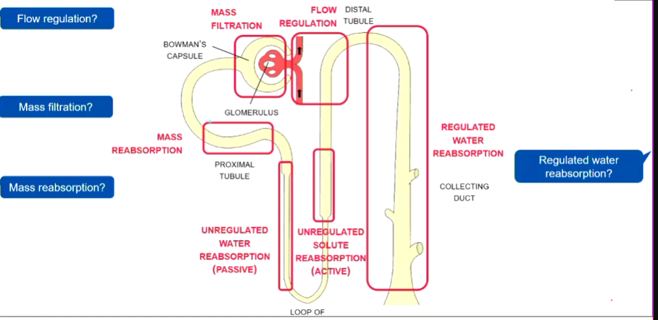
flow regulation - the diameter of the tube, arterioles ( packed with smooth muscle ), ideally suitable - pressure gradients
regulated water absorption - presence of aquaporins
mass filtration - more villi, more membrane, more blood vessels all clustered up tightly knotted = surface area
mass reabsorption - thin membrane, surface, convoluted (windy) tubule = gives surface area, mitochondria as energy is needed to move substances around, leaky gap junctions
renal system 2 - lecture 16
structure and function in the nephron
filtration at the glomerulus
lots of blood vessels connected to BC
filters proteins ( because they are negative + too big ) + cells
glomerular capillaries are very leaky
covered in podocytes
specialized epithelial cells
have “feet” connected by slit junctions - part of filtration barrier
gfr - glomerular filtration rate
determined by:
permeability of the filtration barrier (only changes in disease)
surface area for filtration ( only change during development)
glomerular filtration pressure (changes day to day, variable in the short term)
first two aspects don’t change too much.
forces contributing to filtration pressure
filtration = the movement of fluid from glomerulus → bowman’s capsule
filtration across the glomerulus is determined
hydrostatic pressure (pressure exerted by fluid) - promotes fluid moving into the capsule
oncotic pressure ( pressure exerted by water moving down its concentration gradient)
there is always a net filtration ( fluid movement into nephron) in a healthy kidney
Water pressure is lower in Bowman's capsule than in the capillary due to the following reasons:
Filtration Process: In the glomerulus, blood pressure forces water and solutes out of the capillaries into Bowman's capsule, creating a filtration pressure.
Resistance: The efferent arteriole creates resistance, maintaining higher pressure in the capillaries.
Volume and Space: Bowman's capsule has a larger volume and space, leading to a decrease in pressure compared to the confined space of the capillaries.
This difference is crucial for effective filtration in the kidneys.
bulk reabsorption of the proximal tubule
most reabsorption in the nephron happens here
several factors increase permeability and or transport
dense capillary network nearby
winding structure
extensive microvilli
high numbers of mitochondira
leaky “tight” junctions
many + diverse transporters
Differences between Symport and Antiport:
Symport:
Transports two different substances in the same direction across a membrane.
Example: Glucose and sodium ions in intestinal cells.
Antiport:
Transports two different substances in opposite directions across a membrane.
Example: Sodium-potassium pump (Na+/K+ pump).
Key Points:
Both are types of cotransport mechanisms.
Symport moves substances together, while antiport exchanges them.
introduction to the loop of Henle
enables production of small volume of conc urine
recovers solutes from filtrate
descending - permeable to water but not salt
ascending - impermeable to water + active transport of salt ← needs a lot of energy
whether it stays dilute or more conc depends on the collecting duct
water reabsorption in the descending loop of henle
water moves down its electrochem grad through aquaphorins
water conc depends on conc of all dissolved solutes
water follows the solutes
if there is a higher solute conc outside the tubule there is a higher water conc inside the tubule
water will flow out of tubule
filtrate becomes more conc deeper in the loop of Henle
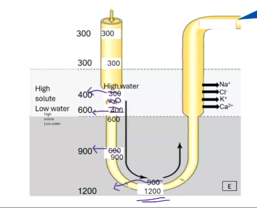
active solute reabsorption in the ascending loop of henle
alot of salt taken out to provide a dilute conc
thick ascending limb expresses mnay transporters Na+ CL from tubule back into the blood
depends on K+ recylcling because - transporter will only will if all ions are present