2026-04-15_GI/Renal Anatomy Introduction
Overview of the Digestive System
Functions:
- Responsible for:
- Mechanical breakdown of food
- Chemical breakdown of food
- Absorption of nutrients
- Elimination of wasteComponents:
- Gastrointestinal (GI) tract
- Also known as the alimentary canal
- A convoluted cylindrical tube
- Allows passage of food material as it is progressively digested
- Lumen of the tract considered external to the body
- Analogy: Think of the lumen as the "donut hole."
- Accessory organs
- Attached through ducts to GI tract
- Secrete substances into the lumen that assist with digestion
Learning Objectives
By the end of this session, students will be able to:
- Describe the organization of the abdominal wall, including:
- Fascial and muscle layers
- Rectus sheath
- Discuss the organization of the inguinal canal:
- Contributions from myofascial layers
- Explain embryological events involved in the development of the abdominal cavity
- Describe the arrangement of the peritoneal lining of the abdominal cavity
- Identify various regions of the alimentary canal and trace the path of a bolus through the digestive tract
- Describe the anatomical features and location of accessory digestive organs and their roles
- Identify major unpaired arteries supplying the digestive system and explain blood flow through portal circulation
Abdominal Quadrants and Regions
Quadrants of the abdomen:
- Right Upper Quadrant
- Left Upper Quadrant
- Right Lower Quadrant
- Left Lower QuadrantAbdominal Regions:
- Right hypochondriac
- Epigastric
- Left hypochondriac
- Right lumbar
- Umbilical
- Left lumbar
- Right iliac
- Left iliac
- Hypogastric
Anterolateral Abdominal Wall Layers
Fascia:
- Camper's Fascia:
- Superficial layer of fat
- Scarpa's Fascia:
- Deep membranous layerMuscular Layers:
- External Oblique:
- Internal Oblique:
- Transversus Abdominis:
- Rectus Abdominis:All three layers receive innervation from intercostal nerves 7-11
Thoracolumbar Fascia
**Layers of fascia (provide support to lumbar spine, act as force transmission between upper and lower limbs)
- Anterior layer
- Middle layer
- Posterior layer
Anterior fascial sheath
Components:
- Anterior Rectus Sheath
- Posterior Rectus Sheath
- Arcuate Line
- Transversalis FasciaPeritoneum:
- Parietal peritoneum: Lines the internal surface
- Visceral peritoneum: Lines many abdominal organs
Inguinal Canal
Key Structures:
- Deep Inguinal Ring
- Superficial Inguinal Ring
- Inguinal Ligament
- Fascia Layers:
- External Spermatic Fascia
- Internal Spermatic Fascia
- Cremasteric FasciaHesselbach's Triangle:
- Bound by the Inguinal Ligament, Transversalis Fascia, and Inferior Epigastric Vessels
Hernias
Types:
- Indirect Inguinal Hernia
- Direct Inguinal Hernia
- Femoral Hernia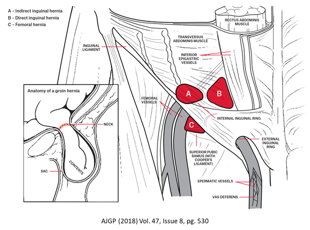
Development of the Abdominal Cavity
Embryological Development:
- Formation of the Gut Tube
- Components:
- Yolk sac
- Amniotic cavity
- Surface ectoderm
- Parietal mesoderm
- Visceral mesoderm
- Formation of Dorsal MesenteryKey Structures Developed:
- Heart, Liver bud, Midgut, Hindgut, Cloacal membrane
Peritoneum and Peritoneal Cavity
Peritoneum:
- Serous membrane lining the abdominal cavity
- Types:
- Parietal peritoneum: Lines the internal surface
- Visceral peritoneum: Lines abdominal organs
- Intraperitoneal organs: Completely enveloped by visceral peritoneum
- Retroperitoneal organs: Situated against the posterior abdominal wall, only lined along their anterior surfaceBilateral Structures:
- Lesser Omentum, Mesentery, Greater Omentum
The Alimentary Canal
Overview:
- Responsible for:
- Mechanical and chemical breakdown of food
- Absorption of nutrients
- Elimination of waste (defecation)Stages of Digestion:
- Ingestion of food
- Propulsion of ingested material
- Digestion (both mechanical and chemical)
- Absorption
- Elimination of wasteStructural Features:
- Convoluted cylindrical tube
- Divided based on anatomical and histological structures
Histology of the Alimentary Canal
Layers:
- Mucosa: (divided into three components)
- Epithelium
- Lamina Propria consists of areolar connective tissue
- Muscularis Mucosa thin layer of smooth muscle
- Submucosa:composed of areolar, dense irregular connective tissue, large concentration of vasculature, ANS supply, lymphatic system
- Muscularis: thin layer of SM, produces the contractions that generate peristalsis, composed of inner circular layer and outer longitudinal layer, autonomic myenteric nerve plexus sandwiches between inner circular and outer longitudinal layerds.
- Adventitia (or Serosa): outer cover of areolar tissue
Sections of the Digestive System
Esophagus:
- Z-line: Visible line where the esophagus transitions into the stomach
- Lower esophageal sphincter
- Made up of non-keratinized stratified squamous epithelium
•Upper 1/3 – 100% skeletal
•Rapid contraction – quick emptying of pharynx to accommodate breathing
•Middle 1/3 – skeletal/smooth muscle blend
•Lower 1/3 – 100% smooth
•Slow contraction – can take 5 – 8 seconds for swallowed food to enter stomach
Stomach:
- Cardiac part: continuous with the esophagus
- Angular notch
- Pyloric canal and sphincter
- Rugae•surrounded externally by 3 layers of smooth muscle
•outer longitudinal layer
•intermediate circular layer
•innermost oblique layer
•Fundus - superior region of stomach; may be compacted in fasted state, but enlarges after feeding to accommodate yet-to-be digested food
•Body - largest portion of the stomach; diameter of tube decreases and gastric rugae increase; main propulsion area
•Pylorus - narrow terminal portion of the stomach
Small Intestine:
- Regions:
- Duodenum (begins at the pylorus)
- Jejunum (begins at the duodenaljejunal junction): longest part of the small intestine
- Ileum
- Features of Jejunum and Ileum:
- Jejunum: Thick walls, prominent plicae circulares
- Ileum: Thinner walls, less prominent plicae circulares
- Duodenal Papilla: Entry point for bile and pancreatic juicesLarge Intestine:
- Features:
- Teniae coli, haustra, semilunar folds, vermiform appendix•large intestine divided into five regions
•cecum - pouch-like proximal region of the large intestine
•recieves digested material from the ileum through the ileocecal valve (superior and inferior frenula close off the valve when the cecum is distended)
•worm-shaped pouch (vermiform appendix) projects from the inferior portion of the cecum; normally wrapped underneath the intestine, but may sit in a variety of positions
•ascending colon - runs superiorly from the cecum to the right (hepatic) flexure
•retroperitoneal structure (reabsorbed during fetal development)
•transverse colon - runs from the right (hepatic) flexure to the left (splenic) flexure
•lies intraperitoneal, surrounded by the transverse mesocolon
•descending colon - runs inferiorly from the left (splenic) flexure to the lower left quadrant
•in this region, the colon once again becomes retroperitoneal
•sigmoid colon - named for its curved appearance; curves medially, then superiorly to form the rectum at the rectosigmoid junction (marked by termination of teniae coli); occurs in the midline at the level of S3
Accessory Digestive Organs
Salivary Glands:
- Parotid: Found superficial to facial bones anterior to eaer
- Submandibular: found inferior to ramus of madible
- Sublingual: found in anterior floor of oral cavityLysozyme: antimicrobial properties to inhibit bacterial growth
Salivary amylase: enzyme that initiates breakdown breakdown of complex carbohydrates
Liver:
Divided into left and right halves by the falciform ligament, terminates at the ligamentum teres
- Structure:
- Right Lobe, Left Lobe (inferior surface of left lobe is two lobes): posterior caudate and anterior quadrate
- Vascular supply:
- Left and right hepatic arteries, hepatic artery proper, common hepatic artery
Gallbladder: Connected to the liver via cystic duct and common bile duct
Pancreas:
- Anatomical regions: Head, body, tail, uncinate process
- Duct system: Accessory pancreatic duct, common pancreatic duct, major duodenal papillaSpleen:
- Structure: White pulp, red pulp, sinusoids, trabeculae
Vascular Supply and Venous Drainage
Arterial Supply to the Digestive System:
- Unpaired arteries:
- Celiac trunk, superior mesenteric artery, inferior mesenteric arteryVenous Drainage:
- Portal vein: It receives blood from the gastrointestinal tract
- Major tributaries include:
- Superior mesenteric vein
- Inferior mesenteric vein
- Splenic vein
Conclusion
The anatomy of the digestive system involves complex structures with various roles in food processing, nutrient absorption, and waste elimination. Understanding the organization and function of these components is essential for the study and practice of medicine in the field of gastroenterology.