PSYCH 261 Week 8: Module 5.2, 5.3, 6.1
VISUAL PROCESSING IN THE BRAIN
okay so first were gonna break down the mammalian visual system
so in the retina, we have rods and cones
these make synapses with both horizontal cells and bipolar cells
horizontal cells make INHIBITORY contact onto bipolar cells, and those ibpolar cells make synpases onto AMACRINE and ganlgion cells
alllllll cells within eyeball
buuut tthen we get axons of the ganglion cells, which form the optic nerve
the optic nerve leaves the retina adn travels along the lower surface of the brain, until optic nerves from both eyes meet at the OPTIC CHIASM
in the optic chiasm, half axons from each eye cross to oppsoite sides of the brain
infomraiton from the nasal half of each eye crosses to the contralateral hemisphere (cross hemispheres when nearing to eye!!)
information from the temporal half tho it goes to the same hemisphere
so axons closest to the nose cross hemispheres, and axons closest to the temporal lobe go to the opposite hemisphere
the percentage of crossover varies from one species to another depending on the location of the species eyes!!!
if theyre on the sides of their heads, most go to opposite sides
lateral geniculate nucleus
THIS IS WHERE MOST OF THE OPTIC NERVE GOES TO
geniculate —> genu (knee), genuflect, bend the knee, shape of it mustve looked like a knee
other axons go from the eye to the superior colliculus (in the midbrain!! imp for eye movemnets), and to hpart of hypothalamus implicated in waking-sleeping schedulate
lateral geniculate sends input to other parts of the thalamus and visual cortex
axons from the cortex go back to the thalamus to modify thalamic activity
STOP AND CHECK
where does the optic nerve start and end?
it starts at the ganglion cells in the retina, which form to make the optic nerve, and it ends at teh optic chiasm? or at the otptic chiasm it becomes the otpic tract so it ends at the lateral geniculate? or acc the optic nerve goes all the way to the primary visual cortex?
PROCESSING IN THE RETINA
the human eye includse a quarter billion receptors
you need to extract meaningful patterns from it
lateral inihbiton
retinas way of sharpening contrasts to emhasize borders of objects
basically what happens is the receptors send emssages to excite nearby bipolar cells, but they also send messages to horizontal cells that slightly INHIBIT Tthose bipolar cells, but also their neighbours
so bcz of the net inhibitoin i nthe middle, we have more contrast between the illuminated area and its darker surround
not sure how this works lol
actually light striking rods and cones DECREAES input, and recptors make INHIBOITRY synapses onto bipoar cells
soooooooooo light actually decreases inhibitory ioutput
decreased inhibiton —> net excitation
so ltes think of receptors output as excitation of the bipolar cells!!!
but they do this through inhibiting net excitation!!
in teh fovea
each cone attaches to just oen bipolar cell
green arrows in diagram tell us whats being excited, and the width of an arrow indicates how much ecitation
ex. receptor 8, excited bipolar cell 8. also excites a horizontal cell that INBHBITS bipolar cells. so biplar cell 8 has NEXT EXCITATION bcz the excitatory snapes soutwegihs the effects of the ohrizontal cells inbition
(like gaining clookies nad losing a smallern umber, gain 5 lose 2, still ahve 3!!)
MODULE 5.3
SPECIALIZED VISUAL PROCESSES
we tend to assume that anyone who sees an object sees everything about it, but one part of brain ses shape, antoher colour, another location, another movements
after localized brain damage, its possible to perceive certain aspects of an object and not others
THE VENTRAL AND DORSAL STREAMS
the primary visual cortex (v1) sends info to teh secondary visual cortex (v2)
v2 processes info further and transmits to additinal areas
receptive fields in v2 longer than those in v1, and v2 has mnay cells that respond to corners, texture,s complex shapes
v2 and v3, some cells highly responsive to colour, others highly responsive to disparity btwn left and right eyes (super important for stereoscopic depth perception)
v4, cluster of cels responsive to specific colours, green, orange, and purple
when you imagine a coloured object, v4 is area that responds most strongly
researchres distinguish btwn vetnral and dorsal stream
ventral stream through TEMPORAL cortex
for the “what” pathway
this is good for identifying and reocgnizing objects
dorsal stream through PARIETAL cortex
its for the ‘where” and “how”
bcz importance of visually guided movements
distinction abased on mri and fmris on animals nadobservaitons of patients iwth brian damage
woman exposed to carbon monixide,caused damage to ventral stream, could not name objects she saw, recognize faces, distinguish square from a rectangle
when asked to put a rectangle through a slot, couldnt say if it was horizontal or vertical but she could put an envelope through it fine
also when asked to guess tehe size of an boject, she was gucci
when asked to pick up object, reached out correctly
man with temporal lobe damage
could not desribe location of objects, but did avoid obstacles as he tawalked!!
ppl with temporal lobe damage
use vision to guide their actions, but ehy have trouble identifying what the objects are (these are the “what” areas, but theyre perfeclty fine with visually giuded omvements)
dorsal stream damge
opposite problem
can read, recognize faces, describe objects at detial, cant reach out to grasp an object!!
can describe what they see, BUNP INTO OBJECTS, OBLIVIOUS OF LOCATION
although tehy can desicribe form memory what furtniure looks like they cant remember where it is in hte house
tehy even seem uncertain of where certain body parts are
one patient had dorsal stream damage only in left hemisphere
demosntrated low accuracy at aiming right arm or leg at object on right side, but accuracy was fine when aiming left arm toward either side, or right arm towadrs left side.
damge ONLY IN LEFT HEMISPHERE
so he had diffuclty for right areas but he as good for left sides (hard time with right arm towards right side, so that makes sense, cause left hemisphere is hurt, but accuracy is the same when aiming left arm toars either side cause thats right, o right arm towards left side, right arm towards left side confuses me cause isnt that eft? 0
for ppl with intact visual cortcies, two systems of ventral nda dorsal WORK TOGEHTER AND SHARE INFO IN MANY EWAYS
STOP ADN CHECK
suppose soemone can describe an object in detail but stumbles and fumbles when trying to walk towards it nad pick it up? which is probably damaged, the dorsal stream or the ventral stream?
wellll
ventral stream —> damage to the TEMPORAL cortex
“what” pathway
damage to temporal lobe, means they cant recognize objects, but are good at orientation
asking “what is that” and tehy go “idk man”
dorsal stream —> damage to the PARIETAL cortex
“where” or “how” stream
its responsible for spatial awareness, motion perception and guiding omvements
damage to the parietal lobe means they ahve trouble iwth spatial navigation adn attention
HEMISPATIAL NEGLECT OCMES WTH THIS TOO!!
so remember hemispatial neglect was due to damage to the parietal lobe!!
sooo if someone can desicrbe an object in detial bu tstumbles and fumbles hwen tryna walk toward it ajnd pitkc it up, its prolly an issue with the dorsal stream, and they have damage to theri parietal area which is implicated in spatial navigation!!
SHAPE PERCEPTION
we encountered simple and complex cells of the primary visual cortex (v1)
as visual info goes from simple cells to coplex cells than other ares, receptive fields get larger and more specialized
think photoreceptors, small areas, then to bipolar cells, bigger areas, than ganglion cells, even bigger
receptive fields getting more specialized is esp true for inferior temporal cortex
INFERIOR TEMPORAL CORTEX
cells in this learn to recognize FAMILIAR OBJECTS
a cellt hat learsn to respond to slight of object initally responds only when it percieves it from the same angle, but after enough exeprience itll respond from other viewpoints nad distances
inferior tempmoral cortex echanges information with the prefrontal cortex with connectiosn that ARE SUPER IMPORTANT FOR IDENTIFYING DIFFICULT OR AMBIGUOUS PATTERNS
prefrontal cortex basically forms hypotheses aboutthe object nad then it feeds htem BACK to the temporal cortex, and the temporal cortex goes “yup this is what it does” or “nah it doesn’t”
and thats how they wrok together for patterns!!
so for people with ADHD do they have like wonderfully working temporal cortexes?
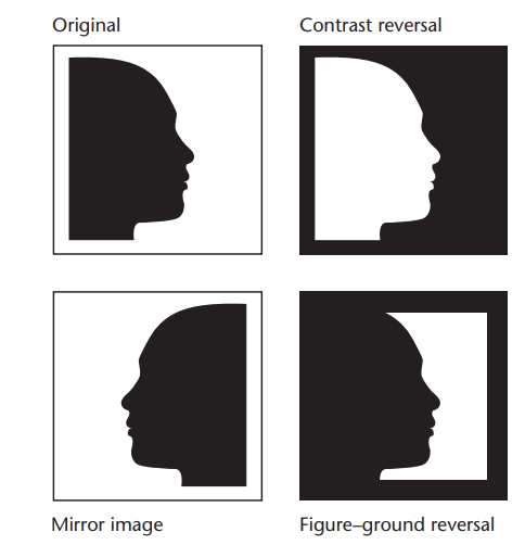
in inferior temporal cortex, cells that respond strongly to the orignal respond about the same to the contrast reversal and mirror image, BUT NOT TO THE FIGURE-GROUND REVRESAL!!!
figure-ground reversal resembles original in pattern of light and darkenss BUT IS NOT PERCEIVED AS THE SAME OBJECT!!
DAMAGE TO VENTRAL PATHWAY (ventral, “what” pathway)
since its the “what” pathway, you have difficulty recognizng objects, despite your vision being fine!!
one of them is called visual agnosia —> “visual lack of knoweldge”
one patient, when shown a key said “idk what it is, perhaps a file or tool of some sort”
when they cant identify things, but then are told what it was tehey go “oh i can see it now” but then when they say “oh what if i told you its not really a pipe” they go “i would take your word for it. maybe its not really a pipe”
within the inferior temporal cortex, several areas detect particular shapes and brain damage can do crazy thing
one eprson, can recognize almost eveveryhting except the numbers 2 through 9, which just seemed like tangles of black lines
isnt that wack!!!!!!
part of the parahippocampal cortex, which is next to the hippocampus, RESPONDS MOSTLY TO THE SIGHT OF PLAAAACESSS
and a part of hte fusiform gyrus responds mainly to FACES
and an area near THAT responds mostly TO BODIES
so all of these are lowkey precise
the brain is really good atdetecting the motion produced by ppl adn animals
yk that video of ppl putting glowy stuff on each like joint of their body, trn the lights off, and then wehn they move its technically only a few spots of light but bcz of where theyre arranged you go “oh its a human”
RECOGNIZING FACES
face recogntion is super important for humans
civilizatoin depends on knowing whether to trust or distrust someone, even tho you havnet seen them in months or years
someday when you attend a high school youll still recognzie them evne tho theyve changed
human newborns come into the world PREDISPOSED TO APY MORE ATTENTION TO FACES, however infants concpet of face is ont like an adults
experimenters recorded infants times of gazing at once face or ther other, an dthey rpeferred a right side up over an upside down regardless of which one was more realistic vs idsotrted
the fusiform gyrus responds strongly to a fac viewed from any angle, as well as meory of it, a line drawing
just IT LOOKS LIKE A FACE, FUSIFORM GYRUS IS ON IT
children accuracy of identifying faces improves as connections get better btwn the right hemisphere fusiform gyrus and part of the occipital cortex known as the occipital face area
ITS CONNECTION BTWN FUSIFORM GYRUS (PARTIUCLAR RIGHT HEMI) AND OCCIPITAL CORTEX, CALLED OCCIPITAL FACE AREA CAUSE IT RECOGNIZES FACES!!!!
cortical cells that respond strongly to faces also respond to face-like objects
in several cases, physicians electrically stimulated the fusiform gyrus during exploratory surgery!! depending on the intensity adn duration of it, the result was either difficulty in perceiving faces or a vivid distortion of them!!!!!!!
like “oh my god your face turned into someobyd else. your face metamorphosed”
PROSOPAGNOSIA
IMPAIRED ABILITY TO RECOGNIZE FACES
this can result from damage to the right fusiform gyrus
if that part of hthe fusirofmr gyrus doens twork or ti doesnt mature right or theres less than normal connections btetwen it and occipital cortex, YOU HAVE TROUBLE RECOGIZNING FACES!!!!!!!1
theres also ppl who have crazy connectiosn between the fusiform gyrus and the occipital lobe and theyre realllllyyyy good at reocgnizng ppl
OLIVER SACKS
famous for writing about others neurological problems SUFFERED FROM PROSOPAGNOSIA HIMSELF :(((((((((
he talked about how he had trouble not only his loved ones but also himself :((((
ppl with prosopagnosia RECOGNIE PEOPLE’S VOICES!!! so its not htat their meomory is funky and they can read so they can see clearly. they can also desribe each ELEMENT of a face, like yes, ears, nose, and faical expresseions as well.
wen they look at two photors of faces side by side, they are allllmost at normal saying theyre same or differnt
the one issue is SEEING A FACE AS A WHOLE, AND RECOGNIZING IT
like for you, think of it as seeing a face upside down
you can recognze oeach of the elemnts but its hard to go “oh yeah thts a face, and its this person’s face” cause its sorta mumbled up
SO THE QUESTION IS, DID WE REALLY EVOLVE A BRAIN MODULE DEVOTED TO FACES????
one hypothesis is we evolved mechanisms for recognzing complex patterns, and faces happen to be the best and most common example
evidence for this is for kids who fuck with pokemon heavy, they look at fusiform gyrus and strong response twhen they see pokemon
chess experts same when they see a chessboard LMFAOOOOOOASKJDFLASDF
children who devoted at least an hour a day to some special interest such as watching soccer or looking at pictures of space travel DMEONSTRATED FUSIFORM GYRUS RESPONSES TO IMAGSE RELATED TO THAT INTEREST. WHATTTTTT
fusifrom gyrus response stronger for kids with ASD who pay even less attentiont o faces
so guys does my fusiform gyrus activate hwen i see my school wrok? when i see a brain?
OTHER ROLES OF THE FUSIFORM GYRUS
detailed visual recognion
many cells in fusiform gyrus respond omre vigorously to faces
those who were blind from birth, elevated actvity in fusiform gyrus when they feel models of faces
resarchers used mri procedure to record brain activty fro 2-9 year olds, found that the fusiform gyrus responded mainly to FACES, just as it does in adults
SO ACTUALLYYYYYY it seems that we do have a module specifically for faces that also dose the same thing for when we see soemething we like!!
MOTION PERCEPTION
any moving object want syour atteniton
you need to know where its going, and how fast, and you need to know this FAST
THE MIDDLE TEMPORAL CORTEX
two areas important for motion perception, MT and MST
MT same thing as V5, and its the middle temporal cortex
MST —> the medial superior temporal cortex
tehse areas receive input mostly from the magnocellular path
the magnocecllular path is implicated in DETECTING OVERALL PATTERNS (much like the system where the prefrontal cortex makes assumptions about an OBJECT, and sends it to the temporal cortex, and then the teporal cortex goes “yup thats a ball” or “nope that is not a ball” and that helps in IDENTIFYING PATTERNS!!!)
so this pathway with the inferior temporal lboe is identifying patterns, and the one where you detect overall patterns, is with the MAGNOCELLULAR path (think magnet, magnets peel away little parts of a big thing that tells us the whole story, so its what you detect patterns from!!)
and this MAGNOCELLULAR PATH (detecting patterns), is COLOUR INSENSITIVTY (it doesnt gaf about colours), the middle temporal cortex is ALSO colour inseesnitivyt
so most cells in the middle temporal cortex respond selectivley weh n something moves iat a particular speed in a particular direction, and ehse cells detect acceleraiton and decceleration as well as aboslute speed, and they respond to MOTION in alll 3 dimensions
MT responds to photographs that imply omveoments as well!!
ppl who have an electrical stimulation of area MT report seeing ivbrations or other movements during stimulation (CAUSE MT IS IMPLICATED IN MOTION PEEERRRRCEPTION. THE BASAL GANLGIA IS IMPLICATED IN MOVEMENT, THE MDIDLE TEMPORAL CORTEX IS IMPLICATED IN MOTION PERCEPTION)
they also become temporarily imparied in seeing somethignt hat is ACTAULLY moving, so in short, middle tmeporal cortex activity is CENTRAL TO SEEING MOVEMENT
cells in the dorsal aprt of the medial superior temporal cortex, respond to more complex stimuli, like the expasion, contraction or rotation of a scene, like what hapens wehn you TILT YOUR HEAD OR MOVE FORWARD OR BACKWARD
IMAGNINE YOURSELF SWINGING
when youre swinging, you start off still, then you go up and things get bigger, and you go back nad they get smaller, and you twirl around they rotate
all those functions when youre swinging are implicated wtih the dorsal part of the medial superior temporal cortex
dorsal parts of MST, more complex stimuli, like the expansion, contraction, rotation
VENTRAL parts of MST, espond briskly if something mvoes relative to backgorund, but little response if object and backgorund both move in the same direction adn speed
BASICALLY THOUGH, THE MT AND MST NEURONS LET YOU DISTINGUISH BETWEEN THE RESULT OF EYE MOVEMNETS AND THE RESULT OF OBJECT MOVEMENTS
cause rmember wtih the ventral part of MST, if they both move at the same time they go “oh the eye just moved” but if one thing moves differntely, it more so goes tot he dorsal side, and you go “oh an OJBECT moved, and it contracted, expanded rotated”, that sorta thing cause MST is like more the specifc stuff, and MT is more magnocellular path, moving thigns, detectimng pattenrs
MOTION BLINDNESS
MT and MST repsond strongly to moving objects BUT ONLY OMVING OBJECTS, what would happen if theres damage to these areas?
motion blindness!!
you can see objects!! but you cant see if theyre moving or not, wha tdirection or how fast
THEYRE BETER AT REACHING FOR A MOVING OBJECT THAN RDESCRIIG ITS MOTION
in all aspects of dealing with visual motion, not great
FOR SOME PEOPLE, (LM) REPROTED WHEN PPL WALKED THEY WERE THERE OR HERE BUT YOU DIDNT SEE THEM MOVING
people would seem to appear to disappear suddenly
wouldnt know what direction theyre going on
she would stop her own walking until the other person was gone, couldnst cross street alone cause she couldnt tell which cars were moving or how fsat, and pouring coffee was hard
people with full colour visoin can imagine what it would be like to be colour deficient, but its hard to imagine motion blindness
SEVERAL PATIENTS WHO REPORTED MOTION BLINDNESS AFTER BRAIN DAMAGE WERE IGNORED
area MT and parts of parietal cortex decrease acitivty during VOLUNTARY eye movements, known as SACCADES
basically brain areas that monitor these saccdes tell MT and pariental cortex “were gonna omve the eye muscles so you can chill out”, and nueral activty adn blood flow in MT decrease 75ms before hte movmenet nad stay suppreseed during movement
SO DURING A VOLUNTERY EYE OVMENT, YOU BECOME MOTION BLID FOR A SPLIT SECOND!!!!!!!!!
SOMEEE PEOPLE ARE BLIND EXCEPT FOR THE ABILITY TO DETECT MOVEMENT
area mt gets input DIRECTLY from teh lateral geniculate nucleus of the thalamus
even after EXTENSIVE damage to v1, enogh to produce blindness, THE MT OVERRIDES THIS SO YOU CAN STILL DETECT MOTION
what would it be like to see motion wtihout seeing the objects that are moving???????
its called blindsight!!! when they say somethings moving they go “oh i must just be guessing” BUT THEY CAN ACTUALLY SEE IT!!!!!!!1
so basically diff parets of brain, diff kidns of visual ifno, possible to develop speicalized kinds of disablity
STOP AND CHECK
when you move yrou eyes, why does it not seem as if the world is moving?
bcz the brain knows youre doing it yourself!! the ventral MST and MT work togethether and when two things are moving at hte asem rate they attribute it to the eye
under what circumstance does someone with an intact brain become motion blind and what accounts for hte motion blindness?
so intact brain is when its a voluntary eye movement, your brain suppresses activity in the MT area so then you are temporarily omtion blind
module 6.1