Signalling II - Electrical Signalling and Neurotransmission
Electrical Signalling
Membrane Potential
Definition: Difference in voltage between the interior and exterior of the cell.
Typically -70 mV at rest but can vary.
Passive Ion Transport
Determined by the electrochemical gradient.
Can be affected by membrane potential (positive inside vs. negative inside).
Ion Gradients and Membrane Potential
Ion Gradients:
The membrane potential arises due to ion gradients and the charges of these ions.
“Fixed anions“ cannot move freely across the membrane. Thus they influence the membrane potential by balancing out the positive charges.
Equilibrium Potential:
Equilibrium potential is the membrane potential at which the net flow of a particular ion across the membrane is zero, meaning that the concentration gradient and the electrical gradient are balanced.
Calculated using the Nernst Equation:
Needs to know: concentration of the given ion in and out of the cell, charge of the ion, temperature in Kelvin, and several constants namely R gas constant, Faraday’s constant.
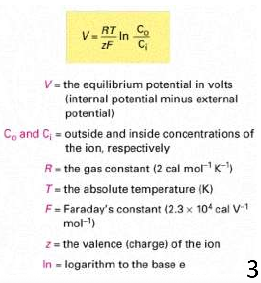
Example:
For K+:
For Na+:
Membrane Potential Calculation
The resting potential (which comprises of the equilibrium potential of all permeable ions) arises mainly due to K+ imbalance (-70mV)
This is because most cells are more permeable to K+ than other ions (“leak channels“). Thus, the resting potential is closer to K+ ion equilibrium potential.
Goldman-Hodgkin-Katz Equation (GHK):
Calculates the membrane potential based on multiple ions.
Expression:
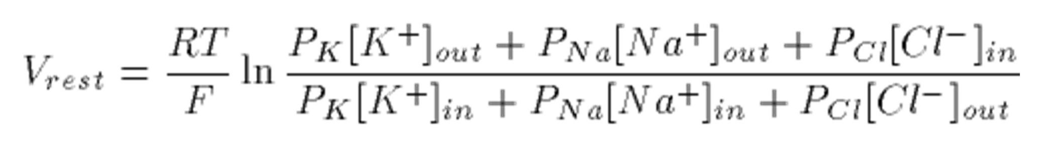
Where permeability ratios are crucial: PK>>PCl>>PNa (at resting potential), with K being the most permeable, followed by Cl, followed by Na.
The Ernest equation is a modification of the GHK equation.
Action Potentials in nerve cells
Characteristics:
Arise from the action of voltage-gated Na+ and K+ channels.
Depolarization initiated by opening Na+ channels, leading to an influx of Na+ ions.
After reaching the peak, K+ channels open leading to repolarization.
Phases of Action Potential:
Rising phase (depolarization): inward flow of Na+, raising the membrane potential towards equilibrium for Na+ which is +60mV
Overshoot (K+ channels open after a delay),
Falling phase (repolarization): K+ channels open following depolarisation, allowing K+ ions to exit and restore to resting potential -70mV.
Neurotransmission
Mechanism:
Action potential is propagated along the axon of the presynaptic cell.
Voltage-gated Ca2+ channels trigger neurotransmitter (e.g. acetylcholine) release at the synapse.
Post-synaptic ligand-gated channels respond to released neurotransmitters. Electrical signalling is enhanced at excitatory synapses and dampened at inhibitory synapses.
Sequential activation of Na+ and K+ channels in the post-synaptic cell allows the propagation of action potential.
Types of ligand-gated Ion Channels:
Ionotropic glutamate Receptors (found at excitatory synapses):
Respond to glutamate, allowing Na+ and Ca2+ into the post-synaptic cell, promoting excitatory signals.
Note that: ligand-gated channels have inverted topology of voltage-gated channels (see figure in Anki) - very similar but inverted.
GABAa receptors are inhibitory
They respond to the neurotransmitter GABA, allowing Cl- ions into the post-synaptic cell, which dampens excitation by leading to a hyperpolarizing effect, ultimately inhibiting neuronal activity.
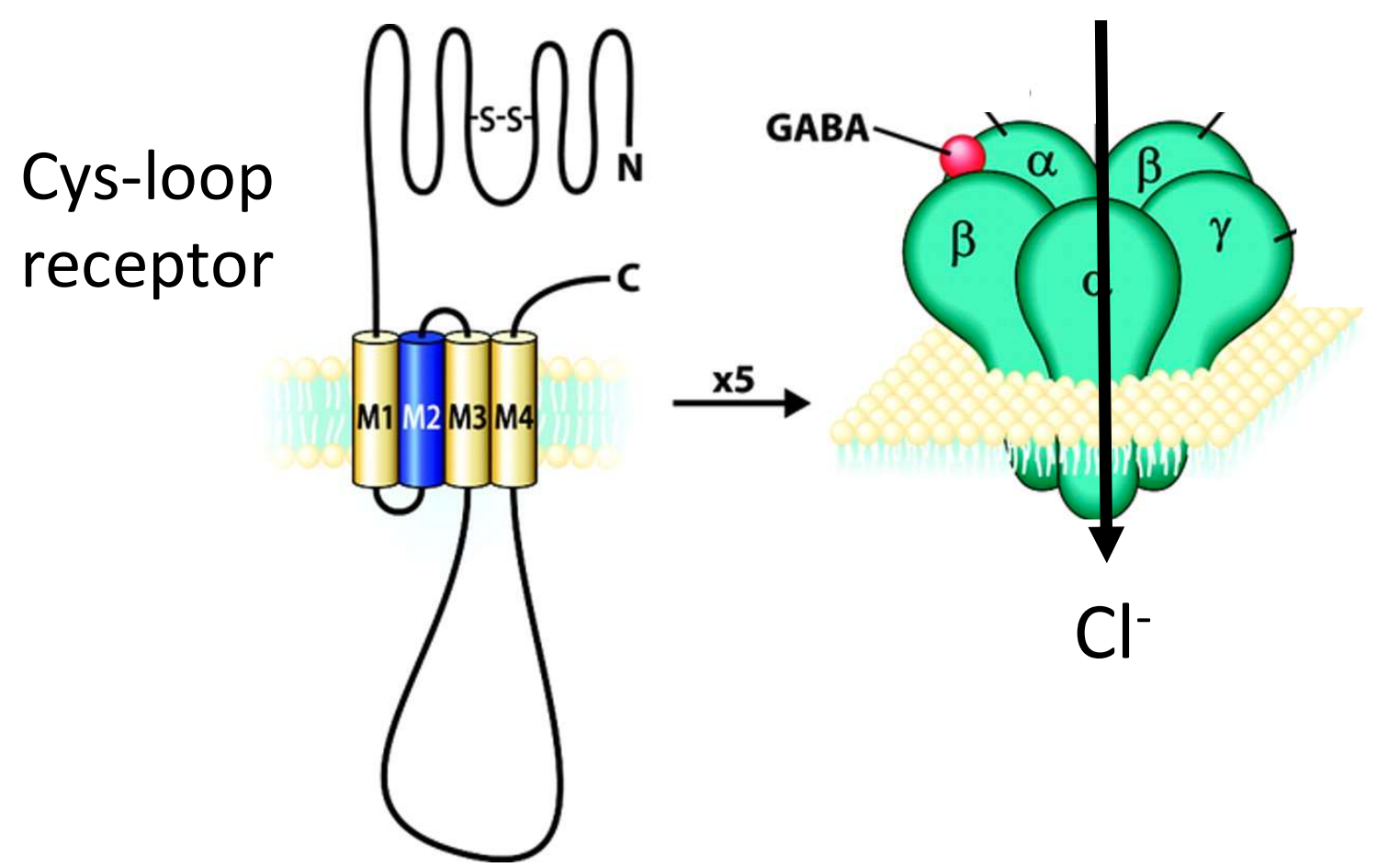
Nicotinic Acetylcholine receptors are excitatory ligand-gated channels. Upon binding of acetylcholine, they open to allow Na+ influx which is critical for both nerve-to-nerve and nerve-to-muscle communication. It also has a cys-loop receptor.
Neuromuscular Junction (NMJ)
After action potential, pre-synaptic neuron releases acetylcholine which is then received by receptors in muscle cells, initiating muscle contraction.
Components:
The NMJ includes voltage-gated Ca2+ channels, acetylcholine receptors (nAChRs), and other voltage-gated channels for ions.
Excitation-Contraction Coupling:
Refers to the process whereby action potential leads to muscle contractions.
Involves calcium release from the sarcoplasmic reticulum, leading to muscle fiber contraction.