ANHB (embryogenesis)
Lecture 1
Introduction to Embryology
Purpose of Studying Embryology:
Understand adult anatomical structures.
Explain unusual anatomical arrangements (e.g., vas deferens route, heart development).
Identify mechanisms when development goes wrong (e.g., folic acid preventing spina bifida).
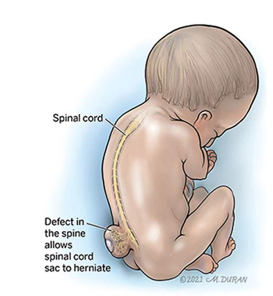
Key Concepts in Embryogenesis
Evolution and Development:
Homologies and phylogenetic constraints.
we all start out looking the same
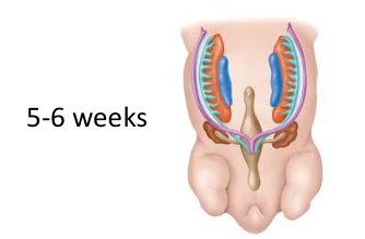
In males the lilac tubes disappear
In females the light blue tubes disappear
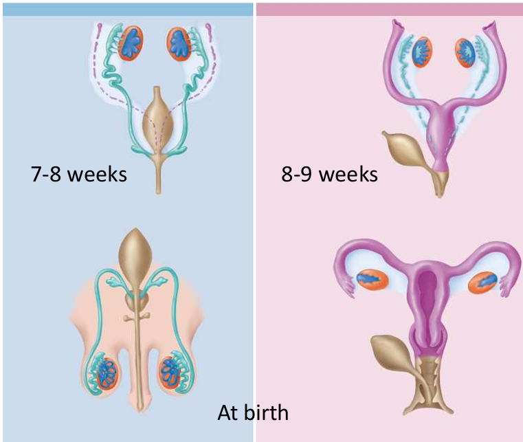
Common ancestry and shared genes.
Cell Biology:
Structure of sperm and ovum.
Inherited organelles.
Genetics:
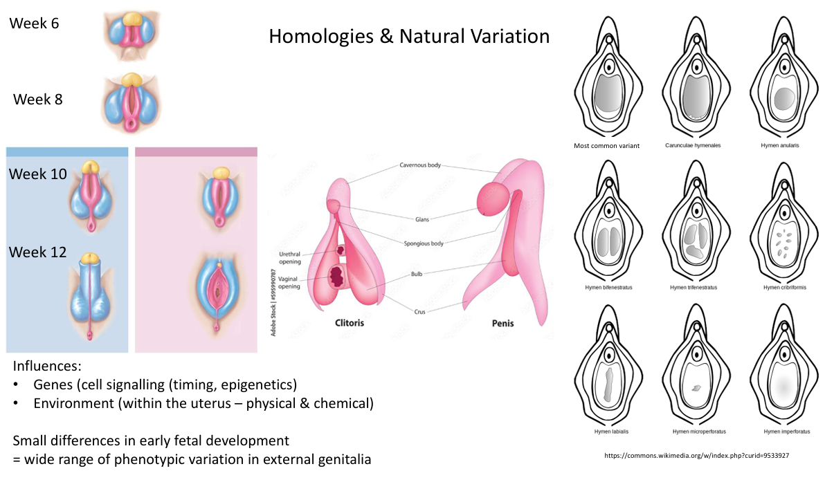
Genotype and cell signaling.
Epigenetics and environmental influences.
Phenotype (interaction of genes and environment).
Reproduction:
Gametes and fertility.
Developmental Stages
Weeks 1 and 2:
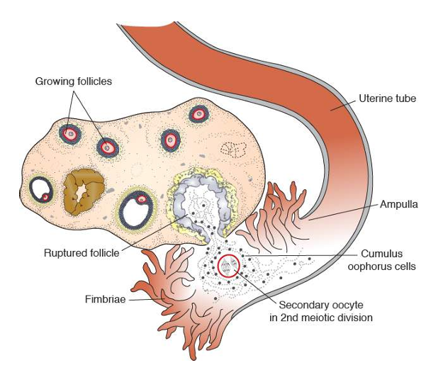
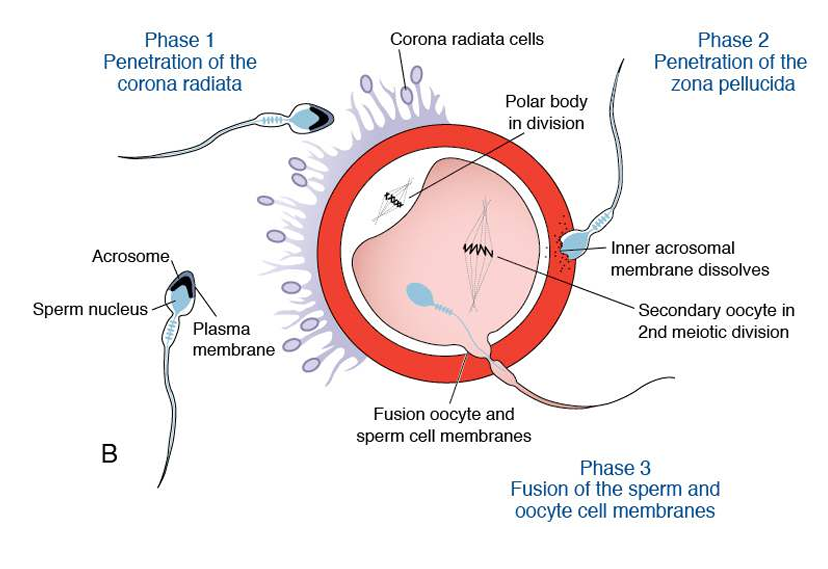
Week 1: Fertilization, formation of single-layered embryo, start of implantation.
Week 2: Formation of bilaminar embryo, body stalk, cavities, completion of implantation, beginning of placental development.
Fertilization and Cleavage
Terminology:
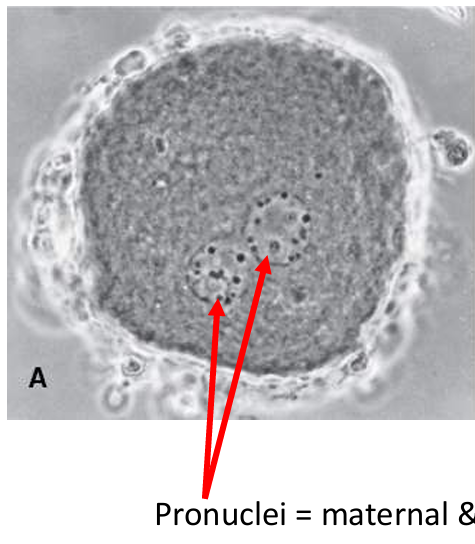
Zygote: Fertilized egg stays a single cell stage (~24 hours). During which the pronuclei will join
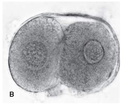
Embryo: From first mitotic division. our zona elucidda still intact
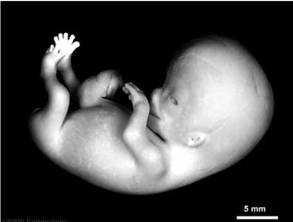
Fetus: Recognizable body parts (~8 weeks).
Process:
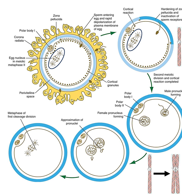
Sperm nucleus forms male pronuclei, tail detaches.
Male and female pronuclei contact, nuclear membranes dissolve.
Pronuclei combine (46 chromosomes), undergo DNA synthesis, mitosis begins.
Maternal and Paternal Contributions to the zygote
Ovum:
23 maternal chromosomes.
Cell membrane, cytoplasm, most organelles (mitochondria).
Sperm:
23 paternal chromosomes.
Centrioles (form mitotic spindles)
critical to bringing maternal & paternal chromosomes tgt
critical for first mitotic division (cleavage)

Week 1 (Cleavage and Blastocyst Formation)
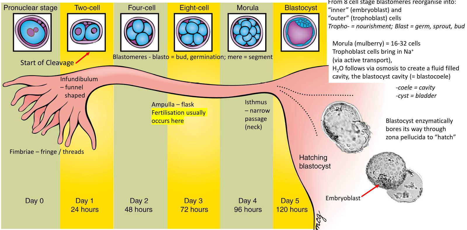
Cleavage:
Blastomeres reorganize into inner (embryoblast) and outer (trophoblast) cells.
Formation of morula (16-32 cells), blastocyst cavity.
Blastocyst:
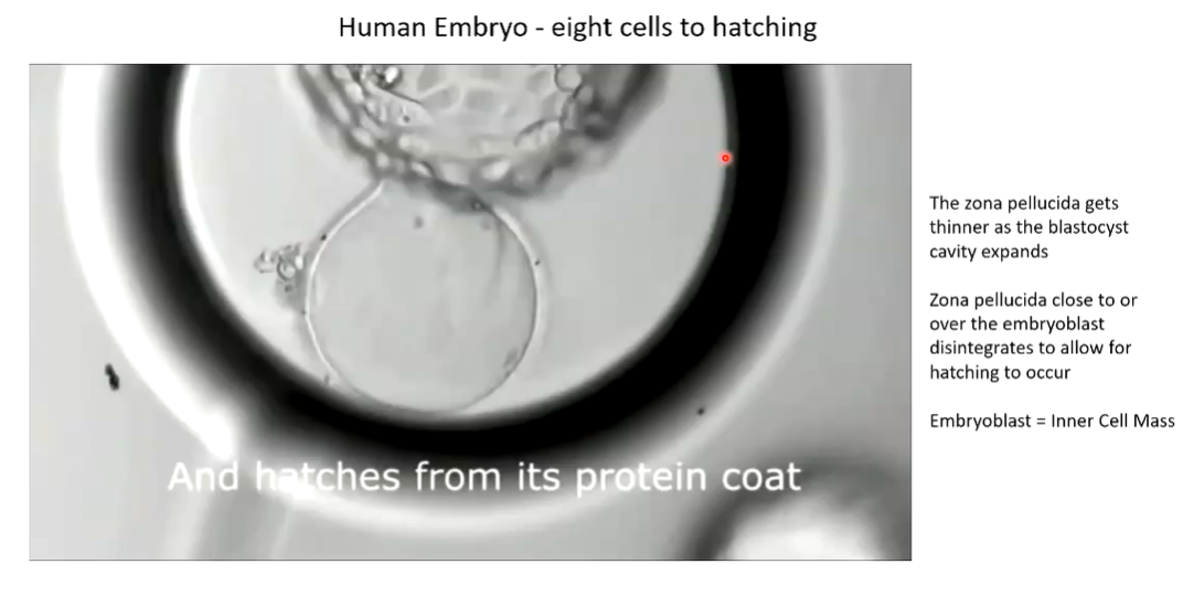
Enzymatically bores through zona pellucida to hatch.
need to rid of zona elucidda to implant
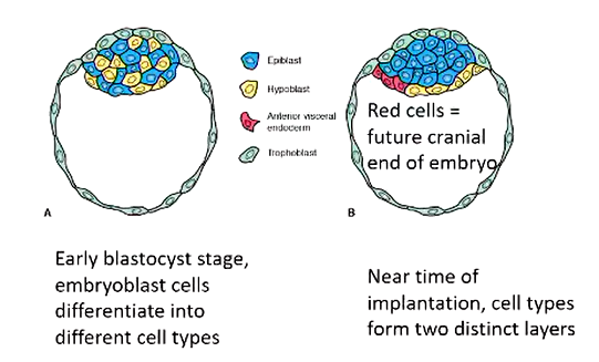
Implantation and Early Development
Week 2:
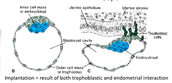
Days 4-5: Trophoblastic and endometrial interaction (needs to occur for implantation)
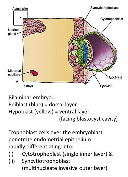
Day 6-7: Formation of bilaminar embryo (epiblast and hypoblast).
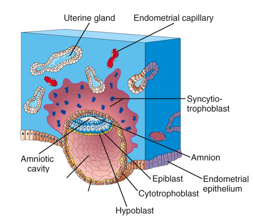
Days 8-9: Bilaminar embryo is implanting. Syncytiotrophoblast is much more developed and erodes endometrium
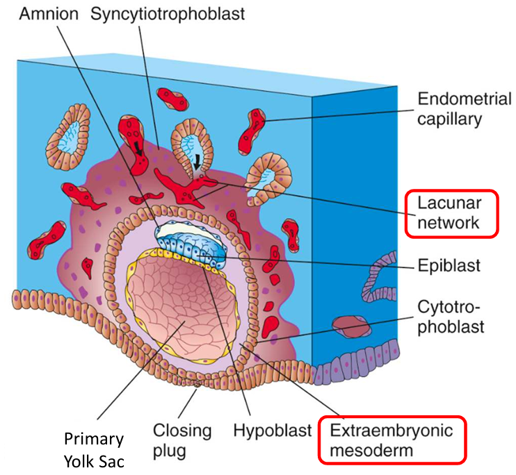
Days 9-10: Bilaminar embryo fully implants. Extraembryonic mesoderm develops. Sysidiotrophoblast erodes maternal blood vessels & uterine glands to create a lacuna network
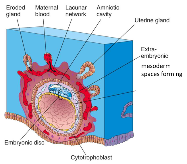
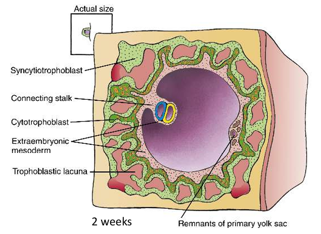
Day 12: Spaces forming in extraembryonic mesoderm—> formation of chorionic cavity
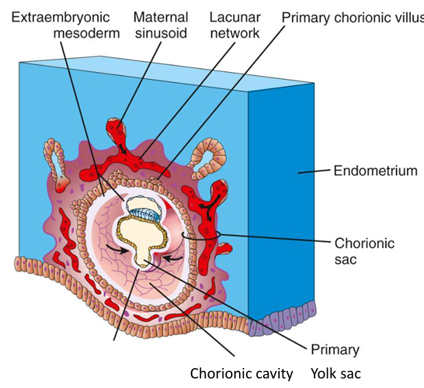
Day 13: Primary yolk sac slowly replaced by secondary yolk sac
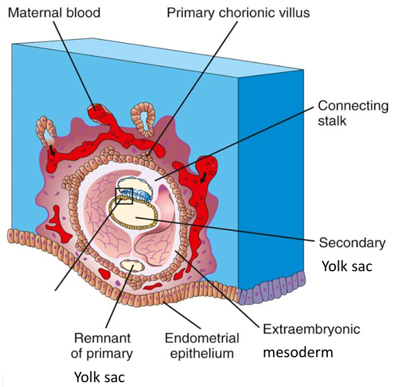
Day 14: development of secondary yolk sac. Connecting stalk develops (evetually becomes part of the umbilical cord)
End of week 2
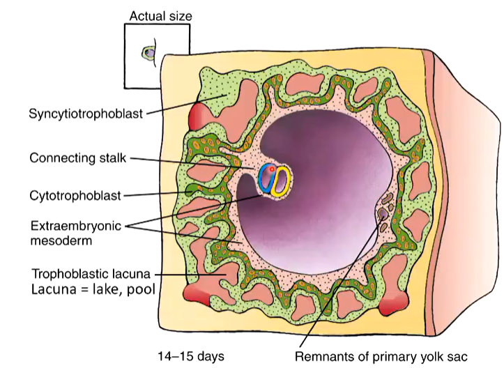
Extraembryonic Structures
Chorionic Cavity (big purple thing):
Space for embryonic growth, fuses with amniotic cavity (weeks 14-16).
Amniotic Cavity (will get bigger):
Provides protection, freedom of movement, space for growth.
Definitive Yolk Sac:
Contributes to digestive tract formation, future gametes, first blood cells production.
Learning Objectives
Cellular contributions of gametes to fertilized ovum.
Processes of cleavage, blastocyst formation, and implantation.
Characteristics of embryonic development during weeks 1 and 2.
Functions of extraembryonic structures during early pregnancy.
Revision Points
Identify: Ovum, zygote, pronuclei, polar bodies, zona pellucida, cleavage stages, morula, blastomere, blastocyst, trophoblast, embryoblast, blastocyst cavity.
Events: Between images H and I.
Lecture 2
Embryogenesis 2:
Formation of the Trilaminar Embryo (Week 3: 14-20 days)
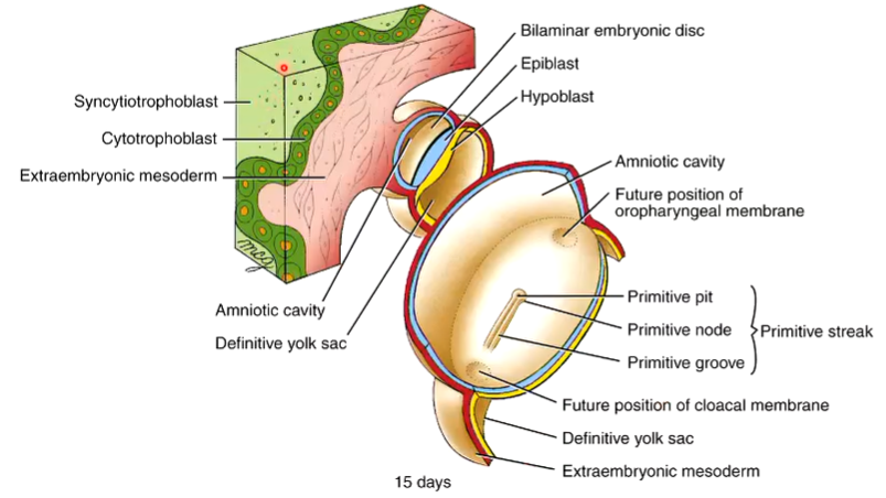
Synidiotrophoblast: our invasive layer uh for the endometrium
Cytotrophoblastic cells: produe the cells that feed into and allow this layer of synidiotrophoblasts to get bigger via mitosis
Primitive Streak:
Divided into 3 parts
Primitive Node
Primitive pit in the centre
Primitive groove that allows things to go through
Directional Terms: Dorsal/back (epiblast), Ventral (hypoblast), Caudal (towards primitive streak), Cranial (cephalic/rostral).
Cloacal Membrane (closer to primitive streak): Future opening for urethra, vagina, and anus.
Oropharyngeal Membrane: Future opening for mouth.
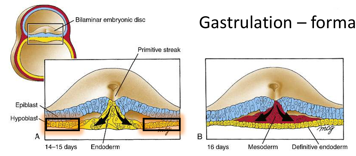
Gastrulation: Formation of three germ layers - ectoderm, intra-embryonic mesoderm, endoderm.
Epiblast cells close to the primitive groove & pit. They are stimulated to migrate through the spaces at the pit and the groove, and they become endodermal cells displacing the hypoblast cells
Intra-embryonic mesoderm is absent in both membranes
Formation of the Notochord:
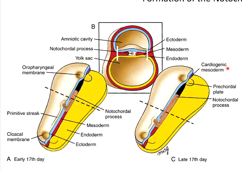
Day 16: Primitive streak is half the length of the embryo.
Notochord Development (Day 17): Critical for nervous system and somite development.
induces the overlying ectoderm to form the neural plate.
Day 22: Primitive streak is 10-20% of the embryo’s length.
Day 26: Primitive streak seems to disappear.
as primitive streak gets smaller, the notochord develops from the mesoderm
The Allantois
allantois etymology = sausage-shaped
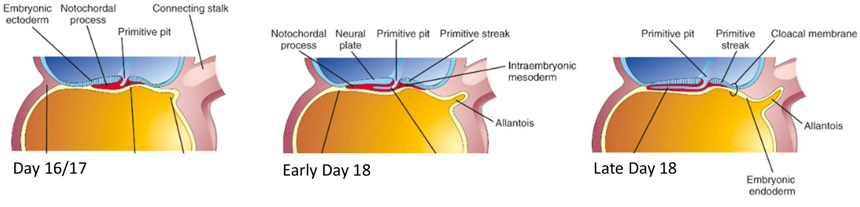
Appears on day 16 as a small outpouching from caudal wall of the yolk sac
projects into the connecting stalk
In birds: respiratory organ & repository for excreta
in humans: give rise to umbilical arteries & veins
Early Formation of Placental Development (up to the end of Week 3)
Embryonic Nutrition:
Uterine Milk: Before implantation.
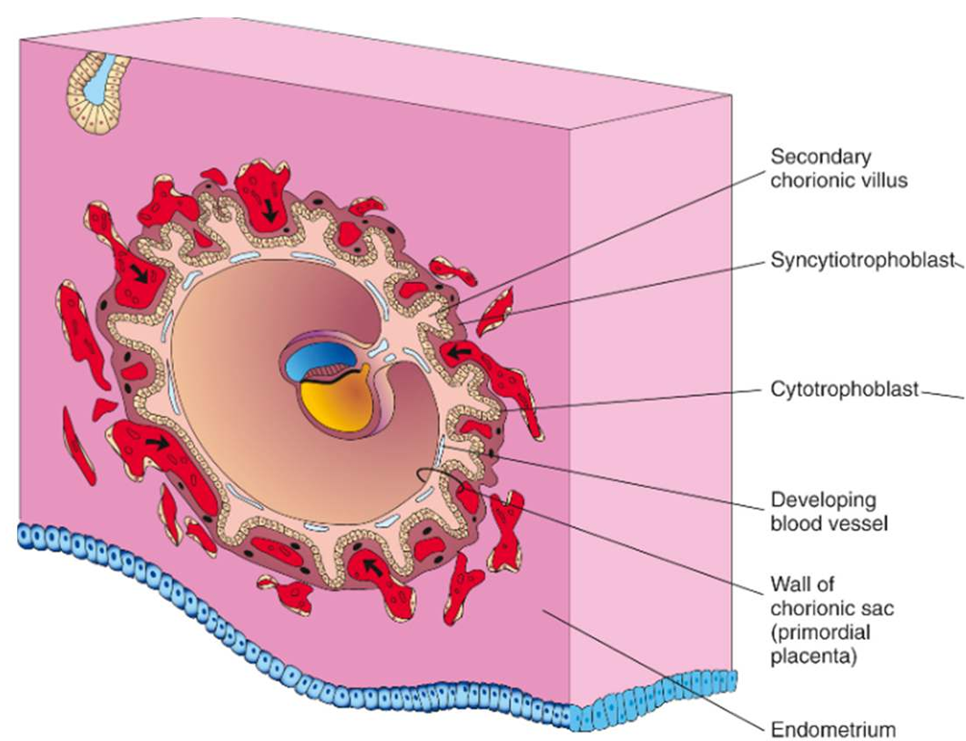
Trophoblastic Nutrition: After implantation.
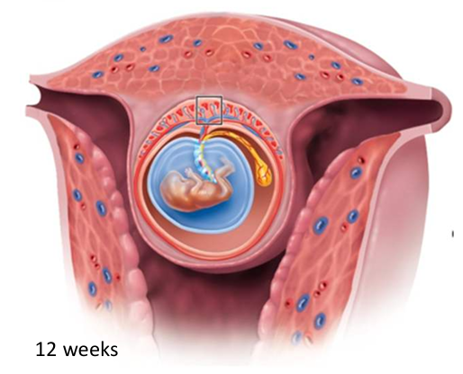
Placenta: Most of pregnancy, source of oxygen, nutrients, and waste removal.
Placental Development (starts midway through week 2):
At first diffusion is sufficient for nourishment during early embryo
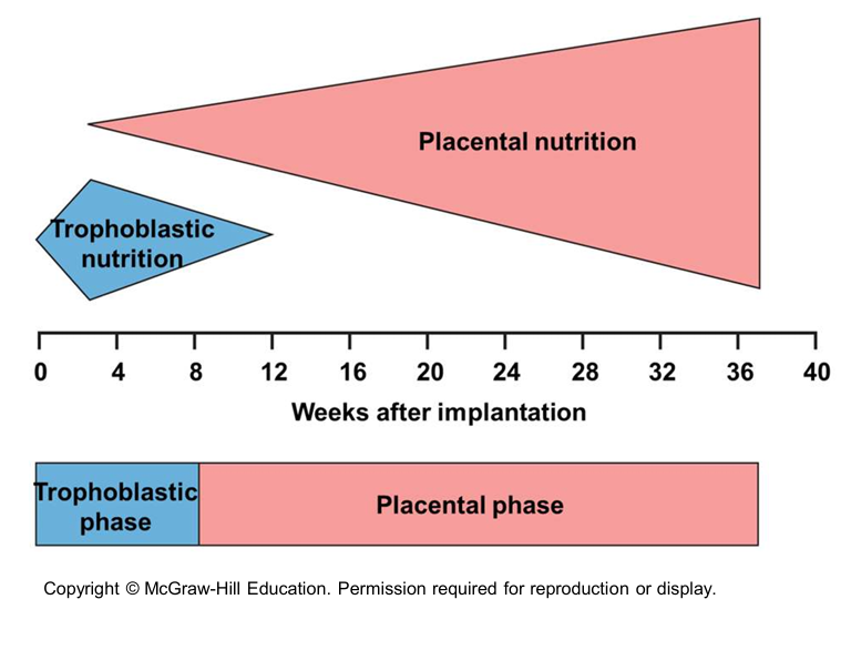
Cardiovascular system develops quickly to support the rapidly growing embryo by connecting to the placenta
By the end of week 3 (Day 22), the placenta is sufficiently developed to start supporting the embryo when the developing heart first starts to beat
By week 12, the placenta is the sole mode of nutritional support until birth
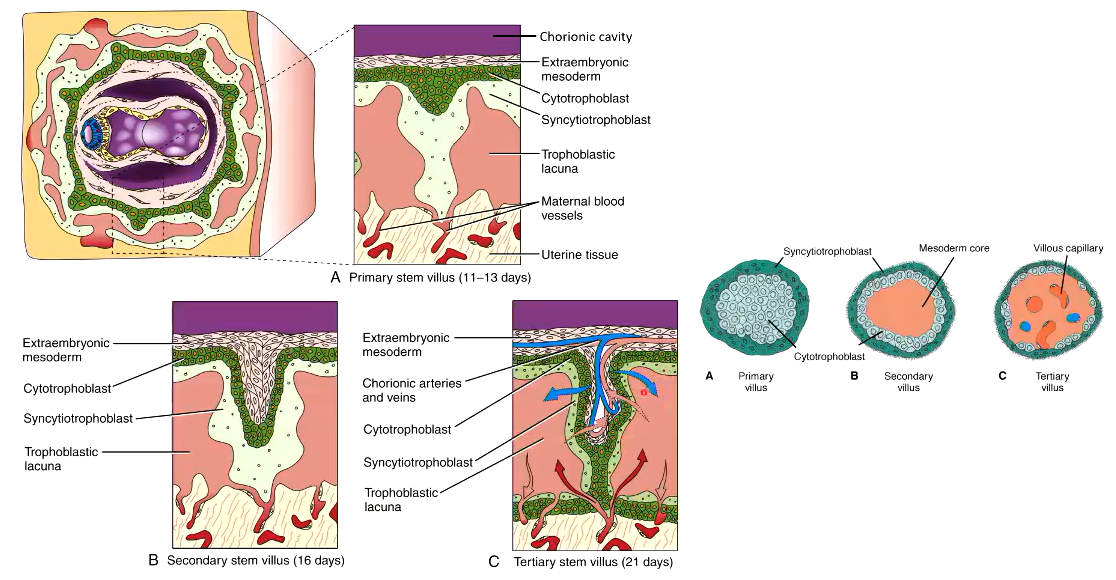
Placenta develops from extra-embryonic tissues:
Extra-embryonic mesoderm
cytotrophoblast
Syncytiotrophoblast
By Day 21: mesodermal core differentiates into connective tissue & blood vessels
Hormones:
Oestrogen— stimulate growth in foetus & mother
Progesterone— suppress follicle development & uterine contractions (prevent 2 pregnancies happening atst & dont rid the endometrium)
human chorionic gonadotrophin (HCG).
Summary of Early “Placental” Development
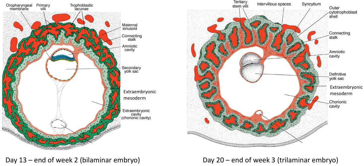
End of week 2 there’s a bilaminar embryo
There’s everything we need to start developing a placenta, and there's that very early placental development; mesoderm invading our cytotrophoblast, which is surrounded by syncytiotrophoblast
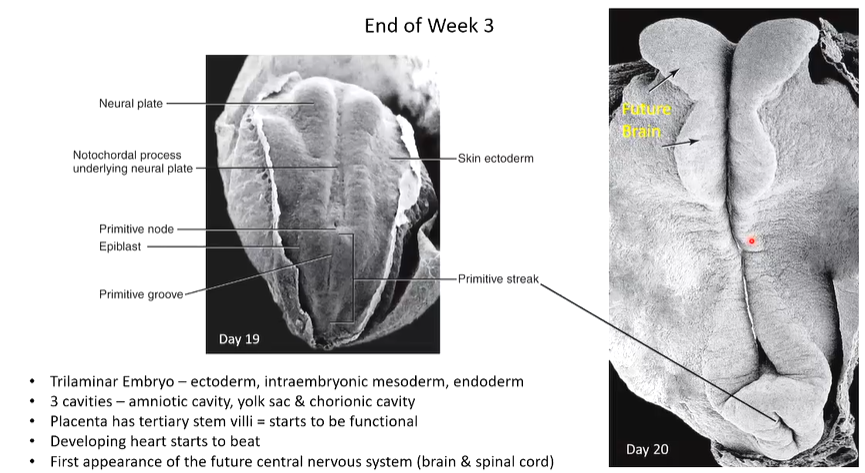
End of week 3 it becomes a trilaminar embryo
Tertiary villi capillaries make contact with capillaries of extra-embryonic mesoderm & connecting stalk which make contact with developing intro-embryonic circulatory system of the embryo
There’s cardiogenic mesoderm which is the precursor
develops cardiovascular system quickly for circulation b/w foetus & placenta
Developing heart starts beating in week 4 (Day 22), the villous system is sufficiently developed to start supporting the rapidly growing embryo
Development of the Future Nervous System (Week 4: 21-27 days)
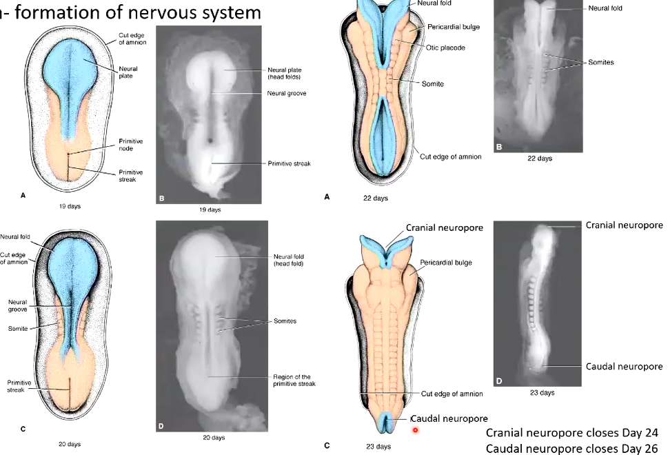
Neural fold is the developing brain
Pericardial bulge is heart development
Somites give rise to skeletal muscle, cartilage, tendons, endothelium, and dermis
Day 23 is now a neural tube
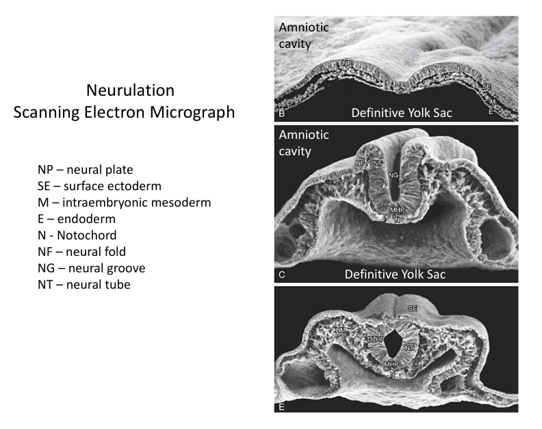
Neurulation:
Formation: Neural plate, neural groove, neural folds.
Shaping: Neural plate shaping.
Bending: Neural folds bending.
Apposition, Adhesion & Fusion: Bringing close together, adhesion, and fusion of neural folds.
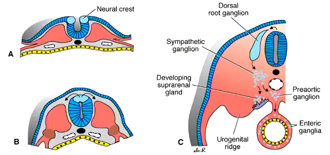
Neural Crest Cells:
Formation: At the tips of neural folds.
Migration (when neural tube closes): Contribute to the formation of structures like the peripheral nervous system, connective tissue of the face and skull, melanocytes of the skin and hair follicles.
If neurulation goes wrong:
Spina bifida— caudal neuropore fails to close
Hydrocephalus (sometimes a consequence of spina bifida)
the fluid is pressing on the developing
brain tissue and if it's not addressed, it can damage it
Anencephaly (cranial neuropore fails to close)
baby does not develop a brain at all
Embryonic Mechanisms of Growth and Development
Proliferation: Increase in cell number.
Cavitation: Formation of internal space.
Differentiation: Alteration of developmental trajectory. (e.g. gastrulation: epiblast → endoderm, intra-embryonic mesoderm, ectoderm)
Migration: Cell movement from one place to another.
Induction: Cells trigger neighbouring cells to differentiate. (e.g. notochord induction of neural plate formation from ectoderm)
Combination: Germ layers combine to form whole organs.
Learning Challenge
Labeling: Neural plate, neural groove, neural folds, neural canal, region of neural crest cell development, ectoderm, mesoderm (intraembryonic), endoderm, primitive streak, amniotic cavity, definitive yolk sac.
Lecture 3
Embryogenesis Week 4 (Days 21-27)
Folding of the Embryo
Craniocaudal (Head-Tail) Folding
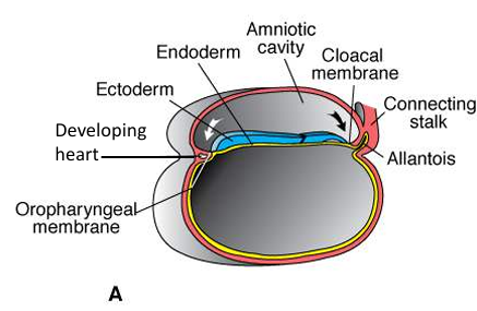
Day 17: Notochord formation begins, gastrulation continues, future heart is cranial to developing brain.
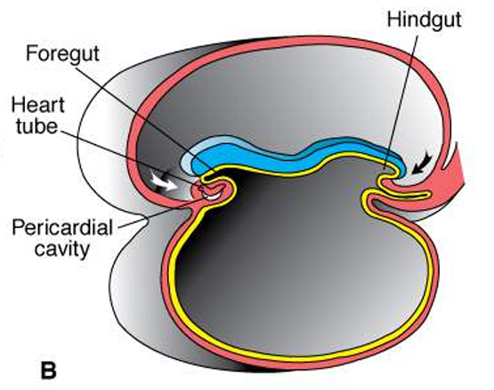
Day 22: Heart tube repositioned ventral to developing brain, starts beating.
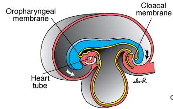
Day 24: Rapid brain development, heart tube positioned caudal relative to brain.
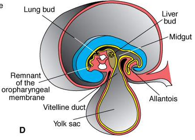
Day 27: Oropharyngeal membrane opens, organs start to form (lungs, gut, liver) hence why it needs to fold to make space.
Lateral (Transverse Plane) Folding
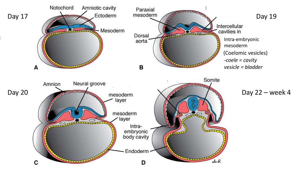
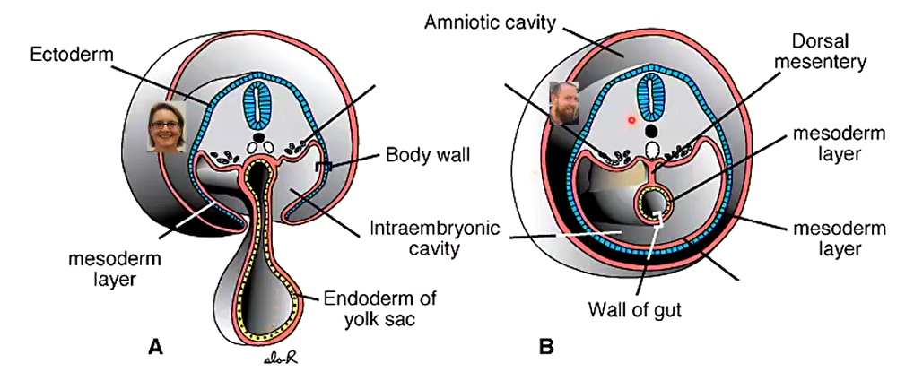
Day 17-22: Formation of intraembryonic coelom (primitive body cavity), spaces for organs to grow (lungs, heart, gut tube).
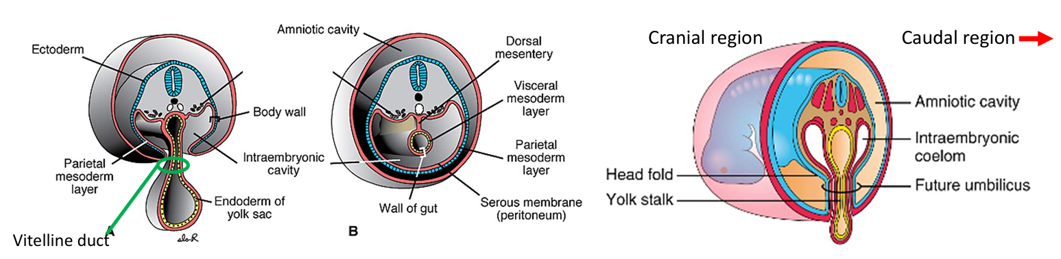
During week 4:
Rapid growth of the amnion surrounds and “pinches” the yolk sac to form the vitelline duct
Last region of the embryo to undergo complete lateral folding = the area occupied by the yolk sac.
In this region, small channels connecting the intraembryonic and extraembryonic coeloms (cavities) persist until the ventral body wall is completely sealed (week 11)
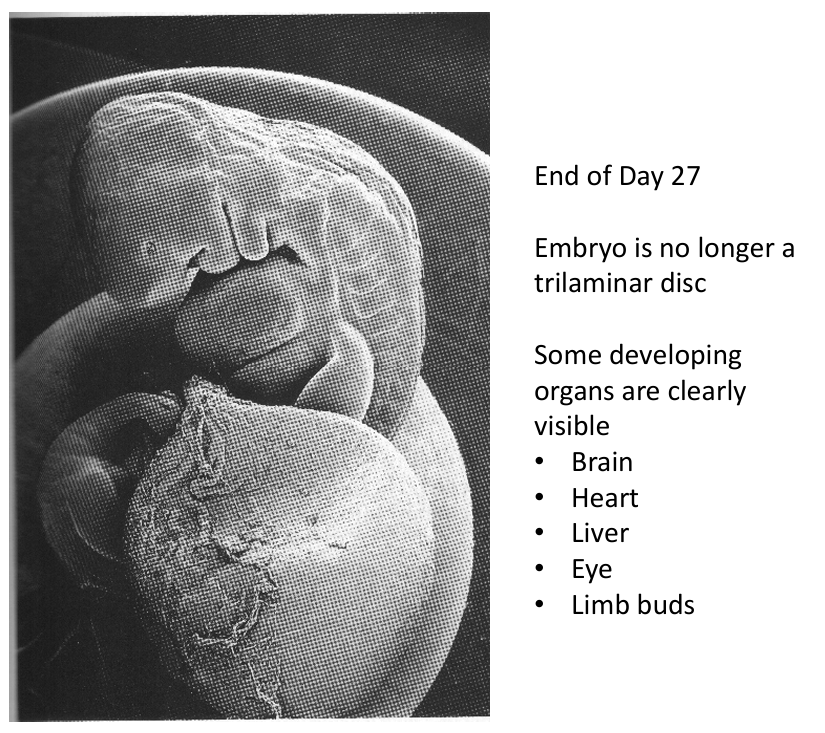
End of Week 4: Embryo is folded into a closed cylinder, major organs visible (brain, heart, liver, eye, limb buds), a sealed system except for the region ard the vitaline duct.
Derivatives of the Primary Germ Layers
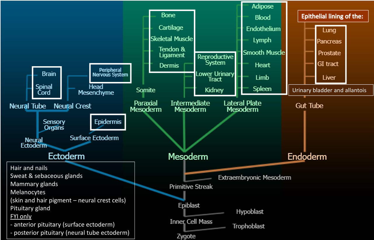
Ectoderm: Epidermis, nerves, hair, nails, sweat & sebaceous glands, mammary glands, melanocytes, pituitary gland.
Mesoderm: Dermis, smooth muscle (e.g., stomach), urinary bladder.
Endoderm: Epithelial lining (e.g., stomach).
Organs are often a combi of germ layers
Stomach:- Epithelial lining is endoderm derived- Smooth muscle is mesoderm derived
Skin:- Epidermis & nerves are ectoderm derived- Dermis is mesoderm derived
Evolutionary Significance of Morning Sickness
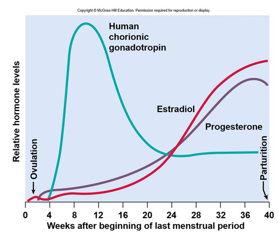
Human Chorionic Gonadotrophin (HCG):
Secreted by blastocyst & placenta, peaks at 10-12 weeks.
Stimulates growth of corpus luteum, which secretes estrogen & progesterone.
Absence of HCG results in corpus luteum & uterus will be expelled
Corresponds to peak period of organogenesis & morning sickness.
Morning Sickness:
Common in first few months of pregnancy, possibly due to HCG secretion.
May protect developing conceptus by encouraging consumption of bland foods, reducing ingestion of teratogens.
Women experiencing morning sickness have fewer infants with birth defects.
Teratogen Sensitivity Periods
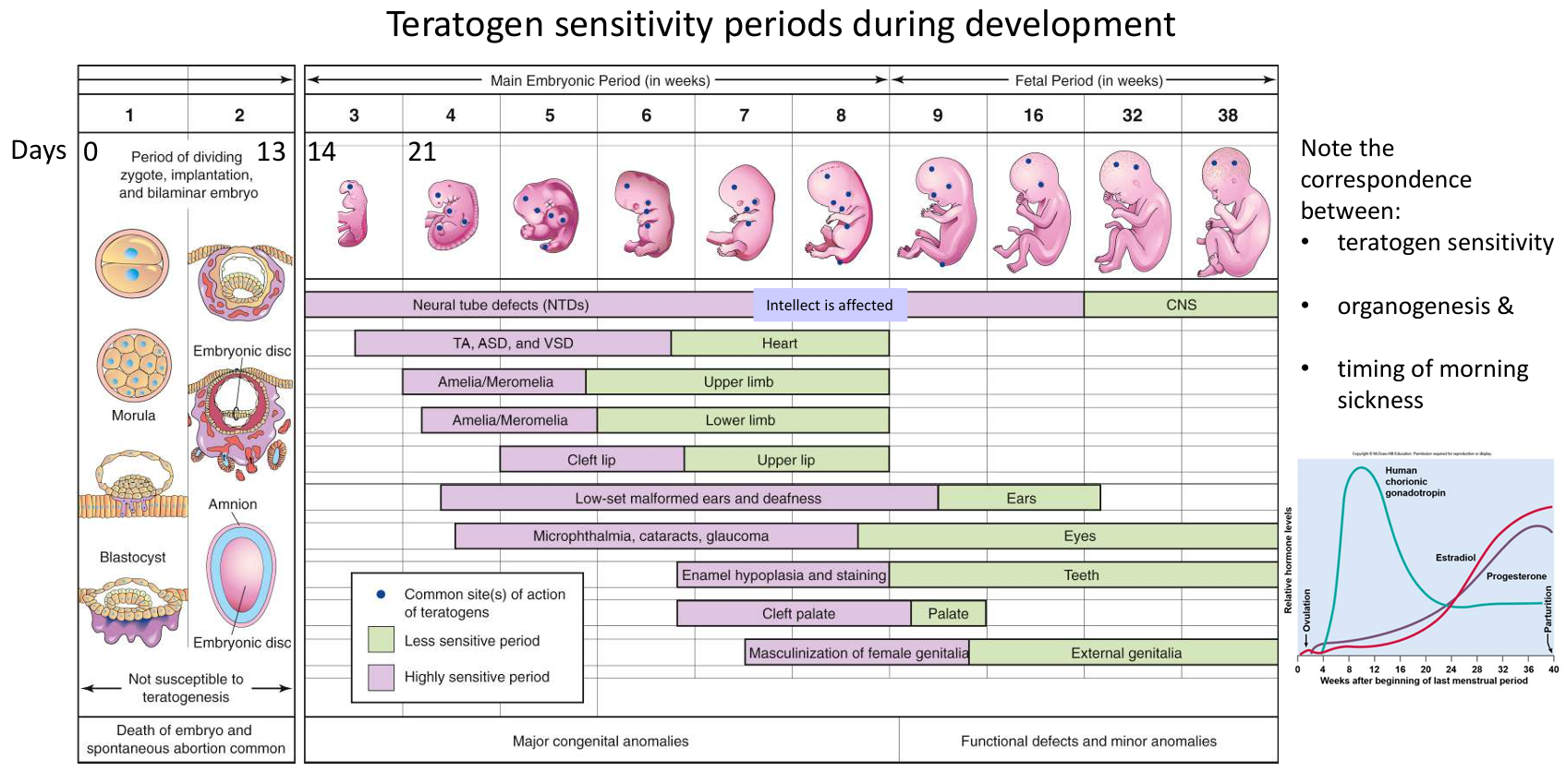
Days 0-21: Correspondence between teratogen sensitivity, organogenesis, and timing of morning sickness.
Examples of Teratogens: Food poisoning (Listeriosis), alcohol, certain drugs, excessive caffeine, tobacco, infections, retinoic acid.