DNA Structure
Lecture Date: 9/17/25
Chargaff
worked on purifying DNA from different molecules
DNA is composed of nucleotides
Took DNA from different organisms and digested them into the four different nucleotides and then compared the ratios of the different nucleotides present
Counted up how many of each nucleotides are in each DNA
Found that there were specific ratios ins which for every Adenine there was a Thymine and for every Guanine there was a Cytosine
This tells us how the nucleotides on either strand of the DNA bind to one another (A binds to T, C binds to G)
Chargaff’s Rules
The base composition of DNA varies from one species to another
Different tissues of the same organism have the same base composition
The base composition of DNA in a given species does not change with an organism over time
In all cellular DNA the number of adenosine residues is equal to the number of thymidine residues and the number of guanosine redisdues is equal to the number of cytosine residues
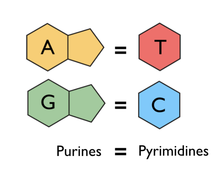
Purines vs. Pyrimidines
Trick: the shorter word (Purine) corresponds to the larger (double ring structure)
Purines contain a six carbon ring and a five carbon ring
Pyrimidines contain only a six carbon ring
Chargaff describes the binding in DNA between the nitrogenous bases
Pauling
Suggested a structure of DNA that was incorrect
Proposed that DNA was a triple helix
This is wrong because the bases do not pair with one another which does not support chargaffs rule and he had the phosphate groups on the inside of the DNA structure which is incorrect because phosphate groups are negatively charged so they would want to be on the exterior because the similar charges cause repulsion and greater repulsion can occur when the phosphate groups are on the exterior and are not confined to a small space
X-ray Crystallography
A method used to determine the arrangement of electrons which can be used to figure out where other atoms are present
Roseline Frankin
This method was needed for Watson and Crick’s research
Hydrogen Bonding
A and T are joined by two hydrogen bonds
G and C are joined by three hydrogen bonds
Since G and C have more hydrogen bonds they are stronger bonds which can influence the structure of DNA
Major vs. Minor Groove
Major Groove: Has more exposed nitrogenous bases which causes this area to have a higher chance of bonding to other molecules
Minor Groove: fewer exposed nitrogenous bases which means that fewer proteins will bind to this area
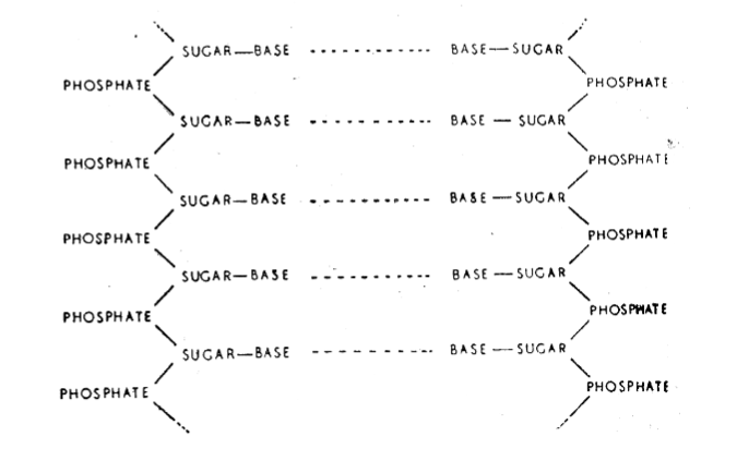
Watson and Crick Summary
DNA is a double helix
Anti-parallel
Hydrogen bonded base pairs on the inside
Sugar-Phosphate backbone on the outside
Each chain runs 5’ to 3’
DNA is negatively charged because of the phosphate groups that have a negative charge
Model hints at how DNA works
Dickerson Dodecamer
Created a molecule of a known sequence that was 12 units long and then analyzed the structure using X-ray crystallography
Results of this differed from the Watson and Crick helix in minor ways
Used a segment that was complementary to itself so that he did not need to synthesize two strands of DNA
Found that the 3D structure of DNA varies with the sequence of DNA
This did not contradict Watson and Crick because it instead suggested that the details can differ from the general structure when observed on a smaller scale in terms of the number of nucleotides present
Drawing DNA Structure For Exam:
Know how to draw the structure of each base as well as how they are oriented in the structure (see below)