2.3.1 hearing
Outline the properties of sounds
Sound waves originate from vibrating objects that cause adjacent particles in a medium to oscillate, thereby propagating the wave.
Pitch
Refers to the frequency of sound waves (the number per second)
higher frequency = higher pitch
Humans can hear between 20 and 20,000 Hertz
Loudness
Refers to the amplitude, or height of the sound wave
Higher amplitude = louder sound (in decibels)
Human threshold of hearing is 0 decibels
Normal conversation = 60 decibels
Sounds >120 decibels cause hearing damage

Identify the structures of the external and middle ear and describe their contribution to the collection of sound.
The primary function of the external and middle ear structures is to amplify sound waves before they enter the fluid-filled inner ear.

External Ear:
Auricle (Pinna): Composed of cartilage and bone; funnels sound waves into the external auditory canal.
External Auditory Canal: Directs sound waves toward the tympanic membrane. The outer one-third is cartilaginous and contains earwax (cerumen) and hair to prevent infection; the inner two-thirds is bony and narrow, concentrating sound waves.
Middle Ear:
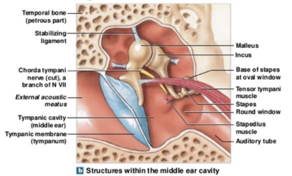
Extends from the tympanic membrane to the oval window (the hollow portion is called the tympanic cavity).
Auditory Ossicles: transfer and amplify vibrations from the tympanic membrane to the oval window
Malleus (hammer)
Incus (anvil)
Stapes (stirrup)
Dampening Muscles: contract to attenuate excessive vibrations
The tensor tympani muscle attaches to the malleus
The stapedius muscle attaches to the stapes
Describe the role of the cochlea in the conversion of sound into neural signals
The inner ear (extends from the oval window) consists of two main structural networks:
Bony Labyrinth: Bone passages in the temporal bone filled with perilymph (a fluid similar to cerebrospinal fluid).
Membranous Labyrinth: Suspended inside the bony labyrinth and filled with endolymph (a fluid similar to intracellular fluid, rich in and low in ).
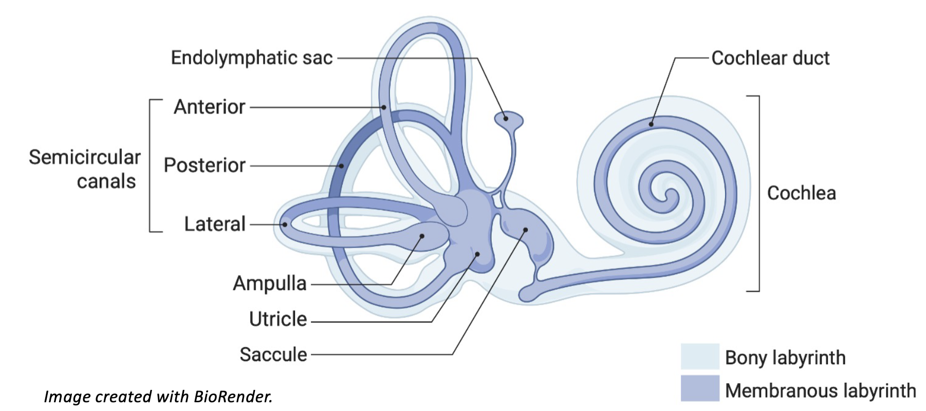

The cochlea contains three distinct fluid-filled channels:
Sound enters the oval window, travels up the scala vestibuli to the apex, and returns via the scala tympani to the round window to equalize pressure.
Scala Vestibuli: Upper compartment (bony labyrinth) filled with perilymph.
Scala Tympani: Lower compartment (bony labyrinth) filled with perilymph.
Scala Media (Cochlear Duct): Middle compartment (membranous labyrinth) filled with endolymph.
The cochlear duct consists of the vestibular and basilar membranes, with the tectorial membrane in between.
Auditory hair cells in the organ of Corti sit on the basilar membrane, with tips embedded in the tectorial membrane.
Perilymph fluid movement vibrates the basilar membrane, while the tectorial membrane does not.
Stereocilia bend, activating the auditory nerve

Describe the process of sound transduction in Hair Cells
Basilar membrane vibration bends hair cell stereocilia, activating mechanoreceptors to convert sound waves into electrical signals (transduction).

Bending of stereocilia opens mechanically gated channels.
Driven by the concentration gradient of high - endolymph, enters the hair cells, causing depolarization.
Depolarization opens voltage-gated channels, triggering influx and the exocytosis of glutamate
Released glutamate excites primary sensory neurons whose cell bodies reside in the spiral ganglion.
Axons of spiral ganglion neurons coalesce to form the cochlear nerve (auditory nerve), which transmits action potentials to the brainstem.

Encoding Sound Intensity and Pitch
Intensity (Loudness) Encoding:
Perception of sound intensity correlates directly with the total number of active neurons and their action potential firing rates.
Higher-amplitude waves cause greater basilar membrane displacement, bend more stereocilia, induce greater glutamate release, and increase neural firing rates.
Frequency (Pitch) Encoding:
Place Theory (Tonotopy): Specific locations on the basilar membrane correspond to specific frequencies. High-frequency tones (e.g., ) trigger maximal hair cell firing at the proximal end near the oval window. Low-frequency tones (e.g., ) trigger maximal activation near the distal apex. Intermediate frequencies (e.g., ) activate middle regions.
Frequency Theory (Phase Locking): Used for low-frequency sound pitch encoding, where the basilar membrane vibrates in direct synchrony with sound waves, causing cochlear nerve axons to fire action potentials at the same frequency.

Identify and describe the auditory pathway, from the cochlea to the auditory cortex

Pathway Sequence:
Spiral ganglion neuron axons form the cochlear nerve and terminate in the cochlear nuclei of the lower brainstem.
Post-cochlear nuclei projections are entirely bilateral across brainstem relays.
Bilateral pathways ascend to the medial geniculate nucleus (MGN) of the thalamus (analogous to the LGN in the visual system).
Auditory radiations project from the MGN to the primary auditory cortex.
Auditory Cortical Structures:
Primary Auditory Cortex: Located in the superior temporal gyrus (Brodmann Area 41). Maintains a columnar tonotopic organization (low frequencies located rostrally, high frequencies located caudally).
Auditory Association Cortex: Located in the middle temporal gyrus; responsible for sound recognition and further processing.
Wernicke's Area: Located in the posterior portion of the superior temporal gyrus; critical for language comprehension.
Broca's Area: Located in the frontal lobe superior to the Sylvian (lateral) fissure; essential for speech production.

Sensorineural Hearing Loss:
Caused by permanent damage to cochlear hair cells or the cochlear nerve from acoustic trauma, illness, tumors, or ototoxic antibiotics.
Unilateral deafness occurs only if damage is confined to the cochlea, cochlear nerve, or cochlear nuclei prior to bilateral brainstem crossover.
Permanent unless managed with a cochlear implant; higher incidence observed in men than in women.
Conductive Hearing Loss:
Caused by mechanical blockages or physical disruptions preventing sound transmission from the outer ear to the cochlea (e.g., cerumen impaction, middle ear fluid, or tympanic membrane perforation).
Neural pathways remain functional; treatable with hearing aids or medical procedures (e.g., grommet tube insertion for fluid clearance).