ECG fundamentals
Chest pain, unexplained collapse extra = ECG
Anatomy and conduction of the heart
Oxygenated blood enters the right atrium via the vena cava
Deoxygenated blood enters the left atrium via the pulmonary vein
SA node in the the right atrium starts the contraction for the top of the R atrium.
The impulse moves from this to the AV node where it is held for 0.1 seconds before the impulse moves to the bundle of his them left and right ventricular bundle branches then the Purkinje fibers for ventricular contraction
Atrial fibrillation (AF) - loss of electrical control of the atria ( less life threating as gravity can bring down the blood)
Cardiac arrests rhythms / need defilation when
Ventricular fibrillation (VF)- loss of electrical control of the ventricles (Shockable) {no patterned squiggle}
ventricular tachycardia ( VT) - fast contractions of the ventricles (Shockable) { patten with big drop }
Pulseless electrical activity (PEA) - absence of a pulse in a patient with electrical activity that would normally be expected to produce a cardiac output (except VT) (Non-shockable)
Asystole - absence of electrical activity in the heart (Non- shockable)
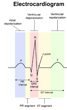
each little square = 0.04s and each big square = 0.2s
P wave = depolarisation of atrial ( atrial contraction)
QRS complex = ventricular depolarisation (ventricles contact)
T wave = repolarisation of the ventricles ( reset of the verticals)
ST segment = Elevation (j point is raised) -STEMI, ischaemia extra ; Depression - SVT extra