Chapter 16
CHAPTER 16
Glycolysis and Gluconeogenesis
Section 16.1 Glycolysis Is an Energy-Conversion Pathway in Most Organisms
• glycolysis = the sequence of reactions that metabolizes one molecule of glucose to two molecules of pyruvate with the concomitant net production of two molecules of ATP
– anaerobic process (does not require O2)
– common to both prokaryotic and eukaryotic cells
• gluconeogenesis = the process by which metabolic products are salvaged to synthesize glucose
- irreversible steps of glycolysis bypassed in gluconeogenesis
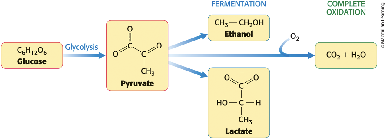
Glucose Can Be Metabolized to Different Products Depending on the Presence or Absence of Oxygen
Glucose Is Generated from Dietary Carbohydrates
• Glucose is typically consumed as starch and glycogen and must be broken down for absorption and transport.
• α-amylase = pancreatic enzyme that digests starch and glycogen
– cleaves α-1,4 bonds, but not α-1,6 bonds
– produces maltose and maltotriose
• α-glucosidase (maltase) = intestinal surface enzyme cleaves maltose, maltotriose, and other α-1,4-linked oligosaccharides into glucose molecules
• sucrase = intestinal surface enzyme that degrades ingested sucrose to fructose and glucose
Glucose is an important fuel for most organisms
mammals use glucose as brain food and red blood cell food
Transport of Monosaccharides
• Monosaccharides are transported into endothelial cells lining the intestine by active transporters.
• Glucose moves passively down its concentration gradient into the bloodstream and passively into cells that will utilize it.
A Family of Transporters Enables Glucose to Enter and Leave Animal Cells
• Glucose transporters (GLUT1 to GLUT5) mediate the thermodynamically downhill movement of glucose across animal plasma membranes.
– have a 12-transmembrane-helix structure
– each member of the family has a distinctive role
Family of Glucose Transporters
TABLE 16.1 Family of glucose transporters

Section 16.2 Glycolysis Can Be Divided into Two Parts
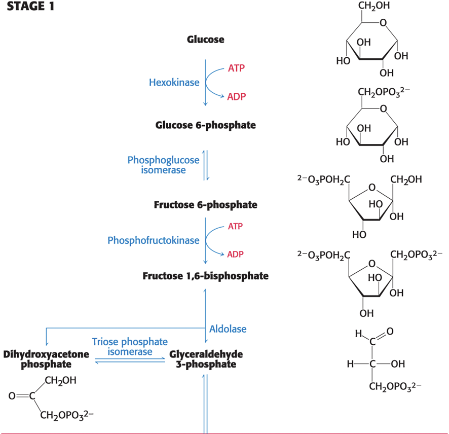
• 10 enzyme-catalyzed reactions make up glycolysis.
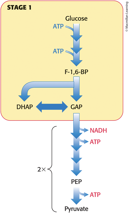
• Stage 1: traps glucose in the cell and modifies it so that it can be cleaved into phosphorylated three-carbon units
– does not generate ATP
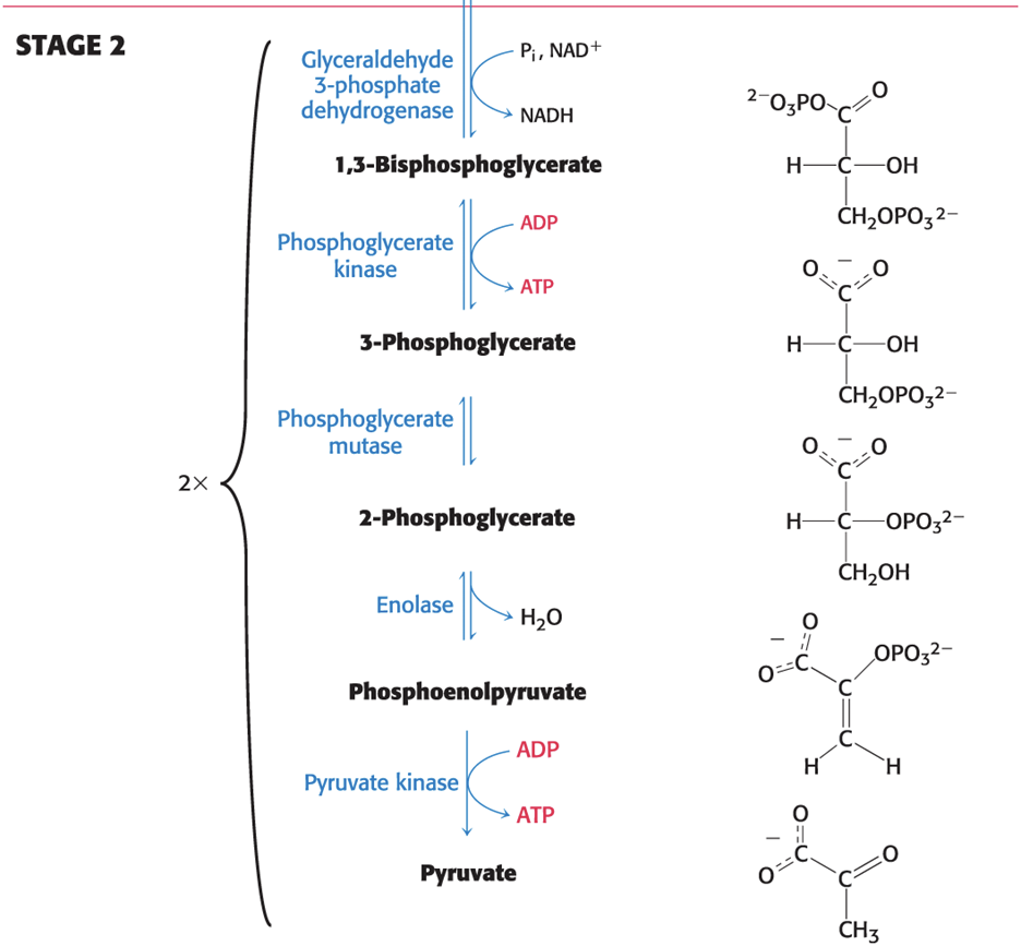
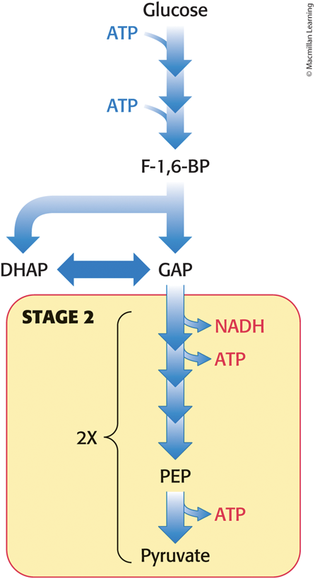
• Stage 2: oxidizes three-carbon unit to pyruvate
– generates 2 molecules of ATP
The Glycolytic Pathway—Stage 1
The Glycolytic Pathway—Stage 2
The First Stage of Glycolysis Uses the Energy from Two Molecules of ATP
Stage 1 Begins: Hexokinase Traps Glucose in the Cell and Begins Glycolysis
• kinases = enzymes that catalyze the transfer of a phosphoryl group from ATP to an acceptor, or to ADP from a phosphoryl donor
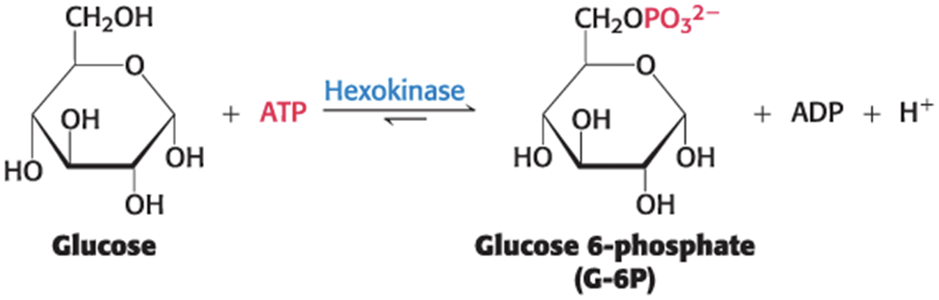
• Hexokinase uses ATP to phosphorylate glucose to glucose 6-phosphate (G-6P).
– requires Mg2+ or Mn2+ as a cofactor
– traps glucose in the cell because G-6P is negatively charged and is not a substrate for glucose transporters
– facilitates the metabolism of glucose into three-carbon molecules with high phosphoryl-transfer potential
The Hexokinase Reaction
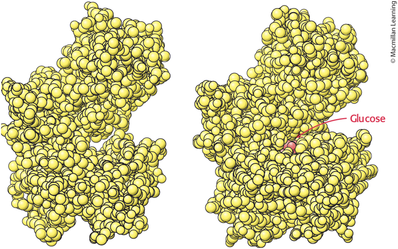
Induced Fit of Hexokinase
• Binding of glucose causes the two lobes of hexokinase to move toward each other.
– makes the environment around glucose more nonpolar favoring the reaction
– excludes water from the active site
Fructose 6-Phosphate Is Generated from Glucose 6-Phosphate
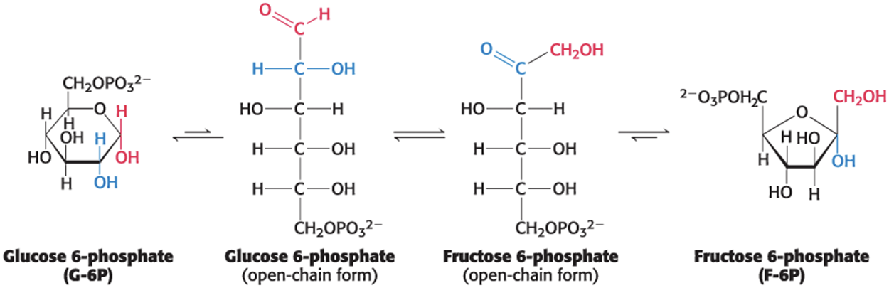
• Phosphoglucose isomerase catalyzes the isomerization of G-6P to the fructose 6-phosphate (F-6P).
– catalyzes a conversion of an aldose into a ketose
– take several steps because the enzyme must open the G-6P ring, catalyze the isomerization, and form the F-6P ring
– prevents the reformation of glucose 6-phosphate
– ensures that two interconvertible 3-carbon fragments results from Stage 1
The Phosphoglucose Isomerase Reaction
Fructose 1,6-Bisphosphate Is Generated from Fructose 6-Phosphate
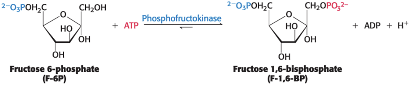
• Phosphofructokinase (PFK) uses ATP to phosphorylate F-6P to fructose 1,6-bisphosphate (F-l,6-BP).
– PFK is an allosteric enzyme that sets the pace of glycolysis.
The Six-Carbon Sugar Is Cleaved into Two Three-Carbon Fragments
• Aldose cleaves F-l,6-BP into glyceraldehyde 3-phosphate (GAP) and dihydroxyacetone phosphate (DHAP).
– this reaction completes Stage 1 of glycolysis
– readily reversible
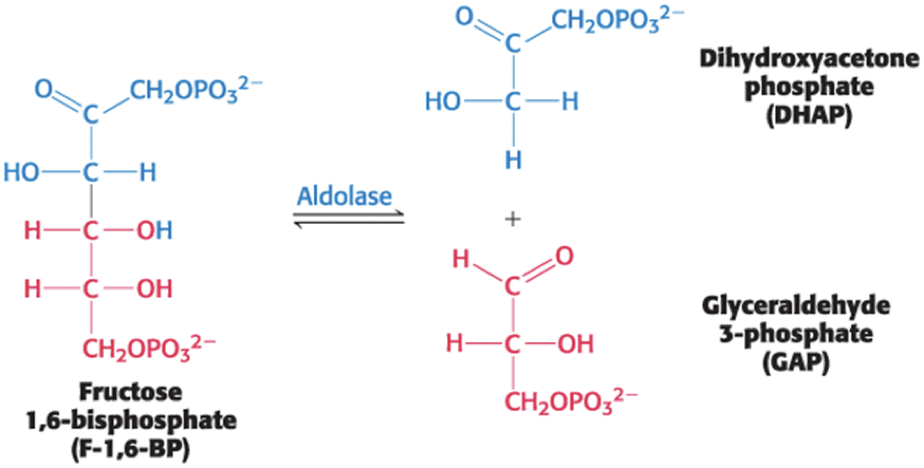
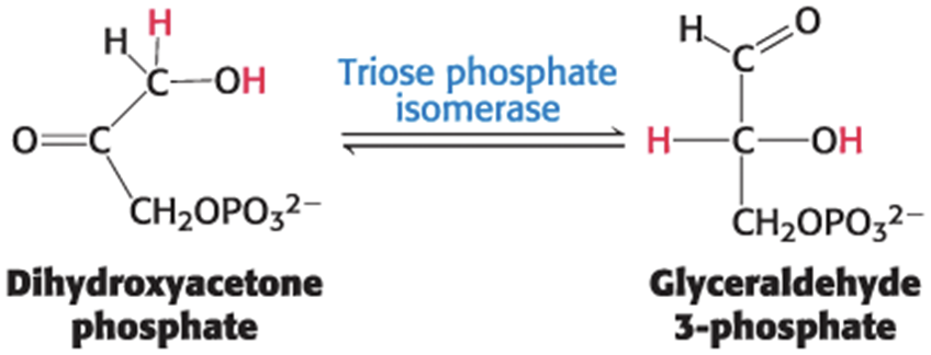
Dihydroxyacetone Phosphate Is Isomerized to Glyceraldehyde 3-Phosphate
• Only GAP can be processed to pyruvate to yield ATP.
• Triose phosphate isomerase (TPI) catalyzes the isomerization of DHAP to GAP.
– readily reversible, but proceeds to GAP because subsequent glycolysis reactions remove GAP
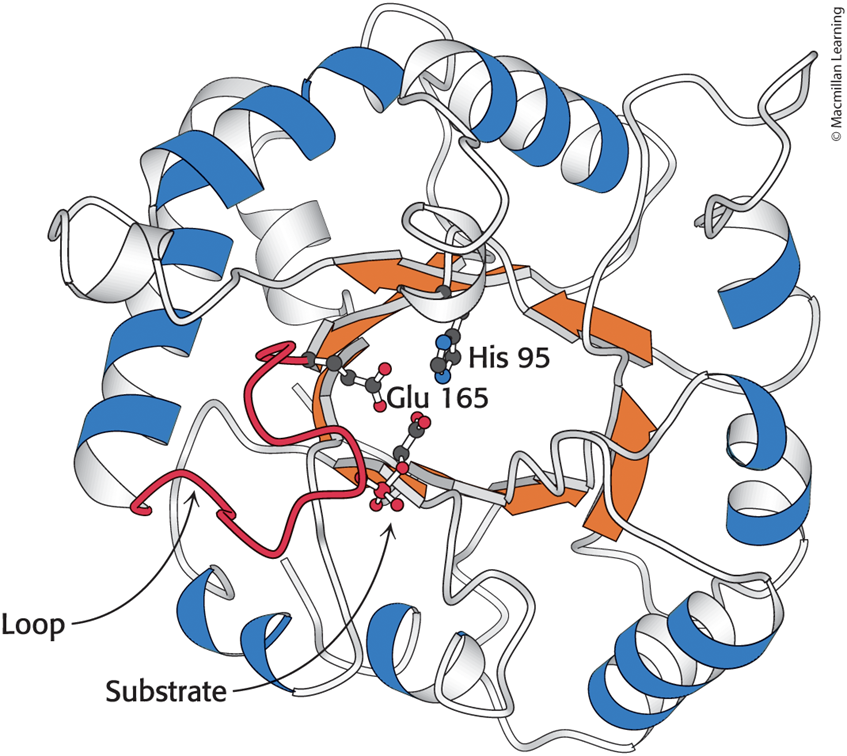
Triose Phosphate Isomerase Is an Example of a Common "Barrel" Motif
• The active site is buried in the center.
Mechanism: Triose Phosphate Isomerase Salvages a Three-Carbon Fragment
• TPI catalyzes the transfer of a hydrogen atom from carbon 1 to carbon 2.
– This is an intramolecular oxidation–reduction.
– Isomerization proceeds through an enediol intermediate.
The Catalytic Mechanism of Triose Phosphate Isomerase
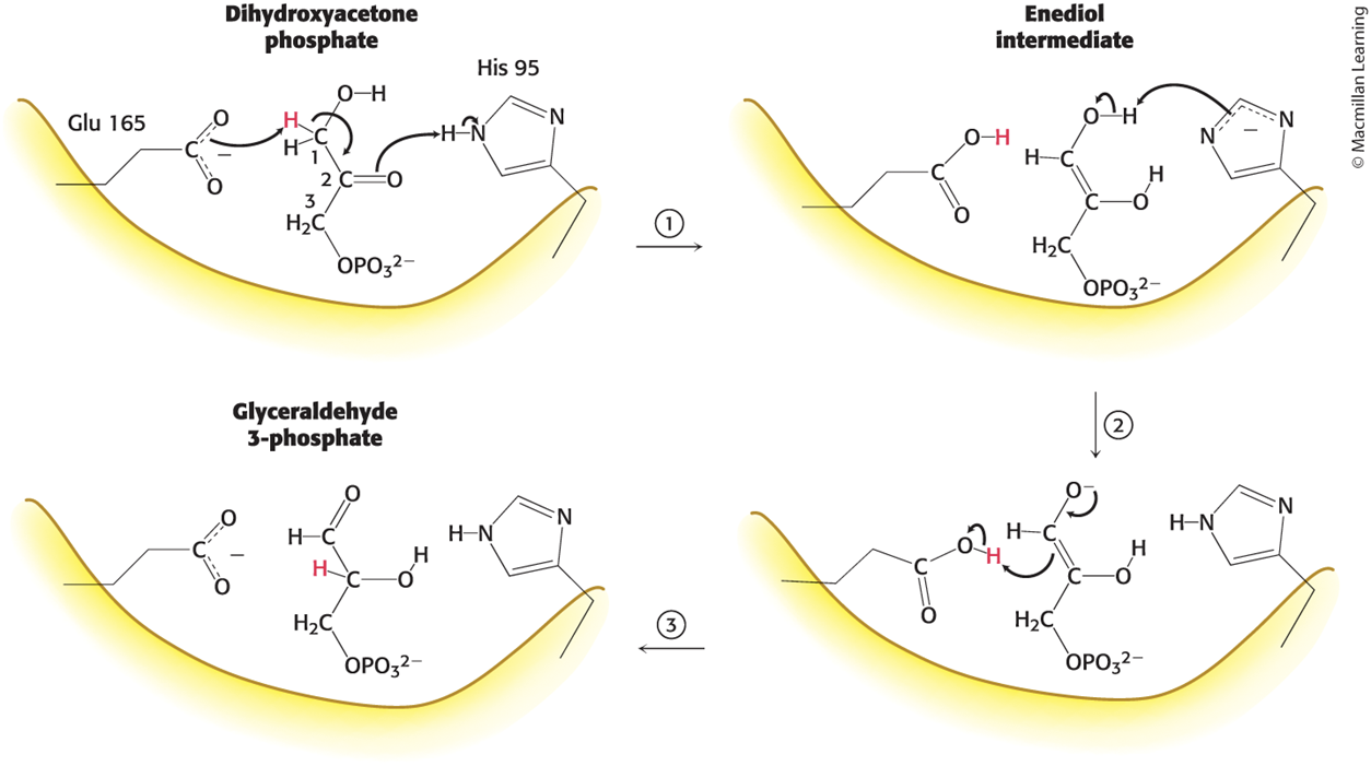
The Roles of Glu 165 and His 95
• Glu 165 plays the role of a general acid–base catalyst.
• His 95 assists catalysis by donating a proton to stabilize the negative charge that develops on the C-2 carbonyl group.
Features of Triose Phosphate Isomerase
• TPI accelerates isomerization by a factor of 1010 compared with a simple base catalyst.
– The kcat /KM ratio for the isomerization of GAP is close to the diffusion-controlled limit.
– TPI is a kinetically perfect enzyme.
• TPI suppresses an undesired side reaction.
The Second Stage of Glycolysis Generates ATP and NADH
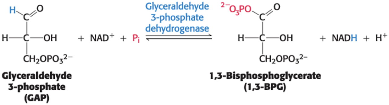
Stage 2 Begins: The Oxidation of an Aldehyde Powers the Formation of a Compound with High Phosphoryl-Transfer Potential
• Glyceraldehyde 3-phosphate dehydrogenase (GAPDH) catalyzes the conversion of GAP into 1,3-bisphosphoglycerate (1,3-BPG).
– 1,3-BPG has high phosphoryl-transfer potential.
The Glyceraldehyde 3-Phosphate Dehydrogenase Reaction Is the Sum of Two Processes
• Process 1: the oxidation of the aldehyde to a carboxylic acid by NAD+
– thermodynamically favorable (∆G°′ of approximately −50 kJ mol−1)
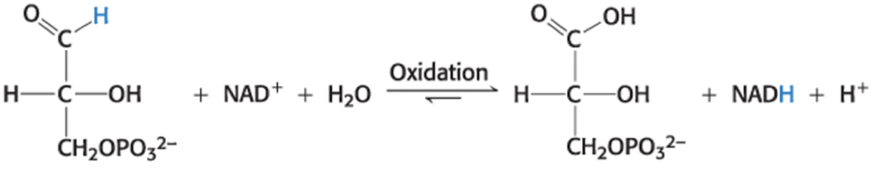
• Process 2: the joining of the carboxylic acid and orthophosphate to form the acyl-phosphate product
– thermodynamically unfavorable (∆G°′ of approximately +50 kJ mol−1)
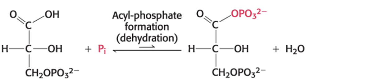
The Glyceraldehyde 3-Phosphate Dehydrogenase Reactions Are Coupled
• The reactions are linked by the formation of an energy-rich thioester intermediate in the active site of GAPDH.
– preserves much of the free energy released in the oxidation reaction
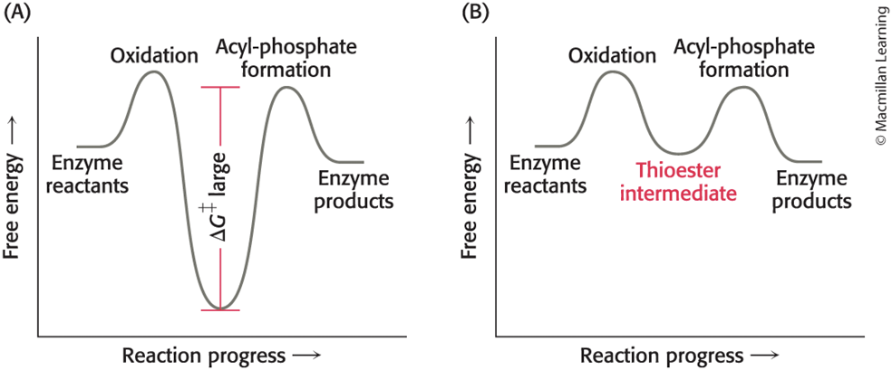
Mechanism: Phosphorylation Is Coupled to the Oxidation of Glyceraldehyde 3-Phosphate by a Thioester Intermediate
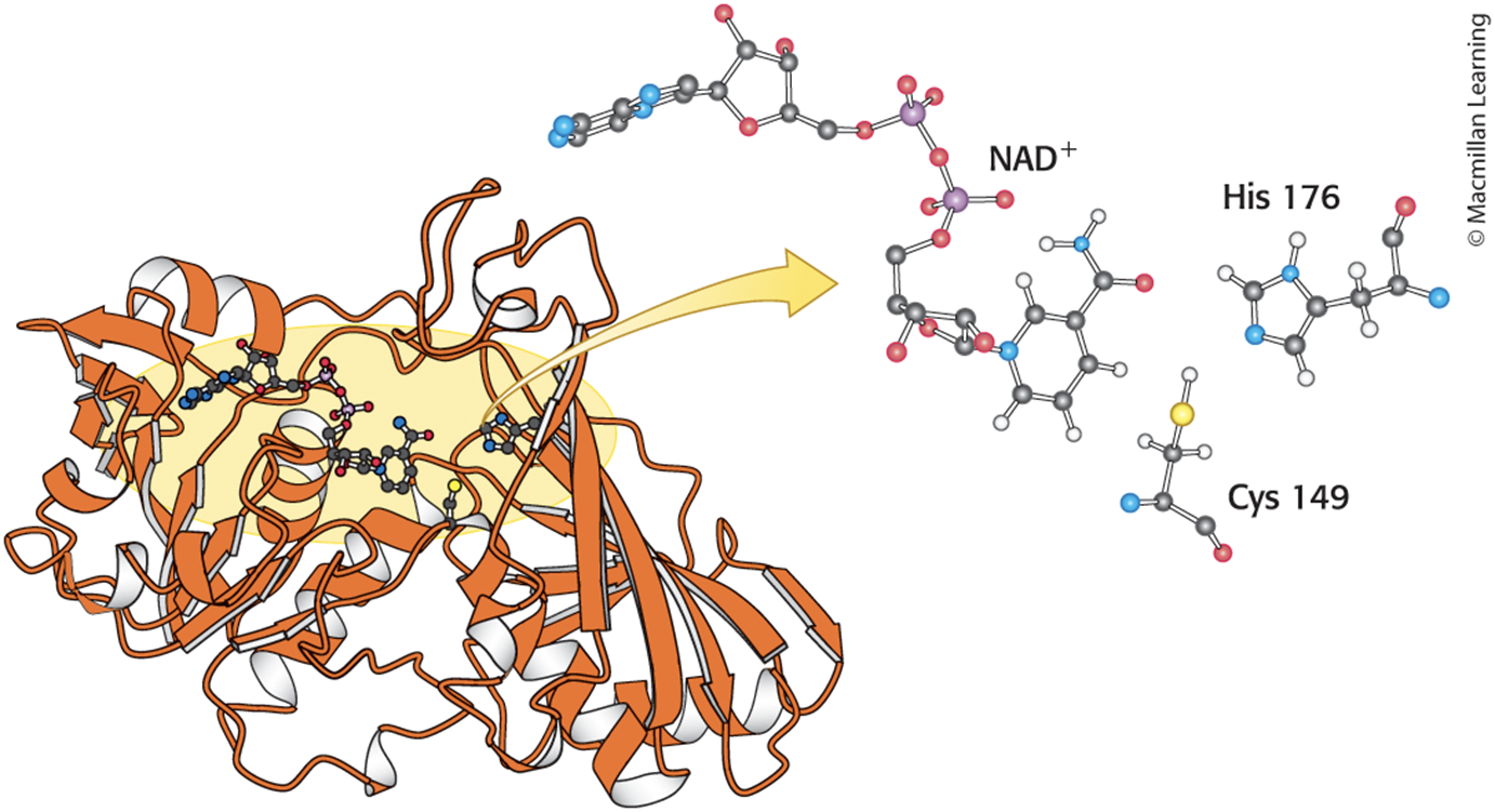
• The active site of GAPDH includes:
– a reactive Cys residue.
– NAD+
– a crucial His.
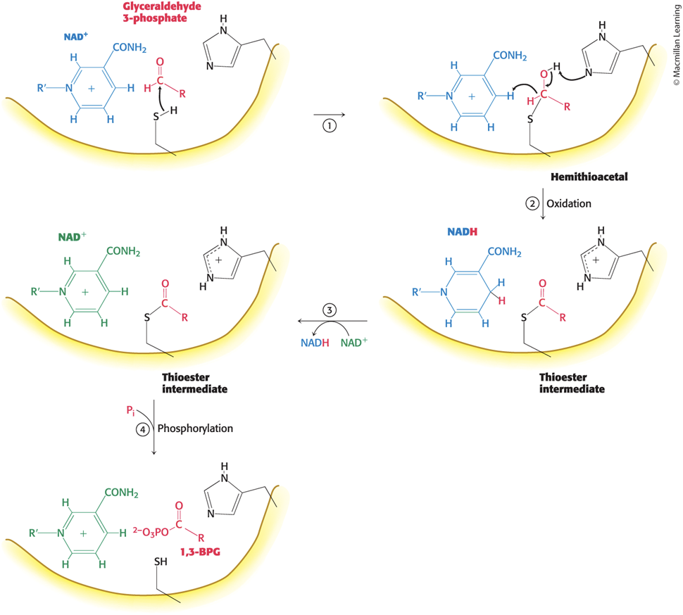
The Four Steps in the GAPDH Reaction Mechanism
• Step 1: the aldehyde substrate reacts with the sulfhydryl group of Cys 149 on the enzyme to form a hemithioacetal
• Step 2: the hydride ion is transferred to NAD+ bound to the enzyme to yield NADH and a thioester intermediate
– favored by the deprotonation of the hemithioacetal by His176
• Step 3: NADH leaves and is replaced by NAD+
– NAD+ polarizes the thioester intermediate to facilitate the attack by orthophosphate
• Step 4: orthophosphate attacks the thioester to form 1,3-BPG and free the cysteine residue
The Active Site of GAPDH
The Catalytic Mechanism of GAPDH
• energy released by carbon oxidation is converted into high phosphoryl-transfer potential
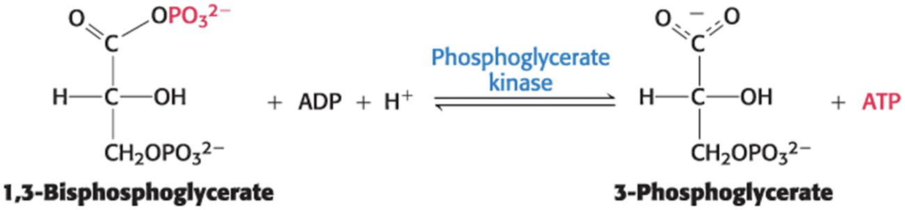
ATP Is Formed by Phosphoryl Transfer from 1,3-Bisphosphoglycerate
• 1,3-BPG has greater phosphoryl-transfer potential than ATP.
– can be used to power the ATP synthesis
• Phosphoglycerate kinase catalyzes the transfer of the phosphoryl group from 1,3-BPG to ADP.
– yields ATP and 3-phosphoglycerate
• ATP is formed by by substrate-level phosphorylation.
Additional ATP Is Generated with the Formation of Pyruvate
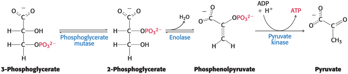
• Phosphoglycerate mutase catalyzes the conversion of 3-phosphoglycerate to 2-phosphoglycerate.
– requires catalytic amounts of 2,3-BPG to maintain an active-site His residue in a phosphorylated form
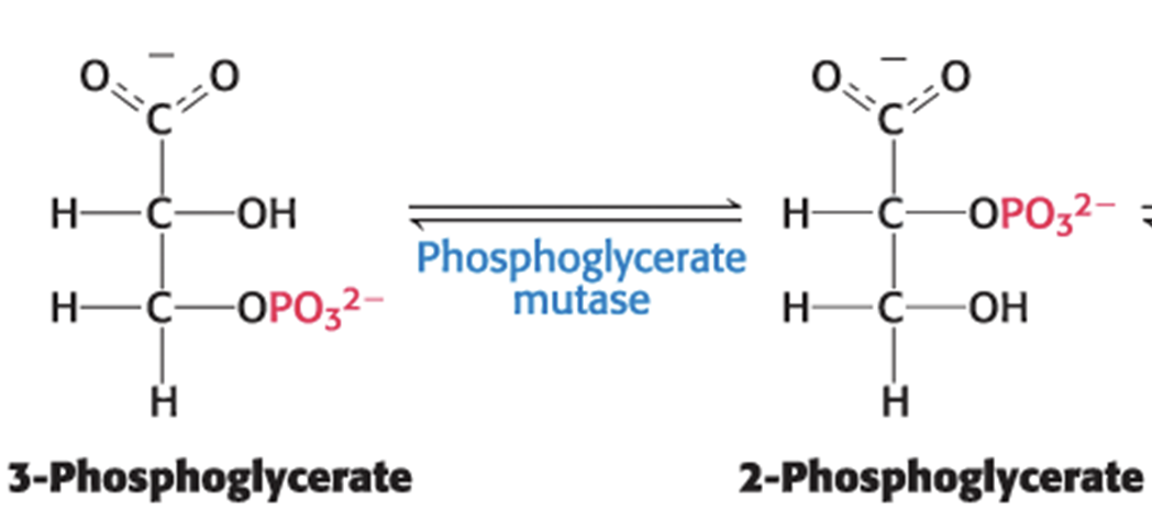
The Phosphoglycerate Mutase Reaction
• Step 1: Phosphoryl group is transferred to 3-phosphoglycerate to form 2,3-BPG

• Step 2: 2,3-BPG is converted to 2-phosphoglycerate
– The mutase retains the phosphoryl group to regenerate the modified histidine.

• The net reaction is

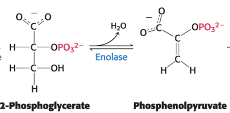
2-Phosphoglycerate Is Dehydrated to Phosphenolpyruvate
• Enolase catalyzes the dehydration of 2-phosphoglycerate to phosphoenolpyruvate (PEP).
• PEP has high phosphoryl-transfer potential because the phosphoryl group traps the molecule in its unstable enol form.
Phosphenolpyruvate Is Converted to Pyruvate
• Pyruvate kinase catalyzes the transfer of a phosphoryl group from PEP to ADP, generating pyruvate and ATP.
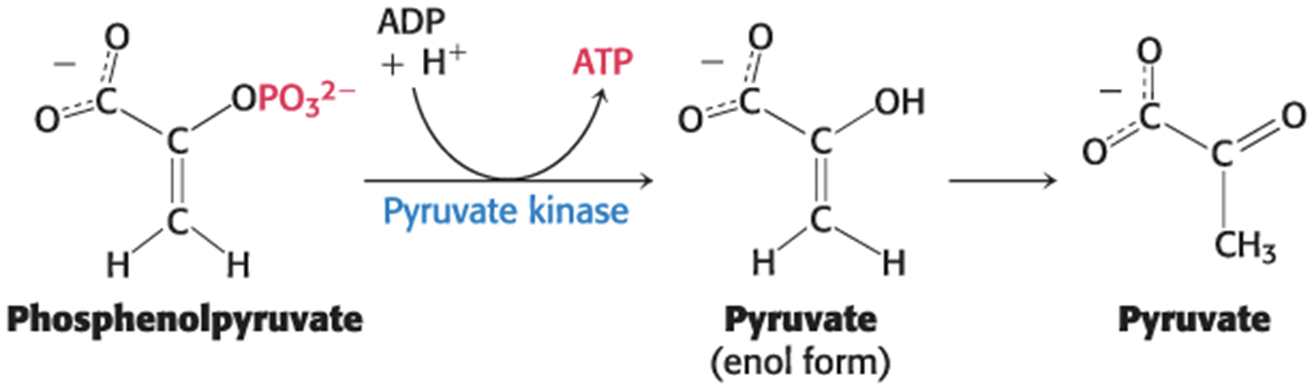
The Final Three Reactions in Glycolysis
Two ATP Molecules Are Formed in the Conversion of Glucose into Pyruvate
• The net reaction in glycolysis is
Glucose + 2 Pi + 2 ADP + 2 NAD+ -> 2 pyruvate +2 ATP + 2 NADH + 2 H+ + 2 H2O
• The energy released in glycolysis is about −90 kJ mol−1
Reactions of Glycolysis
TABLE 16.2 Reactions of glycolysis
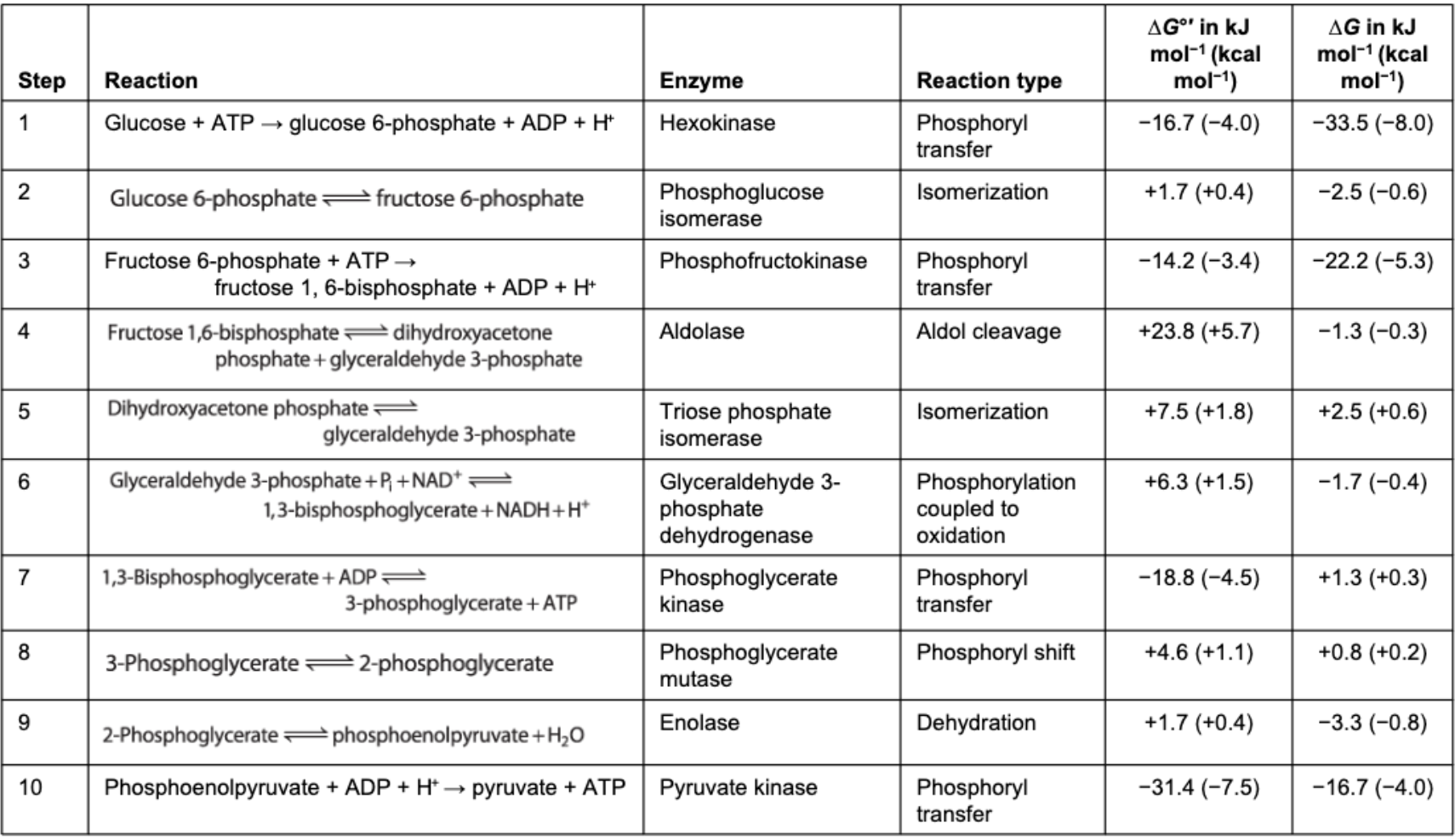 Note: ∆G, the actual free-energy change, has been calculated from ∆G°′ and known concentrations of reactants under typical physiological conditions. Glycolysis can proceed only if the ∆G values of all reactions are negative. The small positive ∆G values of three of the above reactions indicate that the concentrations of metabolites in vivo in cells undergoing glycolysis are not precisely known.
NAD+ Is Regenerated from the Metabolism of Pyruvate
• NAD+ is derived from the vitamin niacin (B3).
• GAPDH reduces NAD+ to NADH.
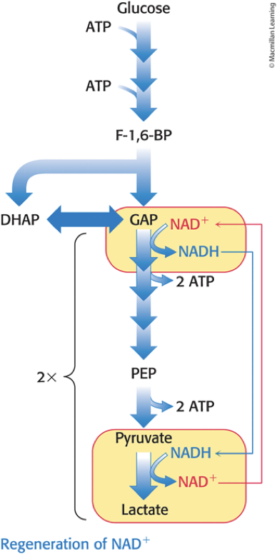
• NAD+ must be regenerated for glycolysis to proceed.
• NAD+ can be regenerated by:
– oxidation of pyruvate to CO2 in the presence of O2.
– fermentation of pyruvate to ethanol or lactate in the absence of O2.
• fermentation = an ATP-generating process in which organic compounds act as both electron donors and acceptors
Pyruvate Has Many Possible Fates
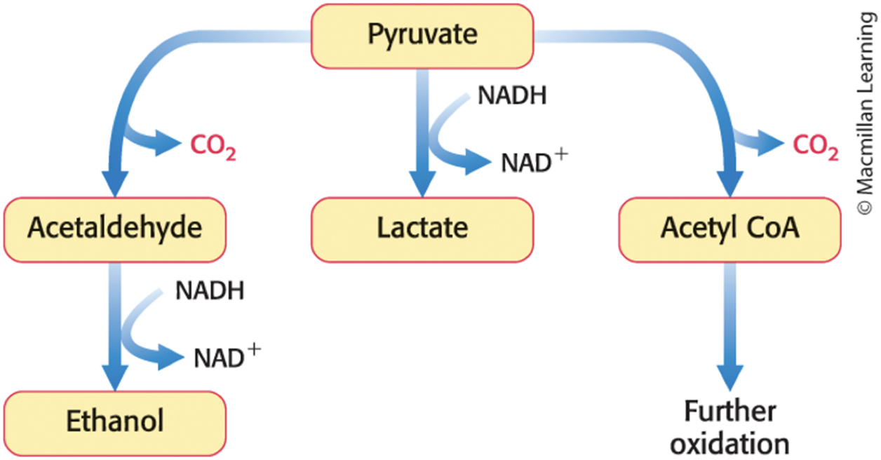
Fermentation
Krebs cycle
Ethanol Fermentation
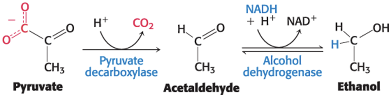
• ethanol fermentation = the conversion of glucose into ethanol in anerobic conditions
– occurs in yeast and other microorganisms
– used for brewing and winemaking
• Step 1: pyruvate decarboxylase catalyzes the decarboxylation of pyruvate
– requires the coenzyme thiamine pyrophosphate that is derived from the vitamin thiamine (B1)
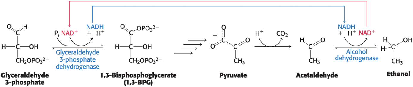
• Step 2: alcohol dehydrogenase catalyzes the reduction of acetaldehyde to ethanol by NADH
– regenerates NAD+
The Reactions of Ethanol Fermentation
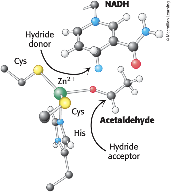
The Active Site of Alcohol Dehydrogenase
• The active site contains a zinc ion coordinated to the sulfur atoms of two Cys residues and a nitrogen atom of His.
• Zinc polarizes the carbonyl group of the substrate to favor the transfer of a hydride from NADH.
Two ATP Molecules Are Formed in the Conversion of Glucose to Ethanol
• The net reaction in this anaerobic process is
Glucose + 2 Pi + 2ADP + 2H+ -> 2 ethanol + 2 CO2 + 2 ATP + 2 H2O
• NADH generated by the oxidation of GAP is consumed in the reduction of acetaldehyde to ethanol.
The NADH Produced in Glycolysis Must Be Reoxidized to NAD+ for the Glycolytic Pathway to Continue
Lactic Acid Fermentation
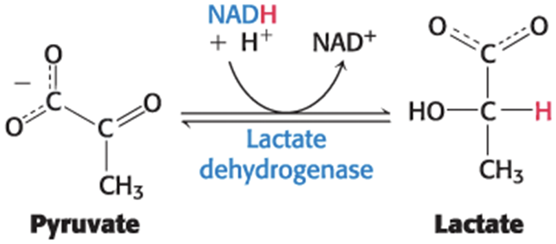
• lactic acid fermentation = the conversion of glucose into lactate in anerobic conditions
– occurs in microorganisms and animal tissues, including skeletal muscles
• Lactase dehydrogenase catalyzes the reduction of pyruvate by NADH to lactase.
Two ATP Molecules Are Formed in the Conversion of Glucose to Lactate
• The net reaction in this anaerobic process is
Glucose + 2Pi + 2 ADP + 2 H+ -> 2 ethanol + 2 CO2 + 2 ATP + 2 H2O
• NADH generated by the oxidation of GAP is consumed in the reduction of pyruvate.
NAD+ Is Regenerated by Lactate Dehydrogenase During Lactic Acid Fermentation
Oxidation by the Citric Acid Cycle and the Electron Transport Chain
• Pyruvate can be metabolized under aerobic conditions through the citric acid cycle and the electron-transport chain.
– forms CO2, H2O, and a great deal of energy
– the transfer of electrons to O2 through the electron-transport chain regenerates NAD+
• Pyruvate dehydrogenase complex catalyzes the oxidative decarboxylation of pyruvate to acetyl coenzyme A (acetyl CoA) in mitochondria
Fermentations Provide Usable Energy in the Absence of Oxygen
• obligate anaerobes = organisms that cannot survive in the presence of O2
– example: the bacterium Clostridium perfringens, the cause of gangrene
• facultative anaerobes = organisms that metabolize glucose aerobically when O2 is present and perform fermentation when O2 is absent
• Microorganisms are capable of generating a wide array of molecules as end points of fermentation.
Examples of Pathogenic Obligate Anaerobes
TABLE 16.3 Examples of pathogenic obligate anaerobes

Starting and Ending Points of Various Fermentations
TABLE 16.4 Starting and ending points of various fermentations
Glucose → Lactate
Lactate → Acetate
Glucose → Ethanol
Ethanol → Acetate
Arginine→ Carbon dioxide
Pyrimidines→ Carbon dioxide
Purines → Formate
Ethylene glycol→ Acetate
Threonine → Propionate
Leucine → 2-Alkylacetate
Phenylalanine→ Propionate
Note: The products of some fermentations are the substrates for others.
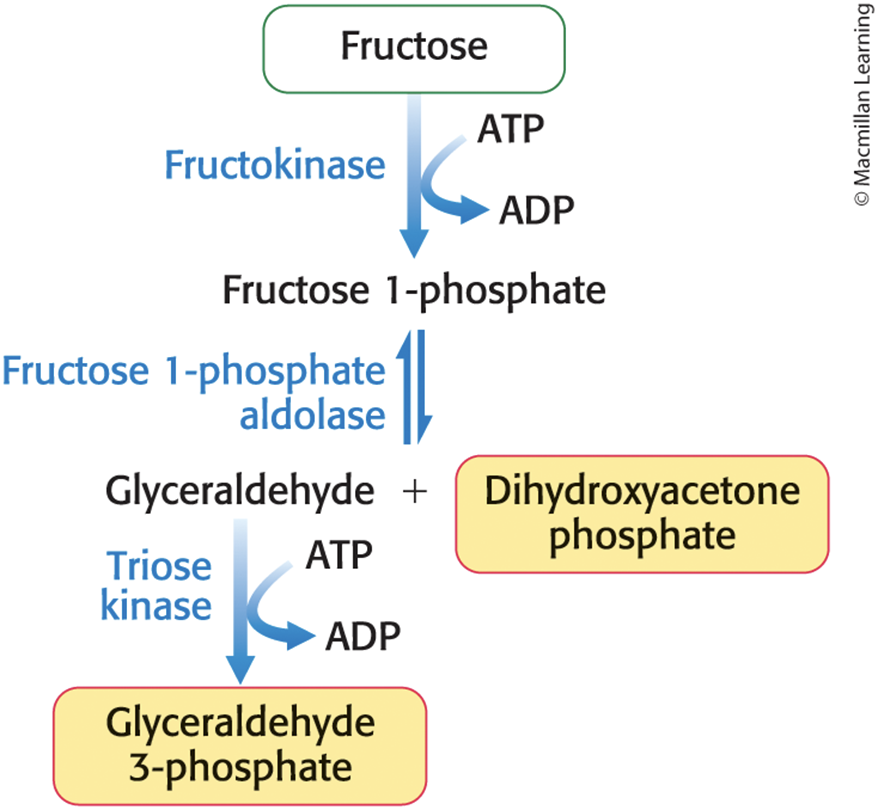
Fructose is Converted into Glycolytic Intermediates by Fructokinase
• Fructose is found in table sugar and high fructose corn syrup.
• To be metabolized, fructose must be converted into a metabolite of glucose.
• The fructose 1-phosphate pathway metabolizes fructose in the liver.
Additional Sugars Can Enter Glycolysis at a Variety of Points
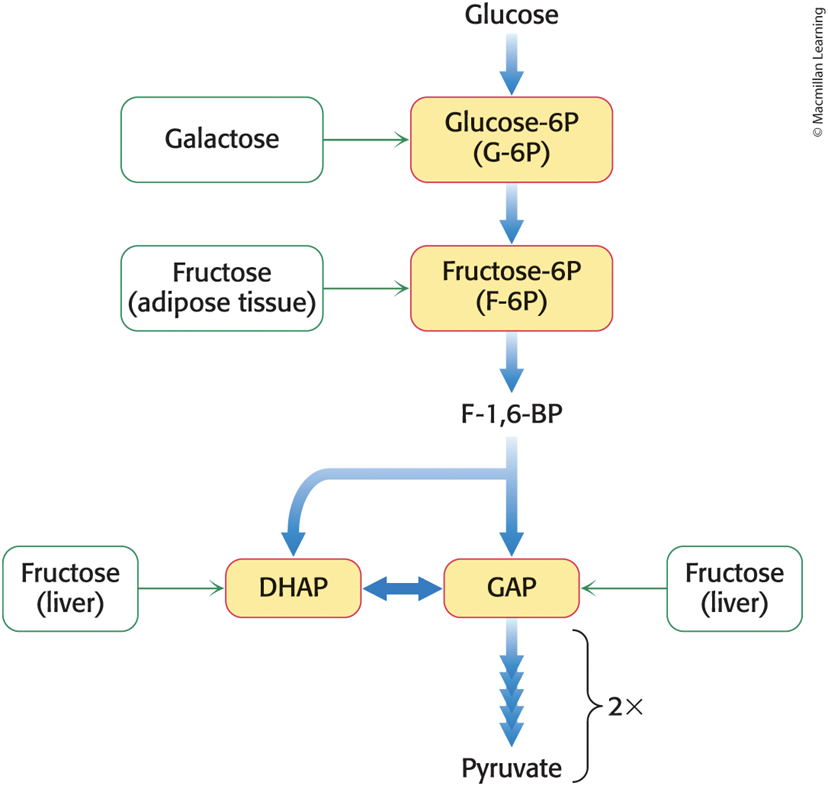
Fructose Metabolism in the Liver
Outcomes of Excessive Fructose Consumption
• Epidemiological and clinical studies have previously linked excess consumption of fructose to fatty liver, insulin insensitivity, obesity, and type 2 diabetes.
– not supported by recent meta-analyses
• Overconsumption of sugars and fats is still strongly associated with these clinical outcomes.
• Excess pyruvate is converted into acetyl CoA and then into fatty acids.
Galactose Is Converted into Glucose 6-Phosphate
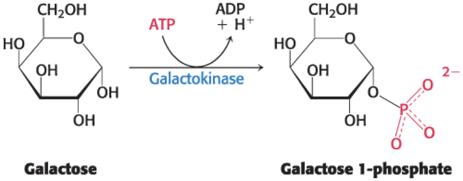
• The galactose–glucose interconversion pathway, a four-step pathway, converts galactose to glucose 6-phosphate.
• Step 1 is the phosphorylation of galactose by galactokinase.
Glucose Is Converted into UDP-Glucose
• Uridine diphosphate glucose (UDP-glucose) is an activated intermediate in the synthesis of carbohydrates.
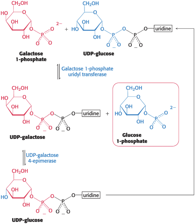
The Galactose–Glucose Interconversion Pathway
• The sum of the reactions of the galactose–glucose conversion pathway is
Galactose + ATP -> glucose 1-phosphate + ADP + H+
• UDP-glucose is not consumed in the conversion of galactose into glucose.
• Phosphoglucomutase isomerizes glucose 1-phosphate to glucose 6-phosphate.

Galactose Can Be Highly Toxic with a Defective Metabolic Pathway
• galactosemias = rare disorders that interfere with the metabolism of galactose
– causes vomiting and diarrhea after milk consumption, enlargement of the liver, jaundice, cataracts, lethargy, and delayed neurological development
– often results from an inherited deficiency in galactose 1-phosphate uridyl transferase activity
• Treatment is to remove galactose and lactose from the diet.
Cataracts Are Evident as the Clouding of the Lens
• A cataract is the clouding of the lens of the eye due to pathological protein aggregation.
Cataract Formation
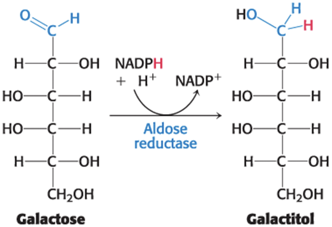
• Aldose reductase catalyzes the reduction of galactose to galactitol in the lens when the transferase is not active.
• Galactitol is poorly metabolized and accumulates.
• Water diffuses into the lens to maintain osmotic balance, triggering the formation of cataracts.
Many Adults Worldwide Are Intolerant of Milk Because They Are Deficient in Lactase
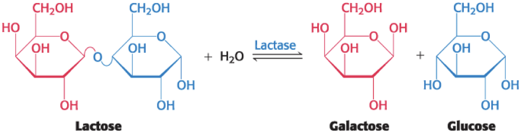
• lactase = an enzyme that cleaves lactose into glucose and galactose
• Lactose intolerance is most commonly caused by a deficiency of lactase.
• treatment involves avoiding lactose products or ingesting lactase with milk products
Lactobacillus Is One Example of an Industrially Useful Anaerobic Bacterium
• Lactose is a good energy source for microorganisms in the colon which ferment it to lactic acid while generating methane (CH4) and hydrogen gas (H2)
– causes gut distension and flatulence
– lactate and undigested lactose are osmotically active and draw water into the intestine, causing diarrhea
Section 16.3 The Glycolytic Pathway Is Tightly Controlled
• Enzymes catalyzing irreversible reactions in metabolic pathways are control sites.
– controlled by allosteric effectors, covalent modification, or by regulating transcription
• In glycolysis, three enzymes catalyze irreversible reactions:
– hexokinase
– phosphofructokinase
– pyruvate kinase
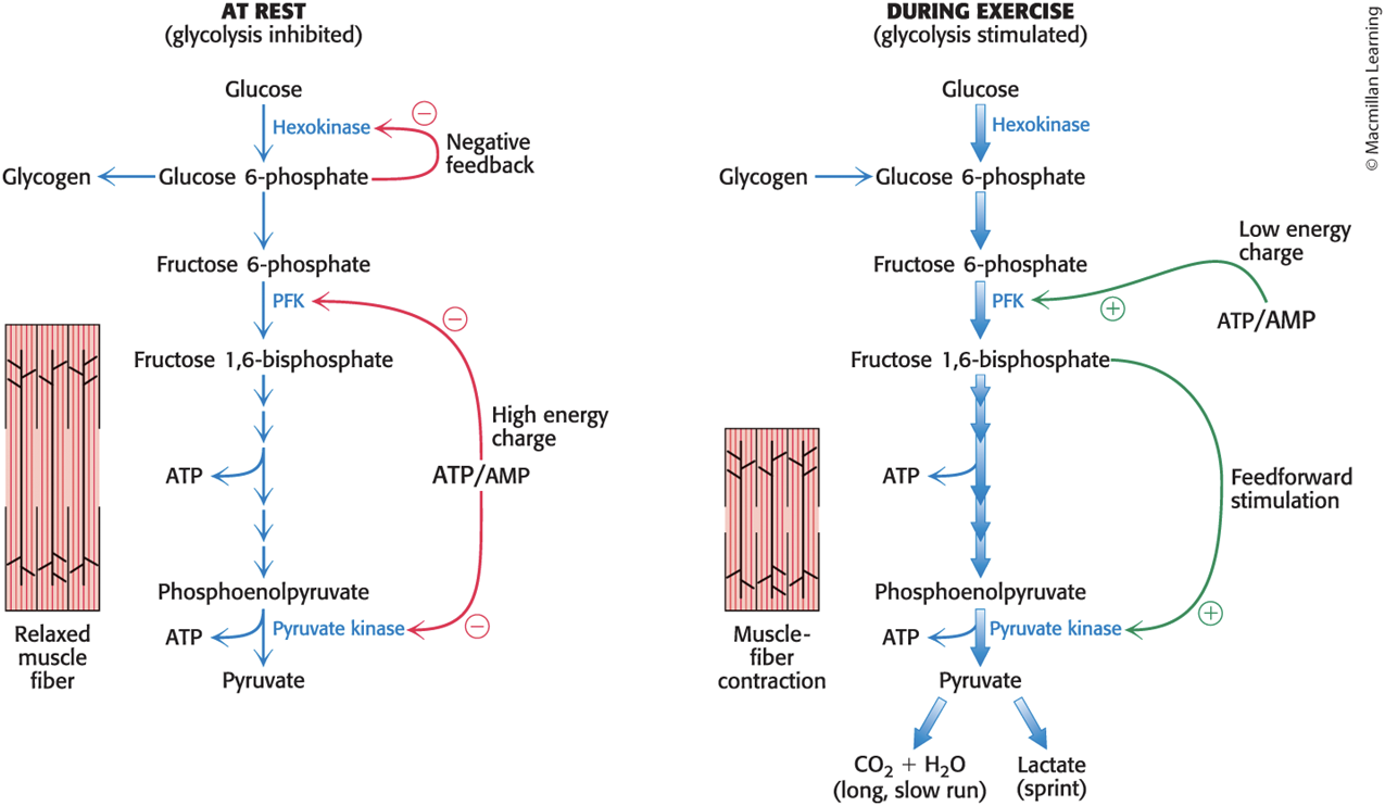
Glycolysis in Muscle Is Regulated to Meet the Need for ATP
• Glycolysis in skeletal muscle provides ATP primarily to power contraction.
• The primary control of muscle glycolysis is the ratio of ATP to AMP.
Regulation of Phosphofructokinase by AMP and ATP in Muscle
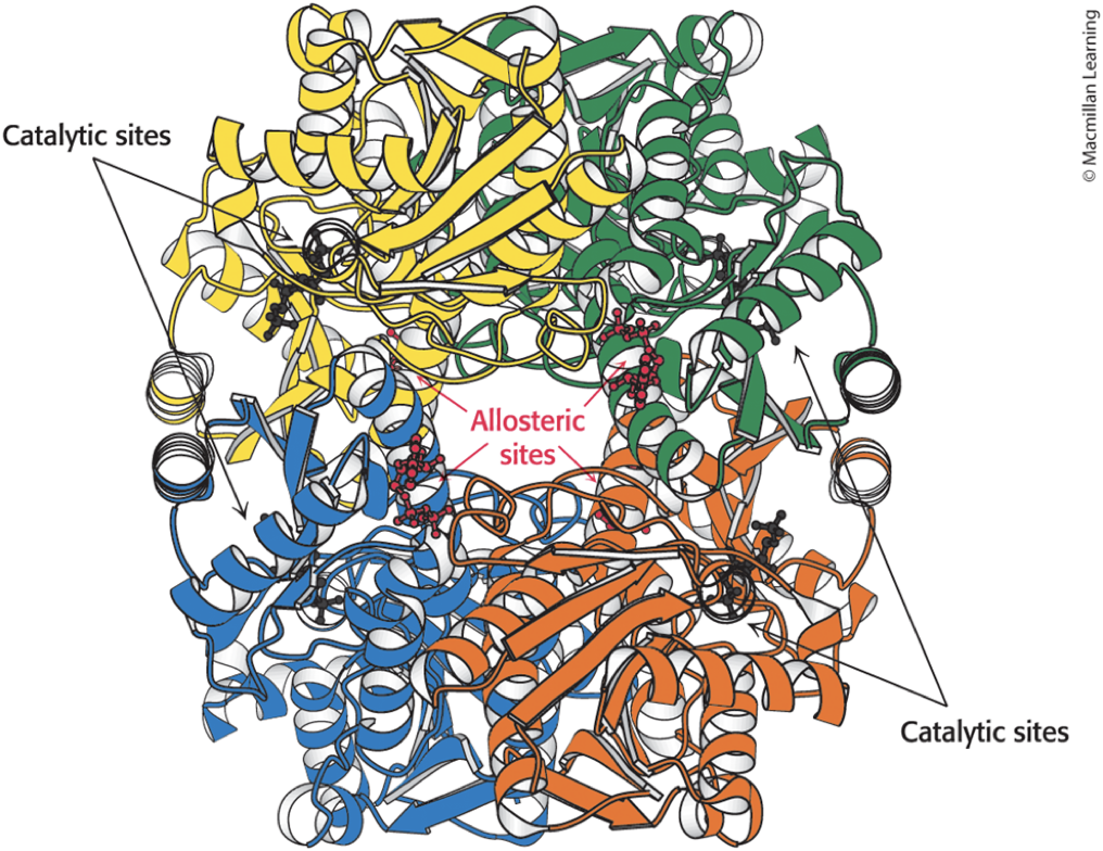
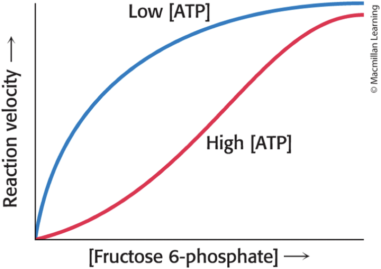
• High levels of ATP allosterically inhibit phosphofructokinase.
– Binding to a specific regulatory site lowers the enzyme's affinity for F-6P.
• AMP reverses the inhibitory action of ATP.
• Enzyme activity increases when the ATP/AMP ratio is lowered.
• committed step = the first irreversible reaction unique to the glycolytic pathway
– example: phosphorylation of F-6P to F-l,6-BP by phosphofructokinase
Structure of Phosphofructokinase
A High Level of ATP Allosterically Inhibits Phosphofructokinase
• High [ATP] converts the hyperbolic binding curve of F-6P into a sigmoidal one.
Regulation of Phosphofructokinase by pH in Muscle
• A decrease in pH inhibits phosphofructokinase activity by augmenting the inhibitory effect of ATP.
– occurs when muscle is functioning anaerobically and producing excessive lactic acid
– prevents muscle damage from too much acid
AMP as a Positive Regulator of Phosphofructokinase
• ADP is not the positive regulator of phosphofructokinase because adenylate kinase forms ATP from ADP

• AMP serves as the signal for the low-energy state.
• AMP as an allosteric regulator provides sensitive control because in the cell [ATP] > [ADP] > [AMP], meaning small changes in [ATP] result in larger changes in [AMP].
Regulation of Hexokinase in Muscle
• Hexokinase is inhibited by its product, G-6P
– high [G-6P] signals that the cell no longer requires glucose for energy or synthesis of glycogen
– leads to glucose remaining in blood
• Inhibition of phosphofructokinase leads to the inhibition of hexokinase because a rise in [F-6P] leads to a rise in [G-6P] as they are in equilibrium.
Regulation of Pyruvate Kinase in Muscle
• Pyruvate kinase is allosterically inhibited by ATP.
– slows glycolysis when the energy charge is high
• feedforward stimulation (feedforward activation) = occurs when products of a preceding irreversible step activate the enzyme
– example: when the pace of glycolysis increases, F-l,6-BP activates the kinase
Regulation of Glycolysis in Muscle
The Regulation of Glycolysis in the Liver Illustrates the Biochemical Versatility of the Liver
• The liver maintains blood-glucose concentration, generates reducing power for biosynthesis, and synthesizes biochemicals.
• Regulation is more complex than in the muscle.
Regulation of Phosphofructokinase in Liver
• Liver phosphofructokinase can be regulated by ATP.
– not an important metabolic signal as the liver does not experience sudden ATP needs
• Liver phosphofructokinase can be regulated by pH.
– not an important metabolic signal as the liver does not produce lactate
• Phosphofructokinase is inhibited by citrate, an early intermediate in the citric acid cycle, by enhancing the inhibitory effect of ATP.
– High citrate means biosynthetic precursors are abundant.
• Phosphofructokinase is activated by fructose 2,6-bisphosphate (F-2,6-BP).
– Binding of F-2,6-BP increases the affinity of the enzyme for F-6P and diminishes the inhibitory effect of ATP.
• High F-6P accelerates the synthesis of F-2,6-BP.
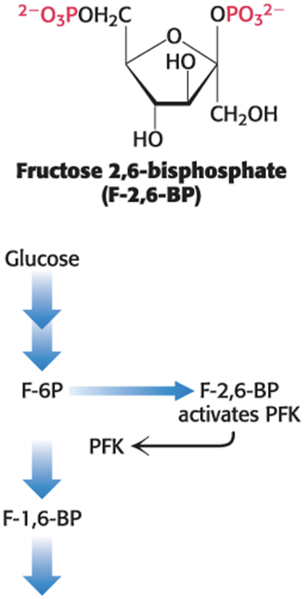
Activation of Phosphofructokinase by Fructose 2,6-Bisphosphate
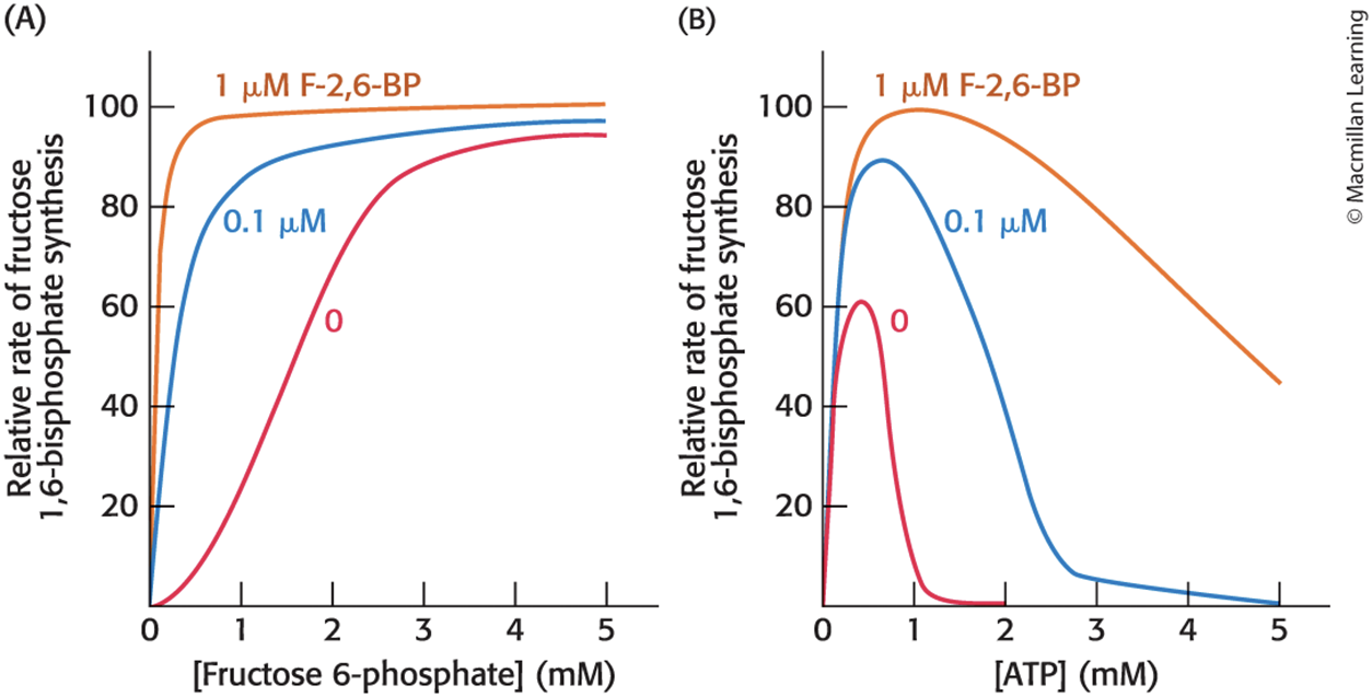
Regulation of Hexokinase and Glucokinase in Liver
• Hexokinase is inhibited by its product, G-6P, as in muscle.
• glucokinase = a specialized isozyme of hexokinase that provides G-6P for glycogen synthesis and fatty acid formation
– not inhibited by G-6P
– functions as a monomer, but displays sigmoidal kinetics
– phosphorylates glucose only when glucose is abundant
– inhibited by the liver-specific glucokinase regulatory protein (GKRP) when [glucose] is low
Regulation of Pyruvate Kinase in Liver
• Several isozymic forms of pyruvate kinase are present in mammals:
– L type = predominates in the liver
– M type = predominates in muscle and the brain
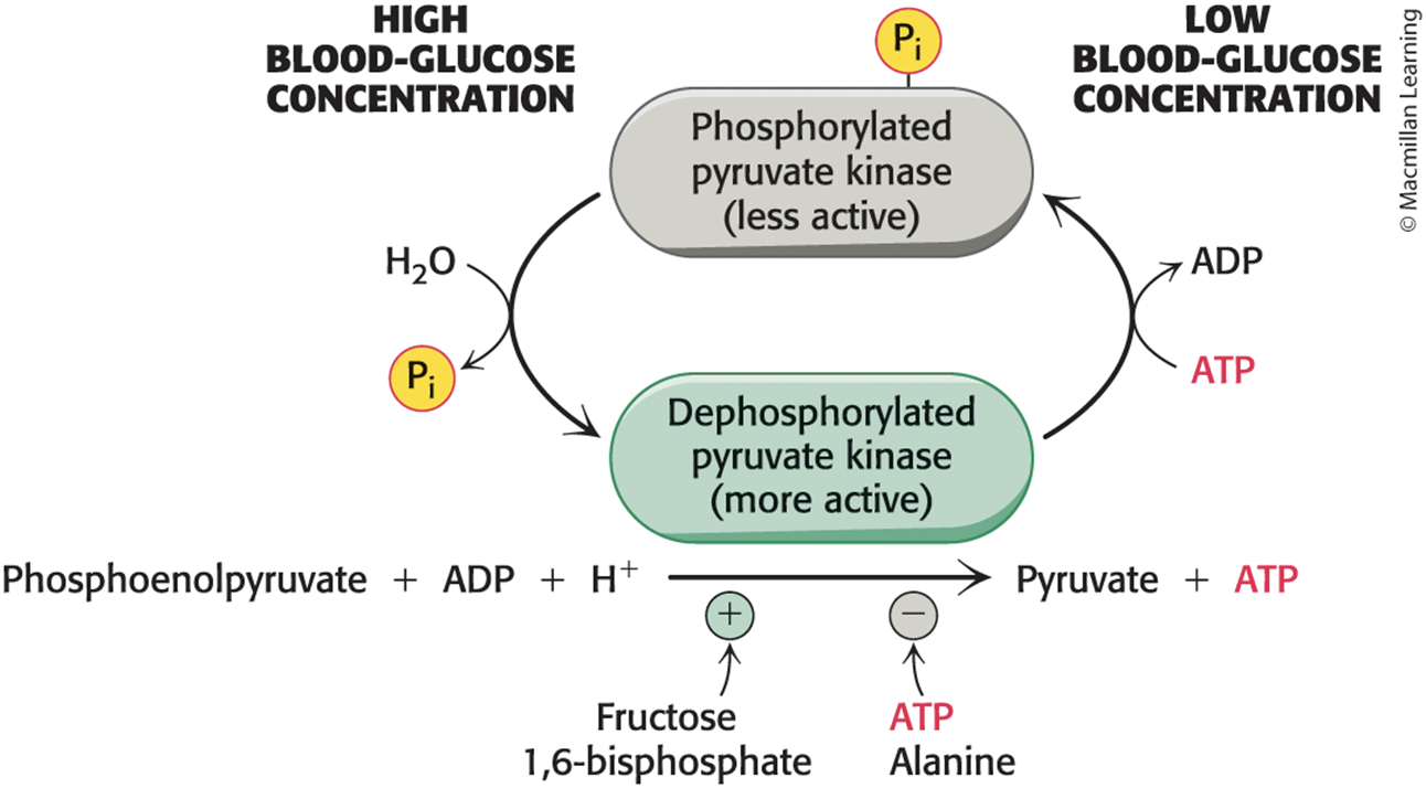
• Pyruvate kinase is allosterically inhibited by ATP and alanine (synthesized in one step from pyruvate).
• L form is controlled by reversible phosphorylation.
– When blood-glucose level is low, the glucagon-triggered cyclic AMP cascade leads to the phosphorylation of pyruvate kinase, which diminishes its activity.
Pyruvate Kinase Activity Is Controlled by Phosphorylation and Dephosphorylation
The Enzymes of Glycolysis Are Physically Associated with One Another
• Enzymes of glycolysis in eukaryotes are organized into complexes.
• substrate channeling = process that facilitates movement of substrates and products between enzymes
– increases enzyme efficiency
– prevents the release of any toxic intermediates
Aerobic Glycolysis Is a Property of Tumor Cells and Other Rapidly Growing Cells
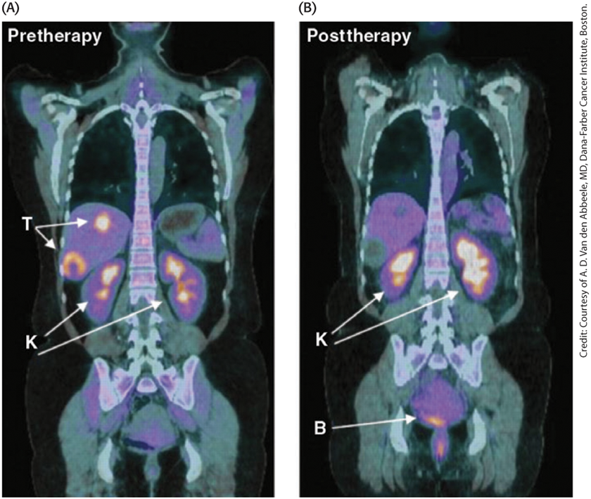
• Tumors display enhanced rates of glucose uptake and glycolysis.
• aerobic glycolysis (the Warburg effect) = process by which rapidly growing tumor cells metabolize glucose to lactate even in the presence of oxygen
• 2-18F-2-D-deoxyglucose (FDG) = a nonmetabolizable glucose analog that is detectable by a combination of positron emission tomography (PET) and computer-aided tomography (CAT)
Tumors Can Be Visualized by FDG and Positron Emission Tomography
Possible Selective Benefits of Aerobic Glycolysis
• Aerobic glycolysis generates lactic acid.
– Acidification of the tumor environment facilitates tumor invasion.
– Lactate impairs the activation of CD8+ T and NK immune system cells that normally attack the tumor.
• Increased glucose uptake and G-6P formation provide substrates for the NADPH-generating pentose phosphate pathway.
• As solid tumors grow, [O2] in their environment falls and the use of aerobic glycolysis reduces the dependence of cell growth on O2.
Cancer and Endurance Training Affect Glycolysis in a Similar Fashion
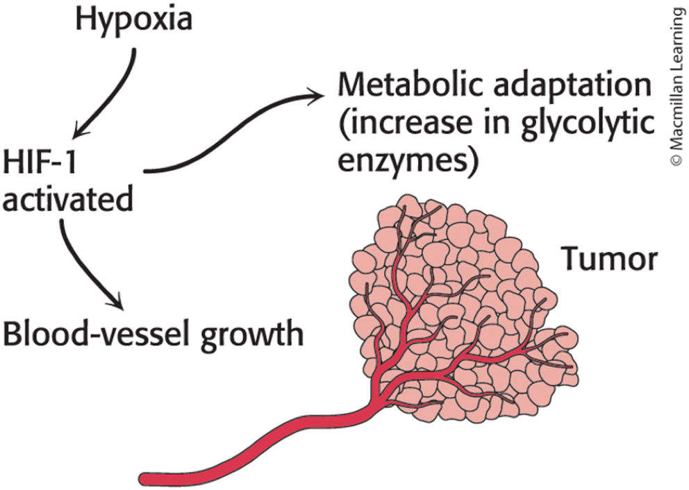
• hypoxia-inducible transcription factor (HIF-1) = transcription factor that facilitates aerobic glycolysis
– increases the expression of glycolytic enzymes, glucose transporters, and signal molecules
– cancer and anaerobic exercise training activate HIF-1
• vascular endothelial growth factor (VEGF) = signal molecule that facilitates the growth of blood vessels that will provide nutrients to the cells
Proteins in Glucose Metabolism Encoded by Genes Regulated by Hypoxia-Inducible Factor
TABLE 16.5 Proteins in glucose metabolism encoded by genes regulated by hypoxia-inducible factor

Hypoxia Alters Gene Expression in Tumors
Section 16.4 Glucose Can Be Synthesized from Noncarbohydrate Precursors
• gluconeogenesis = the synthesis of glucose from noncarbohydrate precursors
– converts pyruvate into glucose
– occurs mainly in the liver, with a small amount in the kidney and other tissues
– important during starvation and fasting as glucose is the primary fuel for the brain and the only fuel for red blood cells
Gluconeogenesis Precursors
• The major precursors for gluconeogenesis are lactate, amino acids, and glycerol.
– are converted to pyruvate or enter the pathway at later intermediates
• Lactate dehydrogenase converts lactate into pyruvate.
• Triacylglycerol hydrolysis in fat cells yields glycerol and fatty acids.
– Animals cannot convert fatty acids into glucose.
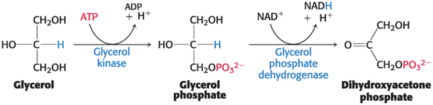
– Glycerol enters either the gluconeogenic or the glycolytic pathway as dihydroxyacetone phosphate.
The Conversion of Glycerol to Dihydroxyacetone Phosphate
Pathway of Gluconeogenesis
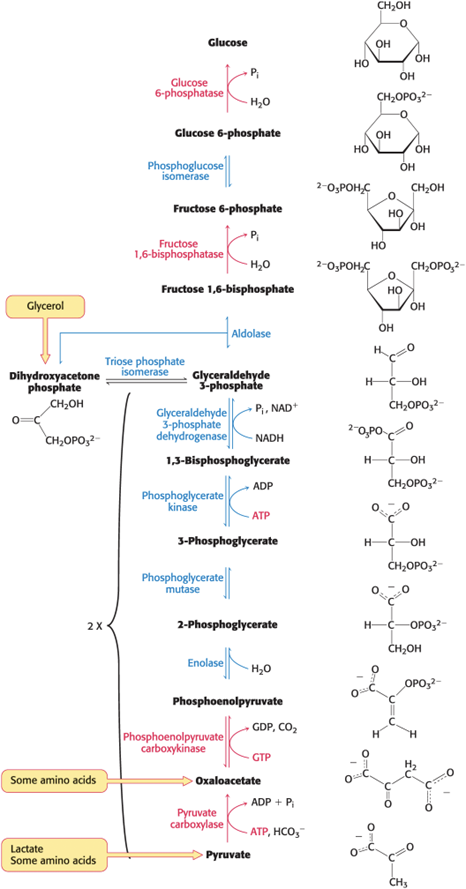
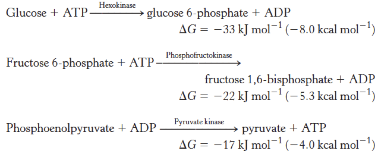
Gluconeogenesis Is Not a Reversal of Glycolysis
• The three irreversible steps in glycolysis must be bypassed in gluconeogenesis.
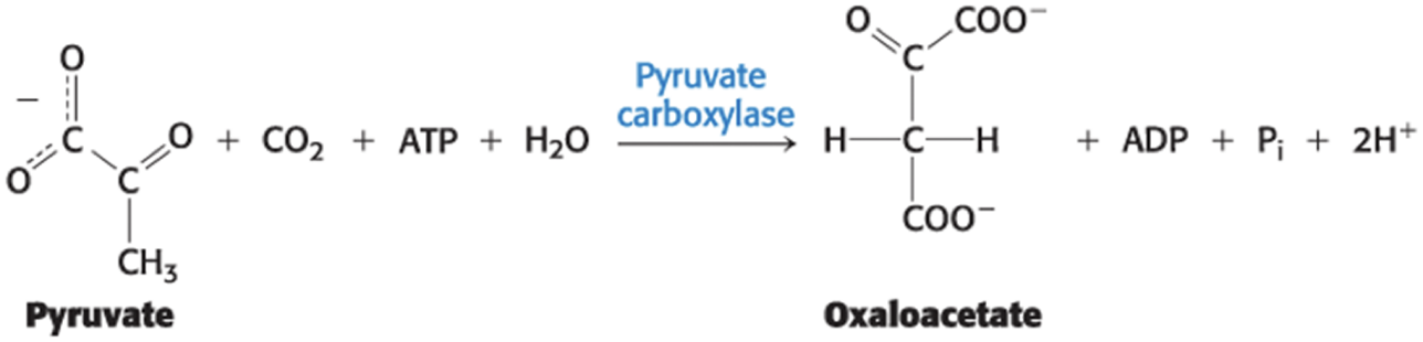
The Conversion of Pyruvate into Phosphoenolpyruvate Begins with the Formation of Oxaloacetate
• Pyruvate carboxylase catalyzes the carboxylation of pyruvate to oxaloacetate using a molecule of ATP.
– occurs in the mitochondria
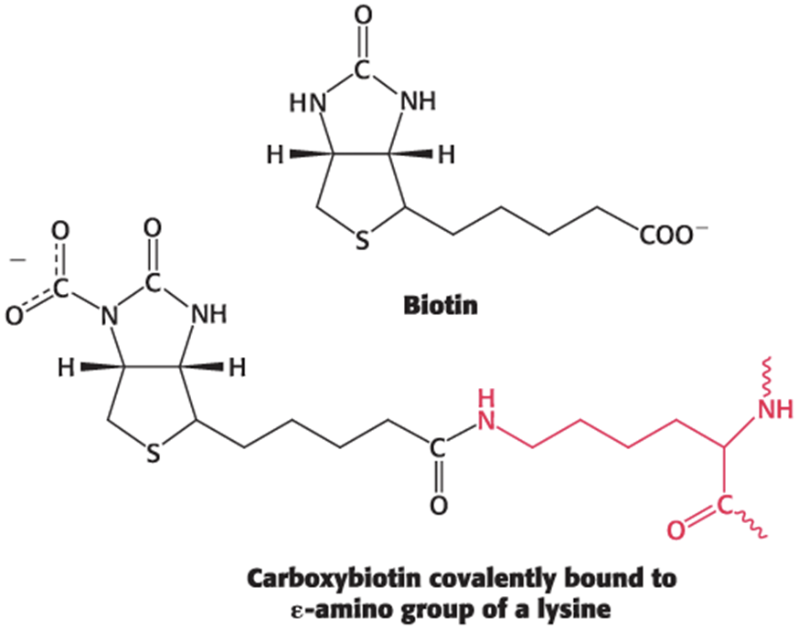
– requires biotin, a covalently attached prosthetic group, which serves as the carrier of activated CO2
Carboxybiotin Linked to Lysine Can Act as a Movable "Arm" Carrying Activated CO2 Groups
• The carboxylate group of biotin is linked to the ε-amino group of a specific Lys residue by an amide bond.
Carboxylation of Pyruvate
• takes place in three stages:

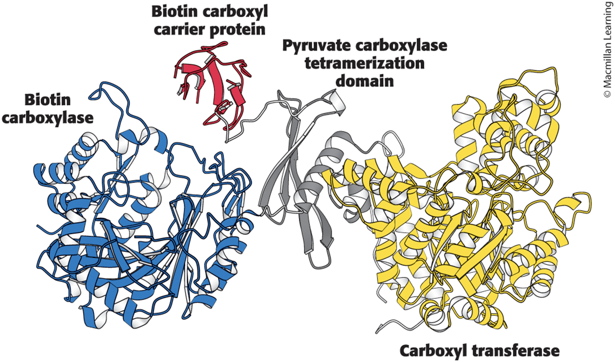
• Pyruvate carboxylase functions as a tetramer composed of four identical subunits, each of which contains four domains.
The Domains of Pyruvate Carboxylase
• biotin carboxylase domain (BC) = catalyzes the formation of carboxyphosphate and the attachment of CO2 to the biotin carboxyl carrier protein domain (BCCP)
• BCCP = swings to the active site of the carboxyl transferase domain (CT)
• CT = transfers the CO2 to pyruvate to form oxaloacetate
• pyruvate carboxylase tetramerization domain (PT) = facilitates the formation of the tetramer and is the binding site for acetyl CoA, a required allosteric activator
A Subunit of Pyruvate Carboxylase Shows the Complexity of the Enzyme
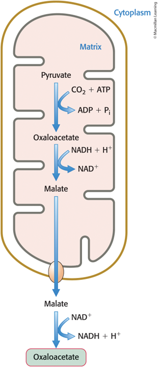
Oxaloacetate Is Shuttled into the Cytoplasm and Converted into Phosphoenolpyruvate
• Malate dehydrogenase catalyzes the reduction of oxaloacetate to malate.
• Malate is transported from the mitochondria to the cytoplasm by a cytoplasmic NAD+-linked malate dehydrogenase and reoxidized to oxaloacetate.
– Reoxidation yields NADH.
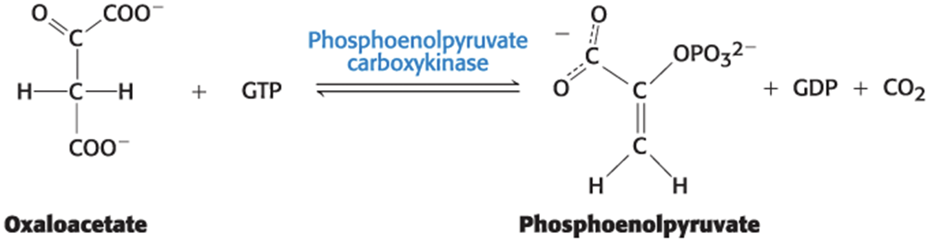
• Phosphoenolpyruvate carboxykinase (PEPCK) decarboxylates and phosphorylates oxaloacetate to phosphoenolpyruvate.
– Phosphoryl donor is GTP.
Oxaloacetate Used in the Cytoplasm for Gluconeogenesis Is Formed in the Mitochondrial Matrix
Oxaloacetate Is Decarboxylated and Phosphorylated to Phosphoenolpyruvate
One ATP and One GTP Molecule Are Expended in the Conversion of Pyruvate into Phosphoenolpyruvate
• The sum of the reactions catalyzed by pyruvate carboxylase and phosphoenolpyruvate carboxykinase is
Pyruvate + ATP + GTP + H2O -> phosphoenolpyruvate + ADP + GDP + Pi + 2H+
• These reactions bypass the pyruvate kinase reaction in glycolysis.
The Conversion of Fructose 1,6-Bisphosphate into Fructose 6-Phosphate and Orthophosphate Is an Irreversible Step
• Phosphoenolpyruvate is metabolized by the enzymes of glycolysis in the reverse direction until the next irreversible step.
• The allosteric enzyme fructose 1,6-bisphosphatase catalyzes the hydrolysis of fructose 1,6-bisphosphate to fructose 6-phosphate and Pi.
Fructose 1,6 bisphosphate + H2O -> fructose 6-phosphate
Fructose 1,6-Bisphosphatase Is a Phosphatase
• phosphatase = an enzyme that catalyzes the hydrolysis of a phosphate to form inorganic phosphate
• The difference between kinases and phosphatases is whether the phosphate is transferred to water or ADP.
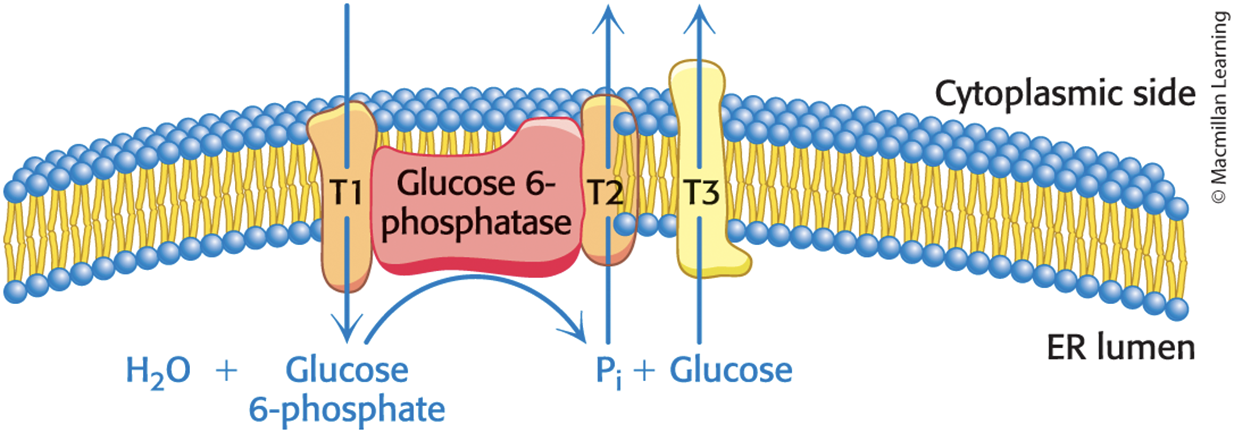
The Generation of Free Glucose Occurs Only in Some Tissues and Is an Important Control Point
• The generation of free glucose occurs primarily in the liver.
• Glucose 6-phosphate is transported into the lumen of the ER.
• Glucose 6-phosphatase hydrolyzes glucose 6-phosphate to glucose.
– This enzyme is bound to the ER membrane.
• Glucose and Pi are shuttled back to the cytoplasm by transporters.
• In most tissues, tissues, glucose 6-phosphate is converted
into glycogen for storage
In the Liver, Free Glucose Is Generated from Glucose 6-Phosphate by Glucose 6-Phosphatase
Six High Transfer-Potential Phosphoryl Groups Are Spent in Synthesizing Glucose from Pyruvate
• The formation of glucose from pyruvate is energetically unfavorable unless it is coupled to favorable reactions.
• The stoichiometry of gluconeogenesis is
• The stoichiometry for the reversal of glycolysis is
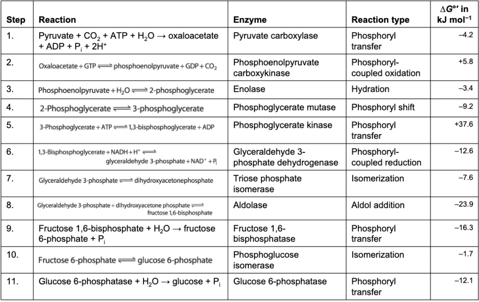
Reactions of Gluconeogenesis
TABLE 16.6 Reactions of gluconeogenesis
Section 16.4 Gluconeogenesis and Glycolysis Are Reciprocally Regulated
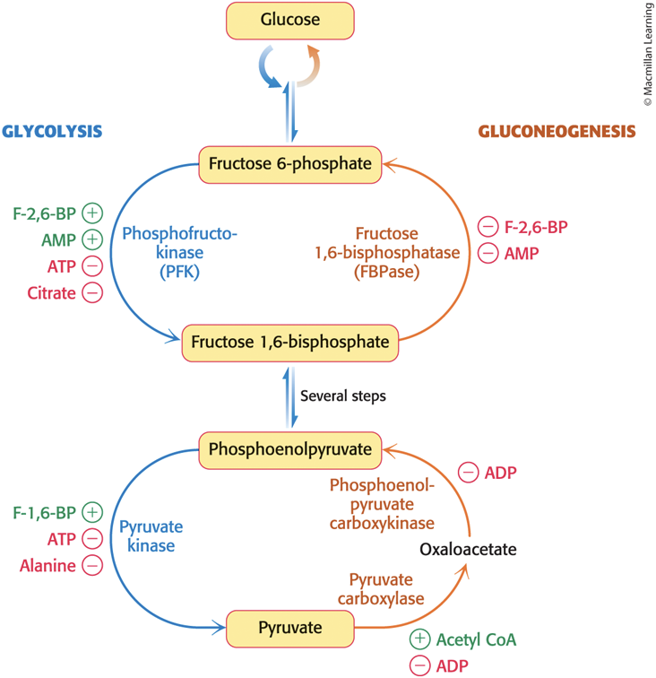
• Gluconeogenesis and glycolysis are regulated within a cell such that one pathway is relatively inactive whereas the other is highly active.
• Glycolysis predominates when energy or glycolytic intermediates are needed.
• Gluconeogenesis predominates when there is a surplus of energy and glucose precursors.
Glycolysis and Gluconeogenesis Are Regulated by Adenosine Nucleotides and Other Metabolic Intermediates
• The interconversion of fructose 1,6-bisphosphate and fructose 6-phosphate is a key regulation site.
• When energy is needed, AMP inhibits fructose 1,6-bisphosphatase and stimulates phosphofructokinase.
– turns on glycolysis and inhibits gluconeogenesis
• When levels of ATP and citrate are high, ATP and citrate inhibit phosphofructokinase, and the decrease in AMP relieves fructose 1,6-bisphosphatase inhibition.
– turns off glycolysis and turns on gluconeogenesis
Reciprocal Regulation at the Interconversion of Phosphoenolpyruvate and Pyruvate
• It occurs in the liver.
• Pyruvate kinase is inhibited by allosteric effectors ATP and alanine.
• Pyruvate carboxylase and phosphoenolpyruvate carboxykinase are inhibited by ADP.
• Pyruvate carboxylase is activated by acetyl CoA.
Reciprocal Regulation of Gluconeogenesis and Glycolysis in the Liver
In Mammals, Glycolysis and Gluconeogenesis in the Liver Are Controlled by Hormones Sensitive to Blood-Glucose Concentration
• The rates of glycolysis and gluconeogenesis are adjusted in the liver to maintain blood-glucose levels.
• F-2,6-BP stimulates phosphofructokinase and inhibits fructose 1,6-bisphosphatase.
• When blood glucose is low, F-2,6-BP loses a phosphoryl group to form F-6P.
– F-6P is not an allosteric effector of PFK.
PFK2 and FBPase2 Are Bifunctional Enzymes
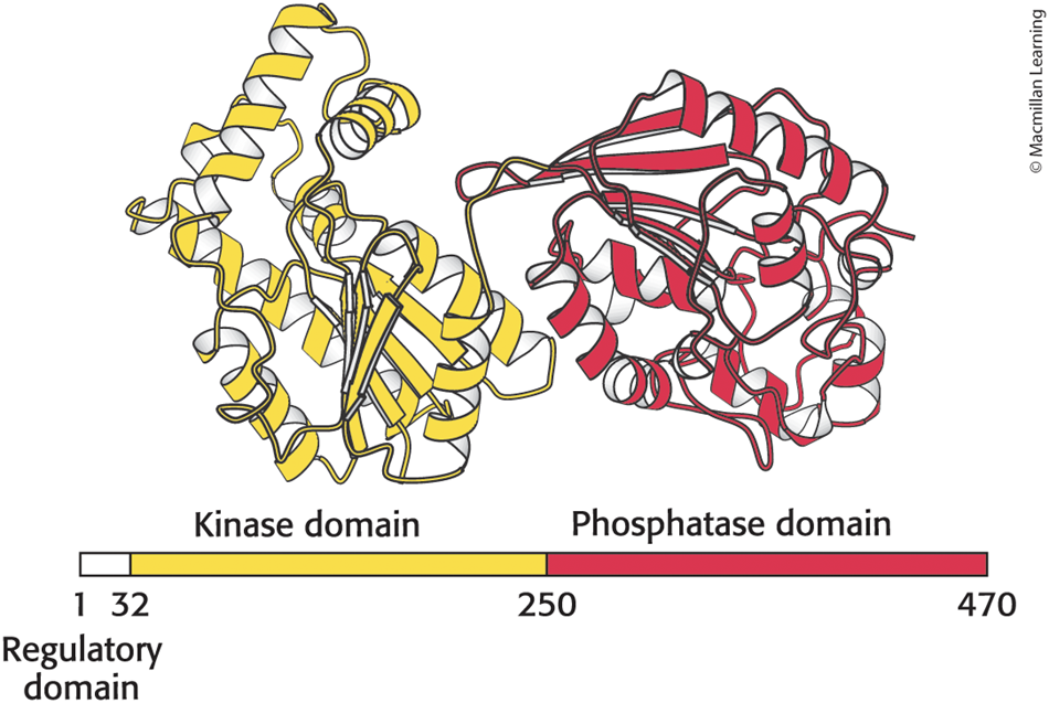
• bifunctional enzyme = contains two enzymes on a single polypeptide chain
• A bifunctional enzyme, containing phosphofructokinase 2 (PFK2) and fructose bisphosphatase 2 (FBPase2), determines F-2,6-BP levels.
• F-2,6-BP concentrations are regulated by two enzymes.
– PFK2 forms F-2,6-BP.
– FBPase2 degrades F-2,6-BP.
The Bifunctional Enzyme Phosphofructokinase 2 Has Two Distinct Domains
• contains an N-terminal regulatory domain, a kinase domain and a phosphatase domain
The Bifunctional Enzyme Is Controlled by Reversible Phosphorylation
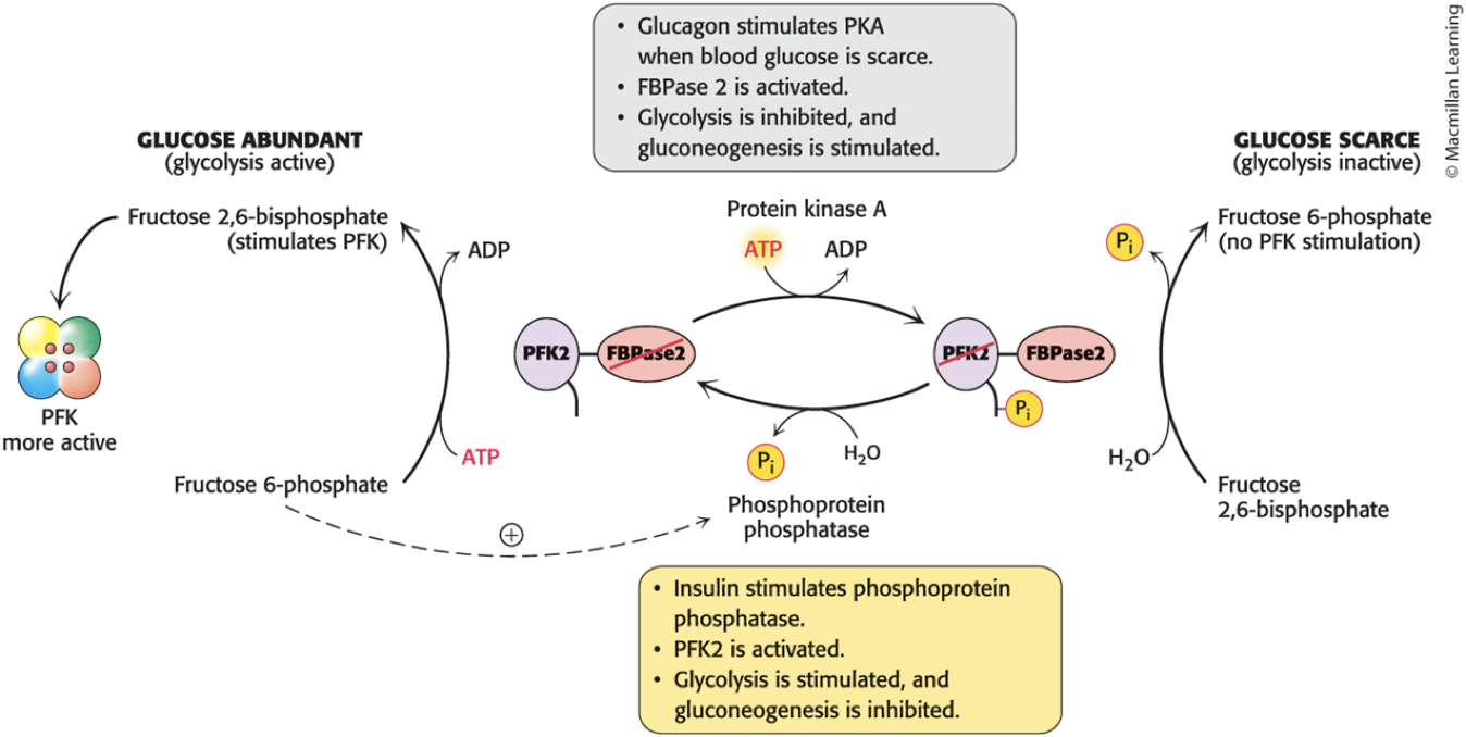
• PFK2 and FBPase2 activities are reciprocally controlled by phosphorylation of a single Ser residue.
• When glucose is scarce, glucagon rises, triggering a cyclic AMP signal cascade and leading to phosphorylation of the bifunctional enzyme by PKA.
– activates FBPase2 and inhibits PFK2 and pyruvate kinase
– lowers F-2,6-BP levels
– gluconeogenesis predominates
• When glucose is high, insulin rises, triggering a signal pathway and leading to dephosphorylation of the bifunctional enzyme by a protein phosphatase.
– activates PFK2 and inhibits FBPase2
– increases F-2,6-BP levels
– glycolysis predominates
The Synthesis and Degradation of Fructose 2,6-Bisphosphate Is Hormonally Controlled
The Bifunctional Enzyme Is Also Under Transcriptional Control by Glucagon and Insulin
• glucagon and insulin altering gene expression by changing the rate of transcription
• To stimulate gluconeogenesis, glucagon:
– inhibits expression of the three regulated glycolytic enzymes.
– stimulates the production of phosphoenolpyruvate carboxykinase and fructose 1,6-bisphosphatase.
• To stimulate glycolysis, insulin stimulates the expression of phosphofructokinase, pyruvate kinase, and the bifunctional enzyme that makes and degrades F-2,6-BP.
Substrate Cycles Amplify Metabolic Signals and Produce Heat
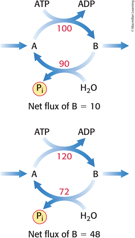
• substrate cycle = a set of reaction in a loop
– example: the phosphorylation of F-6P to F-1,6-BP and its hydrolysis back to F-6P
– reciprocal regulation prevents both reactions from being active at the same time
• futile cycles = substrate cycles with a limited degree of cycling
– may result in pathological conditions, such as malignant hyperthermia
– may be biologically important in enhancing metabolic signals
Substrate Cycles Can Have Advantages
• A small change in the rates of the two opposing reactions can result in a large change in the net flux.
Lactate and Alanine Formed by Contracting Muscle and Peripheral Tissues Are Used by Other Organs
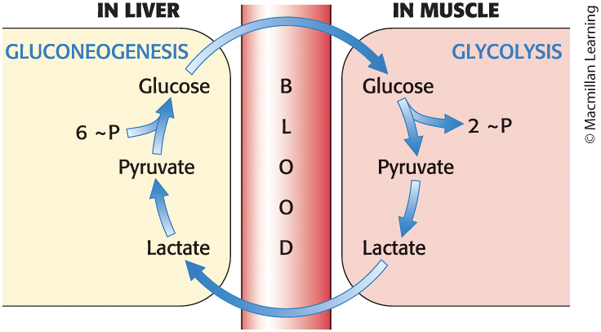
• Cori cycle = a series of reactions carried out by cooperation between the liver and muscle
• Lactate produced by active muscle is released into the blood and converted to glucose by the liver.
• Nitrogens from amino acids used by muscle for fuel are transferred to pyruvate to form alanine.
– The reverse reaction takes place in the liver.
The Cori Cycle
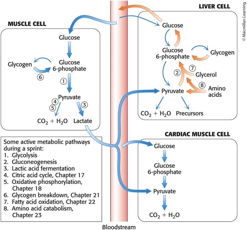
During a Sprint, Cooperation Between Glycolysis and Gluconeogenesis Occurs Within Multiple Tissues
Deficiencies in Glycolytic or Gluconeogenic Enzymes Are Rare Genetic Disorders
• inborn errors of metabolism = genetically inherited deficiencies in the activity or regulation of specific enzymes
– examples: triose phosphate isomerase deficiency and pyruvate carboxylase deficiency
Triose Phosphate Isomerase Deficiency (TPID)
• TPID = a multisystem disorder resulting from dihydroxyacetone phosphate accumulation in cells, especially red blood cells
– presents in early childhood and may lead to death
– symptoms include congenital hemolytic anemia and progressive neuromuscular disorder
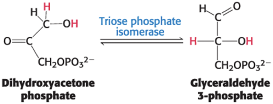
• When triose phosphate isomerase activity is missing, half of the carbons of glucose cannot be metabolized to yield ATP.
– Research suggests disruption of energy metabolism is not the cause TPID symptoms.
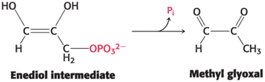
• Instead, buildup of dihydroxyacetone phosphate causes TPID symptoms, as it can be converted into methylglyoxal.
• methylglyoxal = highly reactive molecule that covalently binds to available amino groups on proteins, yielding advanced glycation end products (AGE)
– modifications inhibits protein function
Pyruvate Carboxylase Deficiency (PCD)
• PCD = rare disorder characterized to some extent by hypoglycemia and lactic acidosis
– may lead to death in the first few months of life in severe cases
– symptoms include lethargy and seizures
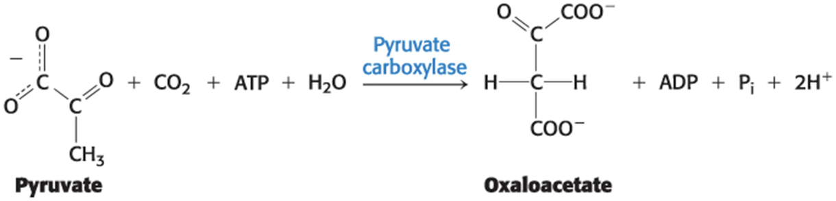
• Pyruvate carboxylase is a key regulatory enzyme in gluconeogenesis, which occurs primarily in the liver.
• When pyruvate carboxylase activity is missing, the liver cannot:
– maintain adequate blood glucose concentration.
– remove lactic acid from the blood and use it as a gluconeogenic precursor.
Glycolysis and Gluconeogenesis Are Evolutionarily Intertwined
• Glycolytic enzymes with similar properties do not have similar amino acid sequences.
– most likely derived independently rather than by gene duplication
• Glycolysis consists of the metabolism of hexoses and the metabolism of trioses.
– Metabolism of trioses is common to glycolysis and gluconeogenesis.
– Enzymes in the metabolism of trioses are present in all species.
– enzymes in the metabolism of hexoses are less conserved