Chap 16: The Brain
Introduction to the Brain
The brain is more complex than the spinal cord, enabling adaptability but leading to slower responses compared to spinal reflexes.
16.1 An Introduction to the Organization of the Brain
Embryology of the Brain
The brain develops from three swellings at the top of the neural tube:
Prosencephalon (forebrain)
Mesencephalon (midbrain)
Rhombencephalon (hindbrain).
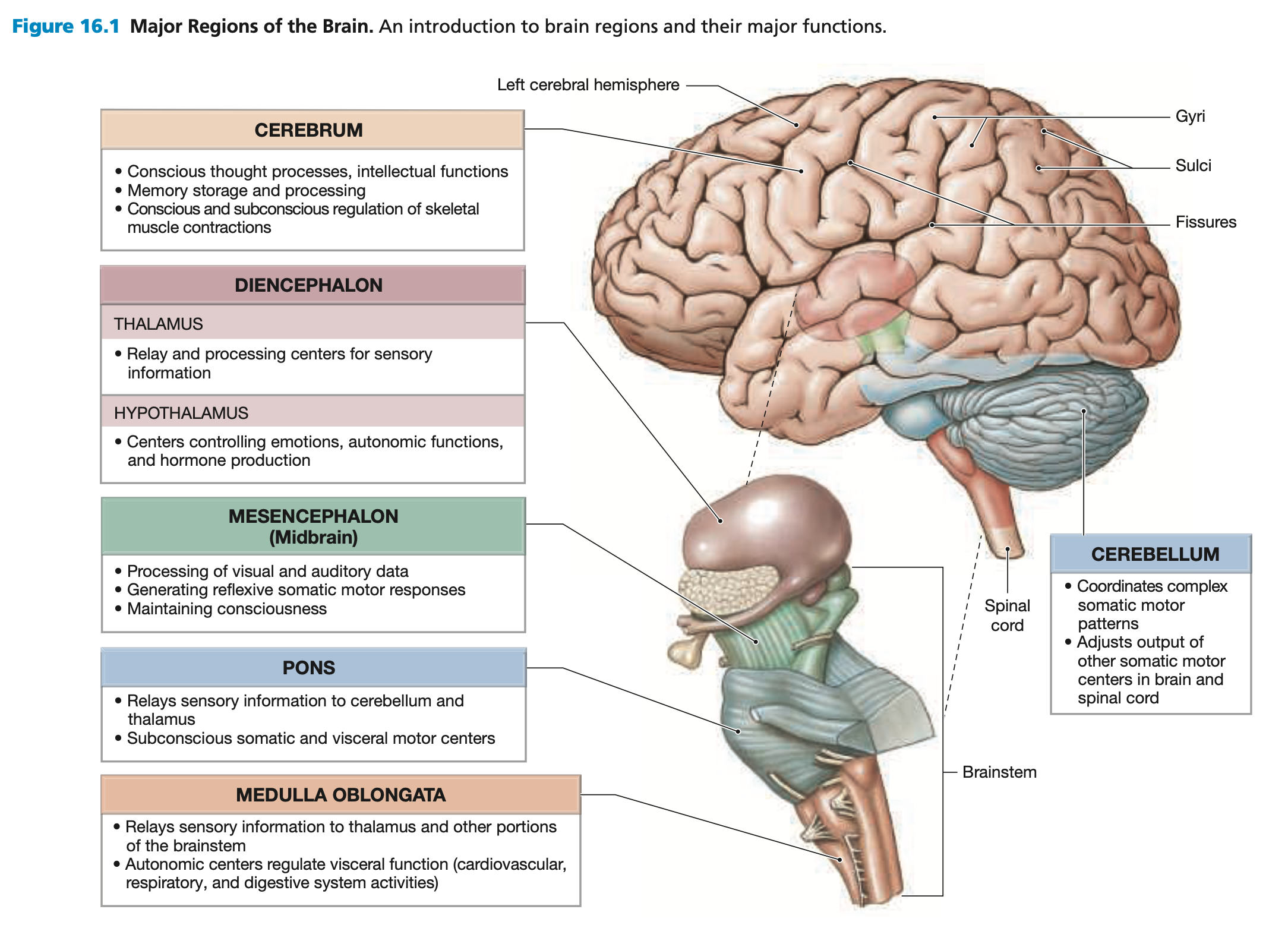
Major Regions and Landmarks
Adult Brain Regions:
Cerebrum: The largest part of the brain, responsible for higher cognitive functions, including thought, memory, voluntary movement, sensory processing, and reasoning. It is divided into two hemispheres and four lobes (frontal, parietal, temporal, and occipital).
Gyri: The raised folds or ridges on the surface of the brain. They increase the brain’s surface area for higher cognitive function.
Sulci: The shallow grooves or indentations between the gyri. They help divide the brain into different functional regions.
Together, the gyri and the sulci form the cerebral cortex or outer layer where the majority of the cell bodies of neurons are located
Gyri & sulci increase the surface are of the brain’s grey matter thereby increasing the number of neuron bodies, which enhances cognitive functions such as memory, attention, and problem-solving.
Diencephalon: Located beneath the cerebrum, it includes the thalamus (sensory relay center), hypothalamus (homeostasis and endocrine regulation), and epithalamus (sleep-wake cycles via the pineal gland).
Epithalamus (roof): Sleep-wake cycle
Thalami (walls): Relay and process sensory data, one thalamus in each hemisphere
Hypothalamus (floor): Involved in emotion, autonomic function, hormone production.
Mesencephalon (Midbrain): Part of the brainstem, processes visual and auditory information; generates involuntary motor responses (reflexes).
Pons: part of the brainstem, acts as a bridge between the cerebrum, cerebellum, and spinal cord. It plays a role in breathing regulation, motor control, and sensory relay.
Medulla Oblongata: Connects to spinal cord; regulates autonomic functions such as heart rate, breathing, and blood pressure and sensory information relay.
Cerebellum: Located at the back of the brain, it coordinates movement, balance, and posture. It fine-tunes motor activity to ensure smooth and precise movements, adjusting motor activities based on sensory data.
The brain consists of neural cortex, gray matter covering white matter.
The central passageway expands to form ventricles filled with cerebrospinal fluid (CSF).
16.2 Protection and Support of the Brain
Cranial Meninges: Surround the brain and provide protection, like an airbag in a car
Dura mater: Outermost later, tough & fibrous
Arachnoid mater: the middle layer, which is web-like and contains cerebrospinal fluid to cushion the brain.
Pia mater: the innermost layer, delicate and closely adheres to the surface of the brain, providing additional protection and support for blood vessels.
The Blood Brain Barrier
The blood-brain barrier (BBB): A selectively permaible barrier that separates the bloodstream from the brain’s extracellular fluid.
Tightly regulates what enters and exits the brain to maintain homeostasis and protect neural tissue from harmful substances.
Cerebrospinal Fluid (CSF)
Functions of CSF:
Cushions neural structures.
Supports brain.
Transports nutrients and wastes.
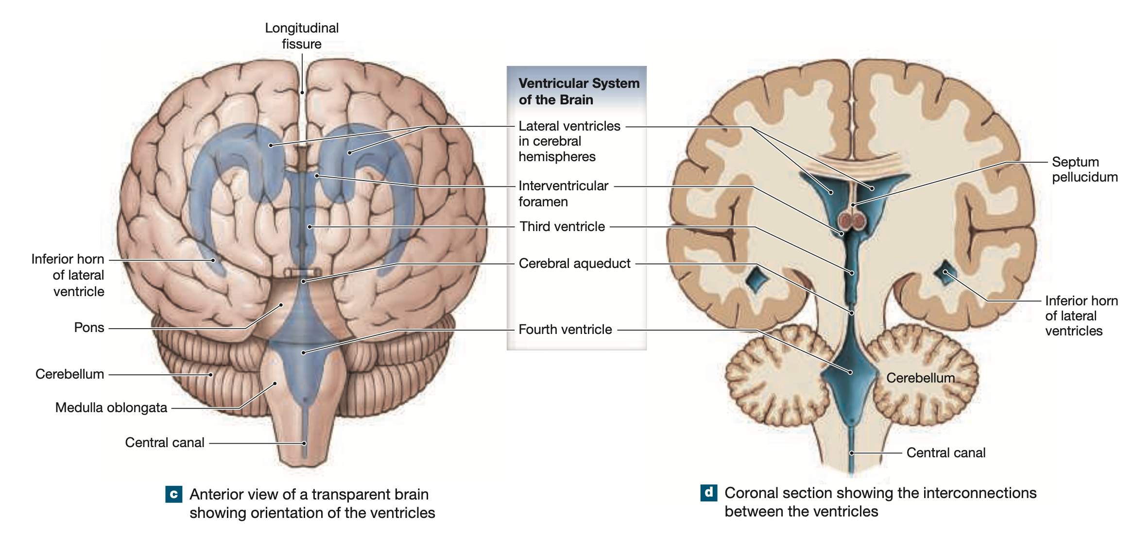
Ventricles: Fluid-filled cavities within the brain that are
Filled with CSF, provides all the materials neurons need to function normally
Lined with ependymal cells play a crucial role in the formation and regulation of CSF
CSF Circulation Pathway
CSF is produced in the choroid plexuses of the lateral ventricles (1 & 2).
Flows through the interventricular foramina into the third ventricle.
Flows through the cerebral aqueduct into the fourth ventricle.
CSF exits the fourth ventricle via the lateral and median apertures into the subarachnoid space, surrounding the brain and spinal cord.
CSF flows around the brain and spinal chord, eventually entering into the venous bloodstream via arachnoid granulations (small protrusions into the superior sagittal sinus, a major venous drainage site).
Blood Supply to the Brain
Blood reaches the brain through internal carotid arteries and vertebral arteries; venous blood leaves primarily via internal jugular veins.
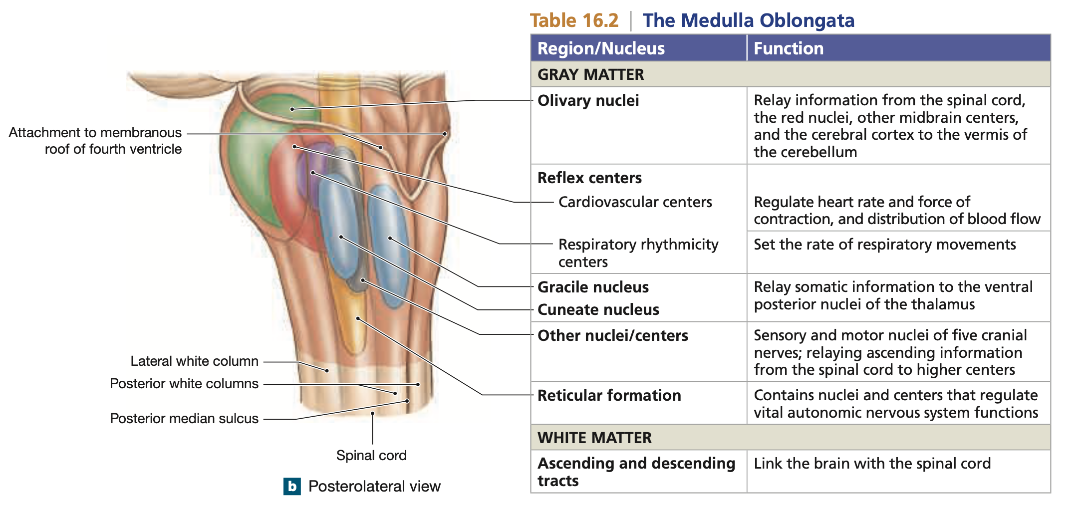
16.3 The Medulla Oblongata (brain stem)
Connects brain to spinal cord; contains:
Gracile nucleus and cuneate nucleus: Processing centers.
Olivary nuclei: Relay information to the cerebellar cortex.
Reflex centers: Control cardiovascular and respiratory functions.
Responsible for visceral or autonomic control
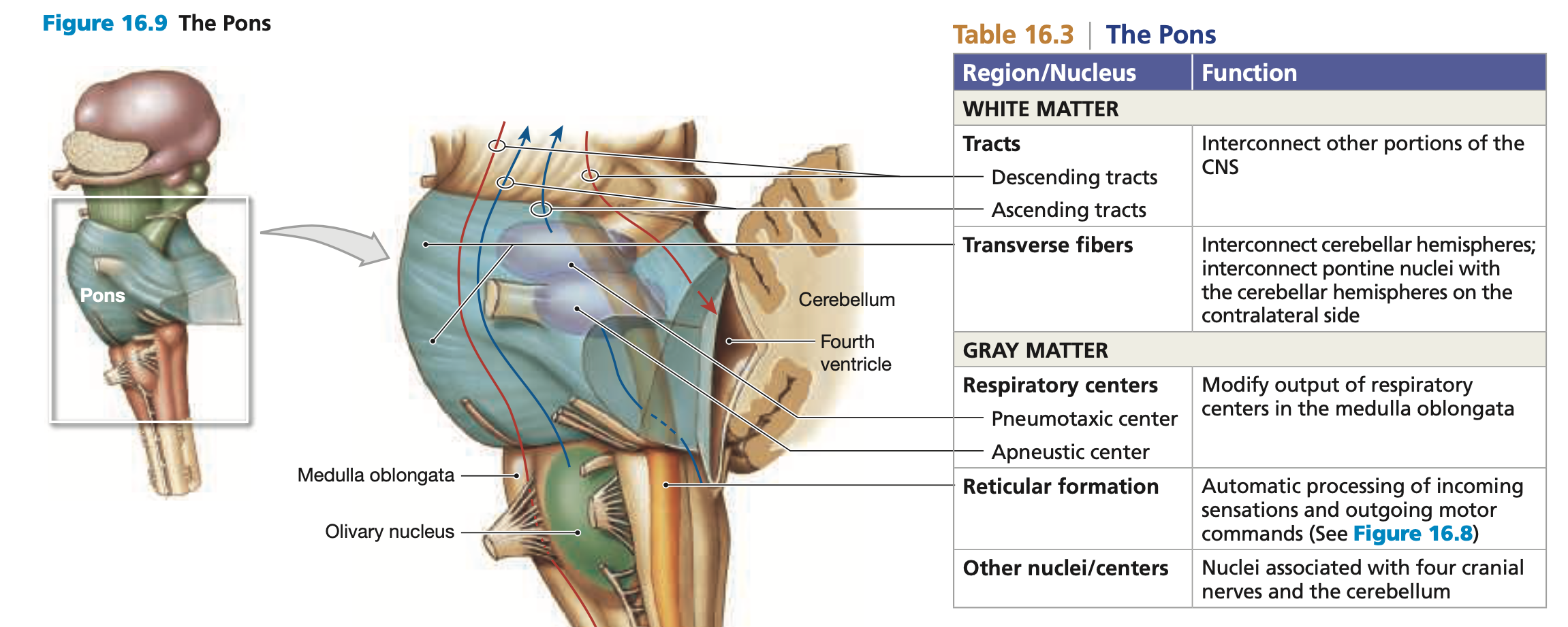
16.4 The Pons
The pons contains sensory and motor nuclei for 4 cranial nerves, nuclei are involved with involuntary control of respiration, and nuclei that process and relay cerebellar signals
Processes and relays cerebellar commands to the spinal cord, facilitating coordination of voluntary movements and maintaining posture.
Contains:
Sensory and motor nuclei for four cranial nerves.
Nuclei for involuntary respiration control.
Nuclei for cerebellar command relay via middle cerebellar peduncles.
Various tracts.
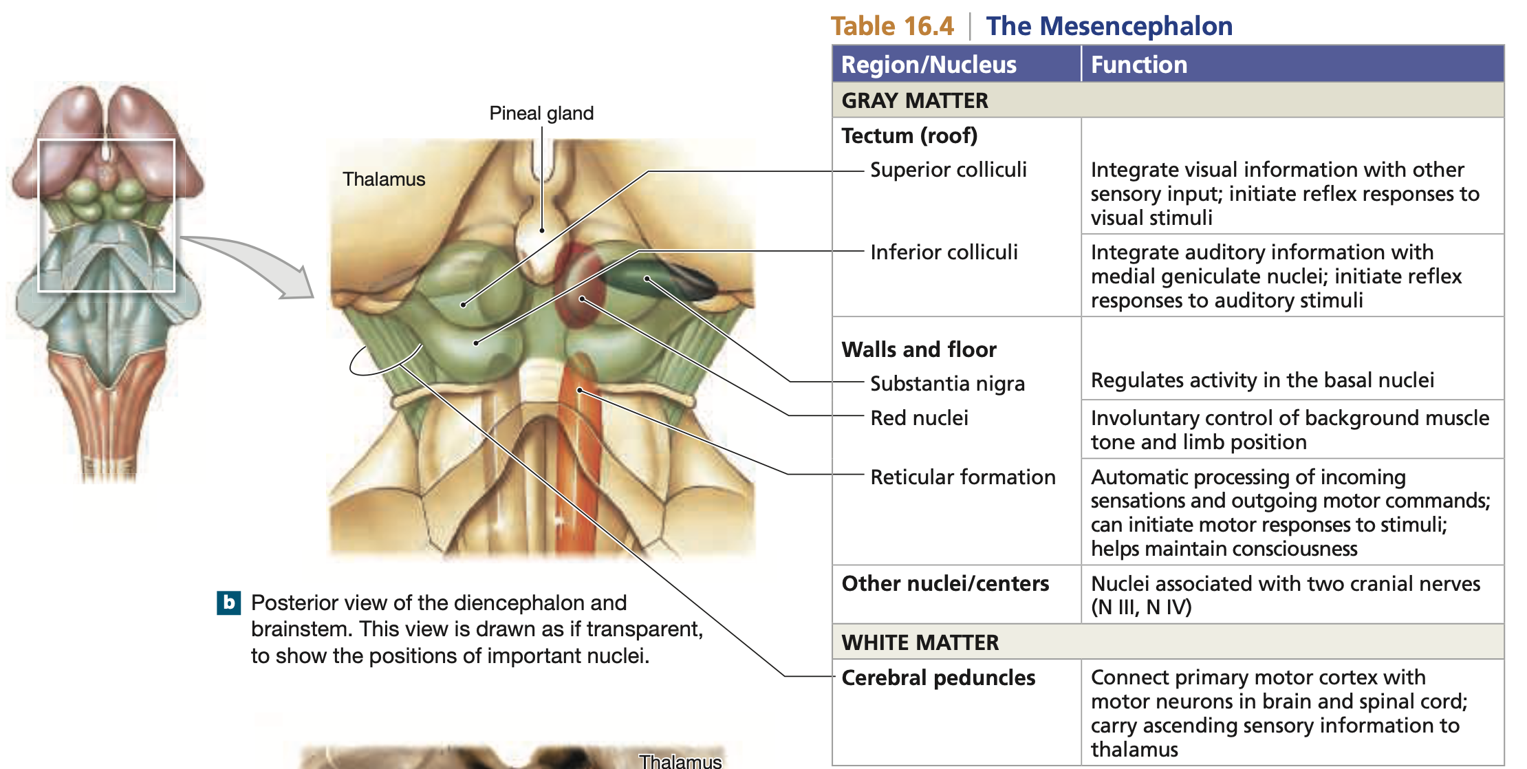
16.5 The Mesencephalon (Midbrain)
The tectum (roof): Contains corpora quadrigemina for visual and auditory processing. Includes:
Superior colliculus (visual inputs).
Inferior colliculus (auditory data).
Collectively called the corpora quadrigemina,
Red nucleus: Integrates cerebrum information; issues involuntary motor commands.
Substantia nigra: Regulates motor output.
Cerebral peduncles: Contains fibers for ascending sensory commands and descending motor commands.
16.6 The Diencephalon
Acts as a relay and integration center for sensory and motor pathways.
The Epithalamus
Forms the diencephalon roof; includes the pineal gland for hormone secretion (melatonin).
The Thalamus
Principal relay point for sensory information; coordinates motor activities. Surrounds 3rd ventricle and acts as walls of the diencephalon.
The Hypothalamus
Contains important control centers (includes autonomic functions, emotions (pain, pleasure, exual arousal), temperature regulation, and hormone secretion). Also connects to the pituitary gland.
16.7 The Cerebellum (motor brain)
Oversees postural muscles and tunes voluntary movements; consists of neural cortex with folds, comprising anterior and posterior lobes.
16.8 The Cerebrum
The Cerebral Hemispheres
Separated into two hemispheres by the longitudinal fissure.
Each hemisphere controls the opposite body side.
Frontal Lobe: Primary motor cortex
Precentral gyrus: The primary motor cortex, directs voluntary movements by controlling somatic neurons in the brain stem/spinal chord
Motor in front
Central sulcus: Boundary between the frontal lobe and parietal lobe
Parietal Lobe: Primary sensory cortex
Postcentral gyrus: The primary somatosensory cortex, responsible for processing sensory information from the thalamus, including touch, temperature, and pain.
Sensory in back
Occipital Lobe: Visual cortex
Primary cortex of conscious perception of visual stimuli
Temporal Lobe: Auditory and olfactory cortex
The primary cortex of conscious perception of auditory & olfactory stimuli
Functional differences between hemispheres exist.
Higher-Order Functions
Characteristics of higher-order functions:
Performed by cerebral cortex.
Complex interconnections and communication.
Involves conscious and unconscious processing.
Not part of programmed wiring.
Wernicke's area: Integrative center receiving sensory information, specifically for understanding language.
Broca’s area: Critical for language production and articulation, allowing individuals to formulate coherent speech.
Prefrontal cortex: Handles learning and reasoning functions.
Central White Matter
Contains:
Association fibers: Interconnect areas within a hemisphere.
Commissural fibers: Connect the two hemispheres.
Projection fibers: Link cerebrum with other brain regions.
The Basal Nuclei
Includes caudate nucleus, globus pallidus, and putamen; controls muscle tone and movement coordination.
The Limbic System (emotions)
A collection of interconnected brain structures responsible for emotion, memory, motivation, and behavior. It also plays a role in linking conscious thought with unconscious autonomic responses, making it crucial for survival instincts.
Hippocampus: Essentail for memory formation and consolidation
Amygdala: Processes emotions like fear, aggression, and pleasure; linking emotions to memory and influencing decision-making.
Fornix: A C-shaped white matter tract that connects the hippocampus to hypothalamus: acts as a major communication pathway.
Mammillary Bodies: Part of the hypothalamus, these small round structures are involved in memory processing and play a role in the recollection of memories, particularly those associated with spatial and episodic information.
The Epithalamus: Forms the diencephalon roof; includes the pineal gland for hormone secretion (melatonin).
The Thalamus: Principal relay point for sensory information; coordinates motor activities.
The Hypothalamus: Contains important control centers (includes autonomic functions, emotion, temperature regulation, and hormone secretion). Also connects to the pituitary gland.
16.9 The Cranial Nerves
12 pairs of cranial nerves attach to the brain near sensory or motor nuclei.
Specific Cranial Nerves
Cranial Nerve | Function | |
|---|---|---|
Olfactory Nerves (I) | Smell sensation. | Sensory |
Optic Nerves (II) | Visual information. | Sensory |
Oculomotor Nerves (III) | Innervate extraocular eye muscles. | Motor |
Trochlear Nerves (IV) | Innervate superior oblique of the eye. | Motor |
Trigeminal Nerves (V) | Mixed nerves for facial sensation and motor functions. | Both |
Abducens Nerves (VI) | Control lateral rectus muscle of the eye. | Motor |
Facial Nerves (VII) | Control facial expressions and sensations. | Both |
Vestibulocochlear Nerves (VIII) | Balance and hearing. | Sensory |
Glossopharyngeal Nerves (IX) | Innervate tongue and pharynx. | Both |
Vagus Nerves (X) | Vital for visceral control. Respiratory, cardiovascular, and digestive functions | Both |
Accessory Nerve (XI) | Controls swallowing and pectoral girdle muscles. | Motor |
Hypoglossal Nerves (XII) | Motor control of tongue movements. | Motor |
Listed most superior to most inferior on the brainstem, they are paired