Comprehensive Study Guide: Human Anatomy, Physiology, Cell Biology, Histology, and Bone Dynamics
Foundations of Anatomy, Physiology, and Homeostasis
Definitions and Scope
- Anatomy: The study of anatomical structures, including their composition, precise location, and associated relationships. It provides the biological blueprint or map of the organism.
- Physiology: The study of how anatomical structures function individually and cooperatively to sustain life.
- Principle of Complementarity: All physiological functions are executed by specific anatomical structures; form dictates function.
Specialties of Anatomy and Physiology
- Gross Anatomy (Macroscopic Anatomy): Examination of large, visible structures.
- Surface Anatomy: Study of general form and exterior body features.
- Regional Anatomy: Detailed study of specific superficial and internal regions of the body.
- Systemic Anatomy: Study of groups of organs that function together in a coordinated manner.
- Developmental Anatomy: Study of structural changes from fertilization (embryology) to maturity.
- Clinical Anatomy: Focuses on anatomical features significant in medical practice.
- Microscopic Anatomy: Examination of structures that cannot be seen without magnification.
- Cytology: The study of individual cells and their internal structures.
- Histology: The study of tissues and tissue structures.
- Specialties of Physiology:
- Cell Physiology: Functions and interactions within and between cells.
- Special Physiology: Physiological processes of specific organs.
- Systemic Physiology: Functions of entire organ systems.
- Pathological Physiology: Effects of diseases on organ or system function.
Levels of Structural Organization

- Chemical or Molecular Level: Atoms in combination form complex protein molecules.
- Organelle Level: Complex molecules form functional protein filaments within cells.
- Cellular Level: Interacting organelles form cardiac muscle cells, the smallest units of life.
- Tissue Level: Specialized cardiac muscle cells aggregate to form cardiac muscle tissue.
- Organ Level: Cardiac muscle tissue combines with other tissue types to form the heart.
- Organ System Level: The heart, blood, and blood vessels form the cardiovascular system.
- Organism Level: Eleven interconnected organ systems maintain the living human individual.
Comprehensive Overview of the 11 Organ Systems
- Integumentary System:
- Major Organs: Skin, hair, sweat glands, nails.
- Functions: Protects against environmental hazards, regulates body temperature, provides sensory information.
- Skeletal System:
- Major Organs: Bones, cartilages, associated ligaments, bone marrow.
- Functions: Provides support and protection, stores calcium and other minerals, forms blood cells.
- Muscular System:
- Major Organs: Skeletal muscles and associated tendons.
- Functions: Provides movement, offers protection and support, generates heat to maintain body temperature.
- Nervous System:
- Major Organs: Brain, spinal cord, peripheral nerves, sense organs.
- Functions: Directs immediate responses to stimuli, coordinates activities of other systems, interprets sensory information.
- Endocrine System:
- Major Organs: Pituitary gland, thyroid gland, pancreas, adrenal glands, gonads (testes and ovaries), endocrine tissues in other systems.
- Functions: Directs long-term changes in other systems, adjusts metabolic activity and energy use, controls structural/functional development.
- Cardiovascular System:
- Major Organs: Heart, blood, blood vessels.
- Functions: Distributes blood cells, water, nutrients, waste products, oxygen, and carbon dioxide; distributes heat.
- Lymphatic System:
- Major Organs: Spleen, thymus, lymphatic vessels, lymph nodes, tonsils.
- Functions: Defends against infection and disease, returns tissue fluids to the bloodstream.
- Respiratory System:
- Major Organs: Nasal cavities, sinuses, larynx, trachea, bronchi, lungs, alveoli.
- Functions: Delivers air to alveoli for gas exchange, provides oxygen to bloodstream, removes carbon dioxide, produces sounds for communication.
- Digestive System:
- Major Organs: Teeth, tongue, pharynx, esophagus, stomach, small intestine, large intestine, liver, gallbladder, pancreas.
- Functions: Processes and digests food, absorbs and conserves water, absorbs nutrients, stores energy reserves.
- Urinary System:
- Major Organs: Kidneys, ureters, urinary bladder, urethra.
- Functions: Excretes waste products from blood, controls water balance by regulating urine volume, stores urine, regulates blood ion concentrations and pH.
- Male Reproductive System:
- Major Organs: Testes, epididymis, ductus deferens, seminal vesicles, prostate gland, penis, scrotum.
- Functions: Produces male sex cells (sperm) and hormones.
- Female Reproductive System:
- Major Organs: Ovaries, uterine tubes, uterus, vagina, labia, clitoris, mammary glands.
- Functions: Produces female sex cells (oocytes) and hormones, supports developing embryo from conception to delivery, provides milk for newborn.
Homeostatic Regulation and Feedback Mechanisms
- Homeostasis: The state of internal balance and physiological equilibrium maintained by bodily systems.
- Mechanisms of Regulation:
- Autoregulation (Intrinsic): Automatic chemical or cellular response within a localized cell, tissue, or organ.
- Extrinsic Regulation: Responses controlled by the nervous system or endocrine system across multiple systems.
- Essential Components of a Homeostatic Regulatory Loop:
- Receptor: A sensor that monitors and detects environmental stimuli or changes.
- Control Center: Processes input from the receptor and sends specific operational commands.
- Effector: A cell, tissue, or organ that responds to the commands to alter the internal environment.
- Negative Feedback Mechanisms:
- The effector response negates or reverses the original stimulus, restoring conditions to normal limits.
- Example (Thermoregulation): An increase in body temperature above the set point (37.0∘C, normal range 36.7∘C−37.2∘C) triggers thermoreceptors in the skin and hypothalamus. The brain's thermoregulatory center commands sweat glands to increase secretion and cutaneous blood vessels to dilate (increasing heat loss). When temperature drops, vessels constrict and sweating decreases.

- Positive Feedback Mechanisms:
- The effector response reinforces, accelerates, or escalates the original stimulus, completing a specific process rapidly.
- Example (Blood Clotting): A break in a blood vessel wall causes bleeding. Damaged cells release chemical clotting factors. Cloting begins, which triggers the release of additional chemicals. This positive feedback loop accelerates clotting until a clot plugs the break and bleeding stops.

- Systems Integration: The coordinated operation of multiple organ systems working together to maintain internal stability. Failure of homeostatic regulation leads to disease or death.
Anatomical Terminology
- Anatomical Position: Standing erect, facing forward, arms at sides, palms facing forward, feet together.
- Body Positions:
- Supine: Lying down, face up.
- Prone: Lying down, face down.
- Anatomical Landmarks and Regions:
- Cephalic: Head (Cephalon); includes Frontal (forehead), Orbital/Ocular (eye), Buccal (cheek), Otic (ear), Nasal (nose), Oral (mouth), Mental (chin), Cranial (skull), Facial (face).
- Cervical: Neck (Cervicis).
- Thoracic: Chest (Thoracis); includes Mammary (breast).
- Abdominal: Abdomen; Umbilical (navel).
- Pelvic: Pelvis; Pubic (anterior pelvis), Inguinal (groin).
- Brachial: Arm (Brachium); includes Axillary (armpit), Antecubital (front of elbow), Antebrachial (forearm), Carpal (wrist), Manual (hand), Palmar (palm), Pollex (thumb), Digital/Phalangeal (fingers).
- Lower Limb: Femoral (thigh), Patellar (kneecap), Crural (leg), Tarsal (ankle), Pedal (foot), Digital/Phalangeal (toes), Hallux (great toe).
- Posterior Regions: Acromial (shoulder), Dorsal (back), Olecranal (back of elbow), Lumbar (loin), Gluteal (buttock), Popliteal (back of knee), Sural (calf), Calcaneal (heel), Plantar (sole).
- Abdominopelvic Quadrants: Formed by two intersecting lines through the umbilicus:
- Right Upper Quadrant (RUQ), Left Upper Quadrant (LUQ), Right Lower Quadrant (RLQ), Left Lower Quadrant (LLQ).
- Abdominopelvic Regions: Nine precise anatomical regions:
- Right Hypochondriac, Epigastric, Left Hypochondriac.
- Right Lumbar, Umbilical, Left Lumbar.
- Right Inguinal, Hypogastric (Pubic), Left Inguinal.
- Directional Terms:
- Anterior (Ventral): The front or belly side.
- Posterior (Dorsal): The back or behind.
- Cranial (Cephalic) / Superior: Toward the head; above.
- Caudal / Inferior: Toward the tail (coccyx); below.
- Medial: Toward the longitudinal axis/midsagittal plane.
- Lateral: Away from the longitudinal axis/midsagittal plane.
- Proximal: Toward an attached base.
- Distal: Away from an attached base.
- Superficial: At, near, or close to the body surface.
- Deep: Farther from the body surface.
- Sectional Planes:
- Frontal (Coronal) Plane: Separates anterior and posterior portions.
- Sagittal Plane: Separates right and left portions (Midsagittal passes through midline; Parasagittal is offset).
- Transverse (Horizontal) Plane: Separates superior and inferior portions.
- Body Cavities and Serous Membranes:
- Ventral Body Cavity: Divided by the diaphragm into the Thoracic Cavity and Abdominopelvic Cavity.
- Thoracic Cavity: Subdivided by the mediastinum into two Pleural Cavities (holding the lungs) and a central Pericardial Cavity (holding the heart).
- Abdominopelvic Cavity: Contains the Abdominal Cavity (digestive glands/organs), Pelvic Cavity (urinary bladder, reproductive organs, distal large intestine), and Peritoneal Cavity.
- Serous Membranes: Line body cavities and coat organs, consisting of a Parietal Layer (cavity wall) and a Visceral Layer (organ surface).
The Chemical Level of Organization
Subatomic Particles and Atomic Structure
- Matter: Anything that occupies space and has mass; composed of atoms.
- Atom: The smallest stable unit of matter with unique elemental identity.
- Subatomic Particles:
- Proton (p+): Positive charge (+1), mass of 1amu, located in nucleus.
- Neutron (n0): Neutral charge (0), mass of 1amu, located in nucleus.
- Electron (e−): Negative charge (−1), negligible low mass, orbits nucleus in electron shells/cloud.
- Atomic Measurements:
- Atomic Number: Equal to the number of protons in an atom; defines the element.
- Mass Number: Total number of protons plus neutrons.
- Isotopes: Atoms of the same element containing identical numbers of protons but different numbers of neutrons.

- Hydrogen Isotopes: Hydrogen-1 (Protium: 1p+), Hydrogen-2 (Deuterium: 1p+,1n0), Hydrogen-3 (Tritium: 1p+,2n0).
Electron Shells and Chemical Bonding
- Electron Energy Levels: Electrons occupy discrete energy shells.
- Shell 1 holds a maximum of 2e−; Shell 2 holds 8e−; Shell 3 holds 8e−.
- Valence Shell: The outermost electron shell. Atoms interact to fill or empty this shell to attain chemical stability.
- Chemical Bonds:
- Covalent Bonds (Strongest): Formed when atoms share electrons to complete their valence shells.
- Non-polar Covalent Bonds: Electrons are shared equally between atoms.
- Polar Covalent Bonds: Electrons are shared unequally, creating partial positive (δ+) and partial negative (δ−) charges across the molecule.
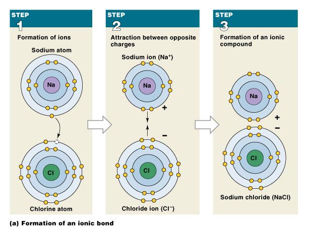
- Ionic Bonds (Intermediate Strength): Electrostatic attraction between oppositely charged ions formed by electron transfer.
- Cations (+) attract Anions (−) to form crystalline ionic structures (e.g., NaCl).
- Hydrogen Bonds (Weakest): Weak attractive forces between a partial positive charge on a hydrogen atom of a polar covalent bond and a partial negative charge on an oxygen or nitrogen atom of another polar molecule. Causes surface tension in water.
Chemical Reactions and Energetics
- Types of Chemical Reactions:
- Decomposition (Catabolism): AB→A+B
- Synthesis (Anabolism): A+B→AB
- Exchange Reaction: AB+CD⇌AD+CB
- Reversible Reactions: Can proceed in either direction depending on conditions.
- Hydrolysis: A—B—C—D—E+H2O→A—B—C—H+HO—D—E
- Dehydration Synthesis (Condensation): A—B—C—H+HO—D—E→A—B—C—D—E+H2O
- Energetics:
- Work: A change in mass or distance.
- Kinetic Energy: Energy of motion.
- Potential Energy: Stored energy.
- Chemical Energy: Potential energy stored within chemical bonds.
- Exergonic Reactions: Release more energy than they absorb.
- Endergonic Reactions: Absorb more energy than they release.
- Heat: Unusable thermal energy produced during energy exchanges.
- Activation Energy and Enzymes:
- Chemical reactions in cells require Activation Energy to initiate.
- Enzymes: Protein catalysts that lower activation energy, allowing vital biological reactions to occur rapidly without being consumed.

Inorganic Chemistry and Water Properties
- Organic vs. Inorganic: Organic molecules are based on carbon and hydrogen backbones; inorganic molecules are not.
- Nutrients: Essential molecules obtained from food.
- Metabolites: Molecules synthesized or broken down by bodily metabolic reactions.
- Properties of Water:
- Solubility: Ability to dissolve solutes in solvent to form aqueous solutions. Polar water molecules surround charged ions forming hydration spheres.
- Reactivity: Participates directly in reactions like hydrolysis and dehydration synthesis.
- High Heat Capacity: Absorbs and retains significant thermal energy, stabilizing body temperature.
- Lubrication: Friction reduction between opposing cellular or tissue surfaces.
- Interaction with Water:
- Hydrophilic: Water-loving; polar molecules and ions that dissolve readily in water.
- Hydrophobic: Water-fearing; non-polar molecules (fats, oils) that do not interact with water.
- Electrolytes: Inorganic ions that conduct electrical currents in solution. Imbalances disrupt nervous, muscular, and cellular functions.
Important Dissociating Body Electrolytes
| Electrolyte | Dissociated Ions Released |
|---|
| Sodium Chloride (NaCl) | Na++Cl− |
| Potassium Chloride (KCl) | K++Cl− |
| Calcium Phosphate (CaPO4) | Ca2++PO43− |
| Sodium Bicarbonate (NaHCO3) | Na++HCO3− |
| Magnesium Chloride (MgCl2) | Mg2++2Cl− |
| Disodium Phosphate (Na2HPO4) | 2Na++HPO42− |
| Sodium Sulfate (Na2SO4) | 2Na++SO42− |
pH, Acids, Bases, and Buffers
- pH Scale: Negative logarithm of hydrogen ion concentration (pH=−log[H+]), ranging from 0 to 14.
- Neutral: pH=7.0 ([H+]=[OH−], e.g., pure water).
- Acidic: pH<7.0 (High [H+], low [OH−]). An acid donates H+ to solution.
- Basic (Alkaline): pH>7.0 (Low [H+], high [OH−]). A base removes H+ from solution.
- Biological Significance: Excess H+ damages tissue, denatures proteins, and halts metabolic functions.
- Buffers: Weak acid/salt compounds that stabilize solution pH by absorbing or releasing excess H+ ions.
- Carbonic Acid-Bicarbonate Buffer System: H2CO3⇌H++HCO3−
- Adding acid shifts equilibrium left (H2CO3←H++HCO3−).
- Adding base shifts equilibrium right (H2CO3→H++HCO3−).
Organic Compounds and Functional Groups
- Carbon Backbone: Carbon chains or rings providing basic molecular structure.
- Functional Groups: Non-carbon functional groups attached to the backbone that perform chemical interactions.
Major Functional Groups
| Functional Group | Structure | Importance | Examples |
|---|
| Carboxyl Group | —COOH | Acts as an acid, releasing H+ to become R—COO− | Fatty acids, amino acids |
| Amino Group | —NH2 | Accepts or releases H+ depending on pH; forms bonds | Amino acids |
| Hydroxyl Group | —OH | Strong bases dissociate to release OH−; links molecules via condensation | Carbohydrates, fatty acids, amino acids |
| Phosphate Group | —PO4 | Links molecules into larger structures; stores energy in high-energy bonds | Phospholipids, nucleic acids, ATP |
Biological Macromolecules
Carbohydrates
- Ratio of Carbon to Hydrogen to Oxygen is 1:2:1. Primary function is an energy source.
- Monosaccharides: Simple sugars containing 3 to 7 carbon atoms (e.g., Glucose C6H12O6).
- Disaccharides: Two simple sugars joined by dehydration synthesis (e.g., Sucrose = Glucose + Fructose).
- Polysaccharides: Long chains of monosaccharides linked together (e.g., Glycogen, stored in liver and muscle tissue).
Lipids
- Hydrophobic molecules composed mainly of carbon and hydrogen (1:2 ratio).
- Fatty Acids: Long carbon chains with a carboxyl group.
- Saturated: No double bonds between carbons; saturated with hydrogen atoms.
- Unsaturated: Contains one or more double bonds (—C=C—), creating kinks in the chain.

- Eicosanoids: Derived from arachidonic acid. Act as local cellular messengers.
- Leukotrienes: Active in immune response regulation.
- Prostaglandins: Local short-chain fatty acid hormones.
- Glycerides: Fatty acids attached to a glycerol molecule.
- Triglyceride: Three fatty acid tails bound to a single glycerol molecule. Used for fat storage, insulation, padding, and energy reserves.
- Steroids: Four-ring carbon structures.
- Types: Cholesterol (cell membrane component), Estrogens and Testosterone (sex hormones), Corticosteroids and Calcitriol (metabolic/calcium regulation), Bile salts (lipid digestion).
- Combination Lipids:
- Phospholipids: Glycerol bound to a nonlipid group + phosphate group + two fatty acid tails.
- Glycolipids: Carbohydrate bound to glycerol + two fatty acid tails.
- Micelles: Spherical aggregates formed in aqueous environments with hydrophilic heads facing out and hydrophobic tails facing inward.
Proteins
- The most abundant organic molecules. Composed of Carbon, Hydrogen, Oxygen, and Nitrogen.
- Seven Major Functions: 1. Structural Support, 2. Movement (contractile proteins), 3. Transport, 4. Buffering (pH regulation), 5. Metabolic Regulation (enzymes), 6. Coordination and Control (hormones), 7. Defense (antibodies).
- Amino Acid Structure: Composed of a central carbon, a hydrogen atom, an amino group (—NH2), a carboxylic acid group (—COOH), and a variable side chain (R group). There are 20 distinct amino acids.
- Peptide Bond: Covalent bond formed via dehydration synthesis between the carboxyl group of one amino acid and the amino group of another.
- Protein Structure Levels:
- Primary Structure: Linear sequence of amino acids along a polypeptide chain.
- Secondary Structure: Hydrogen bonding creating spatial configurations such as Alpha-helices or Beta-pleated sheets.
- Tertiary Structure: Complex 3D folding due to side-chain interactions.
- Quaternary Structure: Combination of multiple tertiary polypeptide subunits forming a functional complex (e.g., Globular Hemoglobin, Fibrous Keratin or Collagen).
- Denaturation: Irreversible loss of 3D protein structure and biological function caused by extreme heat or pH shifts.
- Enzymatic Properties: Enzymes demonstrate Specificity (one enzyme catalyzes one reaction), Saturation Limits (maximum rate of work), and Regulation (allosteric/hormonal control).
- Protein-Carbohydrate Complexes: Glycoproteins (large protein + small carbohydrate; enzymes, antibodies, hormones, mucus) and Proteoglycans (large polysaccharide + polypeptide; increases fluid viscosity).
Nucleic Acids
- Large organic molecules stored in the nucleus that process and store genetic information.
- Building Blocks: Nucleotides, consisting of a pentose sugar (deoxyribose or ribose), a phosphate group, and a nitrogenous base.
- Nitrogenous Bases:
- Purines: Adenine (A), Guanine (G).
- Pyrimidines: Cytosine (C), Thymine (T - DNA only), Uracil (U - RNA only).

- DNA vs. RNA:
- Deoxyribonucleic Acid (DNA): Double-stranded double helix connected by hydrogen bonds between complementary base pairs (A−T, C−G). Determines inherited characteristics, directs protein synthesis, controls metabolism.
- Ribonucleic Acid (RNA): Single-stranded molecule (A−U, C−G). Includes messenger RNA (mRNA), transfer RNA (tRNA), and ribosomal RNA (rRNA).
- High-Energy Compounds: Nucleotides containing high-energy phosphate bonds.
- Adenosine Triphosphate (ATP): Adenosine monophosphate (AMP) + 2 phosphate groups. Energy is released when ATP breaks down into Adenosine Diphosphate (ADP) + Phosphate.
- Metabolic Turnover: Continuous recycling and replacement of cellular chemical components.
The Cellular Level of Organization
Overview of the Cell and Plasma Membrane
- Cell Theory: The cell is the basic structural and functional unit of life. Cells maintain their own homeostasis and originate from the division of pre-existing cells.

- Plasma Membrane Functions:
- Physical Isolation: Forms a barrier between cytosol and extracellular fluid.
- Exchange Regulation: Controls entry of ions/nutrients and exit of wastes/products.
- Environmental Sensing: Detects extracellular chemical signals and fluid composition.
- Structural Support: Anchors cells to adjacent structures and tissues.
- Membrane Structure:
- Phospholipid Bilayer: Hydrophilic phosphate heads face aqueous environments; hydrophobic fatty-acid tails face inward, blocking water-soluble substances. Membrane fluidity is stabilized by Cholesterol.
- Integral Proteins: Embedded permanently within the membrane.
- Peripheral Proteins: Bound loosely to inner or outer membrane surfaces.
- Six Functions of Membrane Proteins: Anchoring (stabilizers), Recognition (identifiers), Enzymes, Receptor proteins (ligand binding), Carrier proteins, Channels (gated or non-gated passage).
- Glycocalyx: Outer carbohydrate coating (glycolipids and glycoproteins) for lubrication, protection, anchoring, locomotion, receptor binding, and immune self-recognition.
Cytoplasm: Cytosol and Organelles
- Cytosol: Fluid intracellular environment containing dissolved nutrients, ions, proteins, and waste products.
- Organelles: Specialized internal structures categorized as nonmembranous or membranous.
Nonmembranous Organelles
- Cytoskeleton: Internal protein framework supplying structure, flexibility, and strength.
- Microfilaments: Thin actin filaments that anchor to plasma membrane, alter cell shape, and interact with myosin for muscular contraction.
- Intermediate Filaments: Durable protein strands (e.g., collagen-like) that strengthen cells and stabilize organelle positioning.
- Microtubules: Large hollow tubulin cylinders extending from the Centrosome. Form structural framework, move vesicles, form the mitotic spindle apparatus, and constitute centrioles, cilia, and microvilli.
- Microvilli: Small, non-motile plasma membrane projections anchored by microfilaments that drastically increase surface area for absorption.
- Centrioles: Microtubule triplets (9+0 array) housed within the centrosome that direct chromosome movement during nuclear division.
- Cilia: Motile projections (9+2 microtubule arrangement) anchored by a basal body. Display a coordinated power stroke and return stroke to sweep fluids and mucus across cell surfaces.
- Ribosomes: Protein synthesis machinery consisting of small and large subunits. Found as Free Ribosomes (cytosolic proteins) or Fixed Ribosomes attached to the Endoplasmic Reticulum (secretory/membrane proteins).
- Proteasomes: Cytosolic complexes containing protease enzymes that disassemble damaged or denatured proteins for recycling.
Membranous Organelles
- Endoplasmic Reticulum (ER): Network of intracellular membranes forming hollow chambers called cisternae. Functions in synthesis, storage, transport, and detoxification.
- Smooth Endoplasmic Reticulum (SER): Lacks ribosomes. Synthesizes lipids and carbohydrates (phospholipids, cholesterol, steroid hormones, glycerides, glycogen).
- Rough Endoplasmic Reticulum (RER): Surface studded with fixed ribosomes. Synthesizes and folds polypeptides, attaches carbohydrates to form glycoproteins, and packages products into transport vesicles.
- Golgi Apparatus: Flattened membranous discs (cisternae) possessing a forming face (cis, receiving transport vesicles from RER) and a maturing face (trans, discharging vesicles).

- Vesicular Outputs:
- Secretory Vesicles: Package products for Exocytosis.
- Membrane Renewal Vesicles: Fuse with plasma membrane to modify surface area and receptors.
- Lysosomes: Transport digestive hydrolytic enzymes to cytosol.
- Lysosomes: Special vesicles containing digestive enzymes.
- Primary Lysosomes: Inactive enzyme vesicles from Golgi.
- Secondary Lysosomes: Formed by fusion of primary lysosomes with endosomes/phagosomes to digest contents and reabsorb nutrients.
- Autolysis: Lysosomal membrane breakdown in damaged cells releasing hydrolytic enzymes, causing programmed self-destruction.
- Peroxisomes: Vesicles loaded with degradative enzymes that break down fatty acids and organic compounds, generating hydrogen peroxide (H2O2) which is decomposed into water and oxygen.
- Membrane Flow: Continuous dynamic movement and exchange of membrane components among ER, Golgi, vesicles, and plasma membrane (excluding mitochondria).
- Mitochondria: Double-membrane energy production centers.
- Outer smooth membrane surrounds a folded inner membrane containing cristae, enclosing fluid matrix.
- Aerobic Cellular Respiration: Glucose is broken down via Glycolysis in cytosol into pyruvic acid. Pyruvic acid enters mitochondrial matrix for the TCA Cycle (Krebs Cycle), releasing CO2 and hydrogen ions. Cristae enzymes synthesize ATP via oxidative phosphorylation using Oxygen (Food+O2→ATP+CO2+H2O).
The Nucleus and Genetic Organization
- Nucleus: The control center directing protein synthesis and cell functions. Enclosed by a double Nuclear Envelope separated by the perinuclear space. Communication occurs via Nuclear Pores.

- Internal Components:
- Nucleoplasm: Fluid matrix containing ions, enzymes, nucleotides, and RNA.
- Nucleolus: Dense nonmembranous region synthesizing ribosomal RNA (rRNA) and ribosome subunits.
- Nuclear Matrix: Structural filament network.
- DNA Organization: Double-helix strands wrap around Histone proteins to form Nucleosomes. In non-dividing cells, DNA forms loose Chromatin. In dividing cells, chromatin supercoils into visible Chromosomes linked at a Centromere bearing Kinetochores and Telomeres.
- Somatic vs. Germ Cells:
- Somatic Cells: Typical body cells containing 46 chromosomes (23 pairs; diploid).
- Germ Cells (Gametes): Sex cells (sperm/oocytes) containing 23 individual chromosomes (haploid).
- Genome: Total DNA instruction set for an organism. A Gene is the functional unit specifying a single protein chain.
Transport Across the Cell Membrane
- Permeability: Determines what substances enter or exit. The plasma membrane is selectively permeable, restricting transport based on molecule size, electrical charge, molecular shape, and lipid solubility.
- Passive vs. Active Transport: Passive processes require no ATP; Active processes require ATP hydrolysis.
Diffusion
- Passive movement of solute molecules down a concentration gradient from high to low concentration.
- Factors Increasing Diffusion Rate: Shorter distance, smaller molecular weight, higher temperature, steeper concentration gradient, favorable electrical charge forces.
- Simple Diffusion: Non-polar, lipid-soluble compounds (alcohol, fatty acids, steroids) and dissolved gases (O2, CO2) diffuse directly through the lipid bilayer.
- Channel-Mediated Diffusion: Water-soluble compounds and small ions (Na+, K+, Cl−) pass through transmembrane channel proteins.
Osmosis
- The net diffusion of water molecules across a selectively permeable membrane toward the solution with the higher solute concentration (lower free water concentration).
- Osmotic Pressure: The force with which pure water moves into a solution as a result of its solute concentration.
- Hydrostatic Pressure: Physical fluid pressure opposing osmotic flow.
- Tonicity: The osmotic effect of a solution on a cell volume:
- Isotonic Solution: Equal solute concentration inside and outside cell; no net water movement.
- Hypotonic Solution: Lower solute concentration than cell cytosol. Water flows into cell, causing swelling and eventual Hemolysis (bursting).
- Hypertonic Solution: Higher solute concentration than cell cytosol. Water flows out of cell, causing cellular shrinkage or Crenation.

- Transport of ions and organic molecules across membranes using integral carrier proteins.
- Characteristics: Demonstrates Specificity, Saturation Limits (maximum rate Vmax when all binding sites are occupied), and Regulation (hormonal or cofactor control).
- Cotransport: Simultaneous movement of two substances in the same direction.
- Countertransport: Movement of two substances in opposite directions.
- Facilitated Diffusion: Passive carrier-mediated transport moving large molecules (glucose, amino acids) down their concentration gradient.
- Primary Active Transport: Uses ATP directly to pump solutes against their concentration gradient.
- Sodium-Potassium Exchange Pump (Na+/K+ ATPase): Countertransports 3Na+ out of the cell and 2K+ into the cell for every ATP molecule hydrolyzed.
- Secondary Active Transport: Transport mechanism where ATP is spent indirectly. Na+ moving down its concentration gradient drives glucose transport into the cell; Na+/K+ pumps subsequently spend ATP to pump Na+ back out.
Vesicular Transport (Bulk Transport)
- Endocytosis: Active packaging of extracellular materials into vesicles.
- Receptor-Mediated Endocytosis: Target ligands bind to specific surface receptors (glycoproteins). Coated pits pinch off into clathrin-coated vesicles (endosomes), which fuse with primary lysosomes to form secondary lysosomes, releasing ligands into cytoplasm. Membrane components recycle to surface.
- Pinocytosis: "Cell drinking"; non-specific endosomes form to gulp extracellular fluid.
- Phagocytosis: "Cell eating"; specialized cells extend pseudopodia around large objects/bacteria, forming phagosomes that fuse with lysosomes for destruction.
- Exocytosis: Reverse of endocytosis; intracellular vesicles fuse with plasma membrane to discharge secretory products or waste into extracellular fluid.
The Tissue Level of Organization
Overview of the Four Basic Tissue Types
- Tissues: Collections of specialized cells and cell products performing specific, limited functions.
- Epithelial Tissue: Covers exposed surfaces, lines internal passageways, forms glands.
- Connective Tissue: Fills internal spaces, provides structural support, transports materials, stores energy.
- Muscle Tissue: Specialized for contraction and mechanical movement.
- Neural Tissue: Specialized for conducting electrical impulses.
Epithelial Tissue
- Epithelia: Layers of cells covering internal or external surfaces.
- Glands: Structures derived from epithelia that produce specialized secretions.
- Five Essential Characteristics:
- Cellularity: Cells bound tightly together by intercellular junctions.
- Polarity: Structural differences between the exposed Apical Surface and attached Basal Surface.
- Attachment: Base attached to an underlying Basal Lamina.
- Avascularity: Lacks blood vessels; nutrients obtained via diffusion.
- Regeneration: Continuous division of stem (germinative) cells near basal lamina.
- Four Primary Functions: Physical protection, permeability control, sensory reception, specialized secretions.
Intercellular Connections and Junctions

- Tight Junctions: Interlocking junctional proteins bound to an adhesion belt and terminal web. Encircles apical region to prevent water and solute passage between cells.
- Gap Junctions: Cells connected by embedded channel proteins called Connexons. Permit rapid ion and small-molecule communication.
- Button Desmosomes: Cell adhesion molecules (CAMs) and intercellular cement anchored to dense areas and intermediate filaments. Resists mechanical bending and stretching.
- Hemidesmosomes: Anchors the basal cell surface to the underlying basal lamina.
- Basal Lamina Layers:
- Lamina Lucida: Thin layer secreted by epithelia; acts as a protein barrier.
- Lamina Densa: Thick layer of network fibers produced by connective tissue; provides mechanical strength and filtration.
Classification of Epithelia
- Layering: Simple (single layer), Stratified (multiple layers), Pseudostratified (appears stratified, but all cells touch basal lamina).
- Cell Shapes: Squamous (flat/scale-like), Cuboidal (cube-shaped/square), Columnar (tall, rectangular).
Epithelial Types, Locations, and Functions
- Simple Squamous Epithelium:
- Locations: Mesothelium lining ventral body cavities, Endothelium lining heart and blood vessels, kidney loops of Henle, cornea inner lining, lung alveoli.
- Functions: Reduces friction, controls vessel permeability, absorption and secretion.
- Stratified Squamous Epithelium:
- Locations: Skin surface (keratinized); lining of mouth, throat, esophagus, rectum, anus, vagina (nonkeratinized).
- Functions: Physical protection against abrasion, pathogens, and chemical attack.
- Simple Cuboidal Epithelium:
- Locations: Kidney tubules, glands, ducts, thyroid gland.
- Functions: Limited protection, secretion, absorption.
- Stratified Cuboidal Epithelium:
- Locations: Rare; lining of sweat gland ducts and mammary gland ducts.
- Functions: Protection, secretion, absorption.
- Simple Columnar Epithelium:
- Locations: Lining of stomach, intestine, gallbladder, uterine tubes, kidney collecting ducts.
- Functions: Protection, secretion, absorption (often bears microvilli).
- Pseudostratified Ciliated Columnar Epithelium:
- Locations: Lining of nasal cavity, trachea, bronchi, portions of male reproductive tract.
- Functions: Protection, secretion; moves mucus via cilia.
- Stratified Columnar Epithelium:
- Locations: Rare; small areas of pharynx, epiglottis, anus, mammary glands, salivary gland ducts, urethra.
- Functions: Protection.
- Transitional Epithelium:
- Locations: Urinary bladder, renal pelvis, ureters.
- Functions: Permits expansion and recoil after stretching without tissue damage.
Glandular Epithelia
- Endocrine Glands: Release hormones directly into interstitial fluid; ductless.
- Exocrine Glands: Discharge secretions onto epithelial surfaces via ducts.
- Modes of Secretion:
- Merocrine Secretion: Released by secretory vesicles via exocytosis (e.g., merocrine sweat glands, salivary glands).
- Apocrine Secretion: Secretion packaged in cytoplasm; apical portion of cell is shed and breaks down (e.g., mammary glands).
- Holocrine Secretion: Secretion accumulates; entire cell bursts, dies, and releases contents. Replaced by stem cell division (e.g., sebaceous glands).
- Types of Secretions: Serous Glands (watery enzyme fluid), Mucous Glands (glycoproteins/mucins), Mixed Exocrine Glands (both serous and mucous).
- Gland Structural Organization:
- Unicellular Glands: Individual scattered cells (Goblet Cells producing mucus).
- Multicellular Glands: Categorized by duct structure (Simple = undivided; Compound = divided) and shape of secretory region (Tubular, Coiled Tubular, Branched Tubular, Alveolar/Acinar, Branched Alveolar, Tubuloalveolar).
Connective Tissue
- Connects epithelium to body, provides framework, stores energy, transports materials. Always enclosed internally, never exposed to external environment.
- Three Components: Specialized cells, solid extracellular protein fibers, fluid ground substance. Fiber + Ground Substance = Matrix.
Classification of Connective Tissue
- Connective Tissue Proper: Connects and protects; variable cell population and fibers.
- Fluid Connective Tissues: Transport cells in watery matrix (blood, lymph).
- Supportive Connective Tissues: Dense matrix for structural support (cartilage, bone).
Connective Tissue Proper
- Eight Cell Types:
- Fibroblasts: Most abundant; secrete protein fibers and hyaluronan (cellular cement).
- Macrophages: Large phagocytic immune cells; fixed (stationary) or free (migratory).
- Adipocytes: Fat cells containing a single large lipid droplet.
- Mesenchymal Cells: Stem cells that divide and differentiate into fibroblasts, adipocytes, etc.
- Melanocytes: Synthesize and store brown pigment melanin.
- Mast Cells: Stimulate localized inflammation by releasing histamine and heparin.
- Lymphocytes: Immune cells migrating through tissue; produce antibodies (plasma cells).
- Microphages: Phagocytic blood cells (neutrophils, eosinophils).
- Three Fiber Types:
- Collagen Fibers: Most common; long, straight, unbranched. Strong and flexible; resists force in one direction (tendons, ligaments).
- Reticular Fibers: Interwoven network (stroma) of strong, flexible fibers; resists force in multiple directions; stabilizes functional organ cells (parenchyma).
- Elastic Fibers: Contain elastin; branched and wavy; stretch and recoil (elastic spinal ligaments).
- Ground Substance: Clear, colorless, viscous fluid that fills intercellular space and slows pathogen movement.
- Subcategories:
- Loose Connective Tissue: More ground substance, fewer fibers.
- Areolar Tissue: Least specialized open framework; subcutaneous layer.
- Adipose Tissue: Dominated by adipocytes. White Fat stores energy, absorbs shock, insulates. Brown Fat is highly vascularized with abundant mitochondria, burning fat to generate heat.
- Reticular Tissue: Complex 3D stroma framework supporting liver, kidney, spleen, lymph nodes, and bone marrow.
- Dense Connective Tissue: Tightly packed fibers, less ground substance.
- Dense Regular: Parallel collagen fibers. Forms Tendons (muscle to bone), Ligaments (bone to bone), and Aponeuroses (sheet-like tendons).
- Dense Irregular: Interwoven network of collagen fibers. Deep dermis, perichondrium, periosteum, organ capsules.
- Elastic Tissue: Dominated by elastic fibers. Spinal ligaments, blood vessel walls.
Fluid Connective Tissues
- Blood: Fluid matrix called Plasma containing dissolved proteins and Formed Elements:
- Red Blood Cells (Erythrocytes): Oxygen transport.
- White Blood Cells (Leukocytes): Immune defense (Neutrophils, Eosinophils, Basophils, Monocytes, Lymphocytes).
- Platelets: Cell fragments involved in clotting.
- Lymph: Extracellular fluid collected from interstitial spaces into lymphatic vessels, filtered by immune system, and returned to venous blood circulation.
Supportive Connective Tissues: Cartilage
- Matrix composed of proteoglycans (derived from chondroitin sulfates) and ground substance proteins. Cells called Chondrocytes occupy small chambers called Lacunae.
- Avascularity: Chondrocytes secrete antiangiogenesis factor, preventing blood vessel growth.
- Perichondrium: Outer fibrous layer (structural attachment) and inner cellular layer (growth and maintenance).
- Cartilage Growth Mechanisms:
- Appositional Growth: Stem cells in inner perichondrium layer divide, differentiate into immature chondrocytes, secrete matrix, and become incorporated as mature chondrocytes.
- Interstitial Growth: Chondrocytes within lacunae divide; daughter cells secretes new matrix, moving apart and expanding cartilage from within.
- Three Cartilage Types:
- Hyaline Cartilage: Stiff, flexible support; reduces joint friction. Translucent matrix without prominent fibers. Found in synovial joints, rib tips, sternum, trachea.
- Elastic Cartilage: Resilient, flexible support containing dense elastic fibers. Found in external ear, epiglottis.
- Fibrocartilage: Extremely durable; resists compression and prevents bone-to-bone contact. Dense collagen matrix. Found in knee menisci, intervertebral discs, pubic symphysis.
Tissue Membranes
- Physical barriers consisting of an epithelium bound to underlying connective tissue.
- Four Types:
- Mucous Membranes (Mucosae): Line passageways opening to exterior (digestive, respiratory, urinary, reproductive). Kept moist by mucus or fluids. Supported by areolar tissue called Lamina Propria.
- Serous Membranes: Line sealed internal cavities. Consist of mesothelium supported by areolar tissue. Secrete watery Transudate to eliminate friction.
- Pleura (pleural cavities/lungs), Peritoneum (peritoneal cavity/abdominal organs), Pericardium (pericardial cavity/heart).
- Cutaneous Membrane: Skin covering body surface. Stratified squamous epithelium + areolar tissue + dense irregular connective tissue. Thick, waterproof, dry.
- Synovial Membranes: Line moving joint cavities. Lack a true epithelium; secretes Synovial Fluid to lubricate joint articulations.
Muscle Tissue
- Specialized for contraction using actin and myosin protein filaments.
- Three Types:
- Skeletal Muscle: Composed of long, thin multinucleated cells called Muscle Fibers. Displays Striations (banded appearance). Controlled voluntarily. Does not divide; repaired by stem cells (Satellite Cells).
- Cardiac Muscle: Cells called Cardiocytes. Striated, branching network connected at specialized gap junction regions called Intercalated Discs. Single nucleus, involuntary control regulated by Pacemaker Cells.
- Smooth Muscle: Small, spindle-shaped/tapered cells. Nonstriated, single nucleus, involuntary control. Located in walls of hollow organs (blood vessels, bladder, respiratory, digestive, reproductive tracts). Can divide and regenerate.
Neural Tissue
- Specialized for conducting electrical impulses and processing information.
- Two Basic Cell Types:
- Neurons: Nerve cells that perform electrical signaling.
- Cell Body (Soma): Contains nucleus, nucleolus, and organelles.
- Dendrites: Short branching processes that receive incoming signals.
- Axon (Nerve Fiber): Long single process that conducts outgoing signals to destination cells.
- Neuroglia: Supporting cells that maintain physical tissue structure, repair framework after injury, perform phagocytosis, supply nutrients to neurons, and regulate interstitial fluid composition.
Osseous Tissue and Bone Structure
Functions of the Skeletal System
- Support: Structural framework for attachment of soft tissues and organs.
- Storage of Minerals: Calcium reserve (99% of body calcium) and phosphate ions.
- Storage of Lipids: Yellow bone marrow stores fat energy reserves.
- Blood Cell Production: Red bone marrow produces red cells, white cells, and platelets.
- Protection: Encloses delicate organs (brain, spinal cord, heart, lungs).
- Leverage: Bones act as levers to alter magnitude and direction of muscle forces.
Bone Classification
- Bones are classified by shape, internal tissue arrangement, and surface markings.

Six Bone Shapes
- Long Bones: Relatively long and slender. Found in arm, forearm, thigh, leg, palms, soles, fingers, toes (e.g., Humerus, Femur).
- Flat Bones: Thin, parallel surfaces. Provide protection and extensive muscle attachment. Found in skull roof (parietal bone), sternum, ribs, scapula.
- Sutural Bones (Wormian Bones): Small, flat, irregularly shaped bones between the flat bones of the skull along suture lines.
- Irregular Bones: Complex shapes with short, flat, notched, or ridged surfaces (e.g., Spinal vertebrae, pelvic bones).
- Short Bones: Small and boxy (e.g., Wrist carpals, ankle tarsals).
- Sesamoid Bones: Small, flat, sesame seed-shaped bones that develop inside tendons near joints of knees, hands, and feet (e.g., Patella).
Bone Surface Markings
| General Description | Anatomical Term | Definition |
|---|
| Elevations and Projections | Process | Any projection or bump |
| Ramus | An extension of a bone making an angle with the rest of the structure |
| Processes for Tendon/Ligament Attachment | Trochanter | A large, rough projection |
| Tuberosity | A smaller, rough projection |
| Tubercle | A small, rounded projection |
| Crest | A prominent ridge |
| Line | A low ridge |
| Spine | A pointed process |
| Processes for Articulation | Head | Expanded articular end of an epiphysis, separated from shaft by a neck |
| Neck | Narrow connection between epiphysis and diaphysis |
| Condyle | A smooth, rounded articular process |
| Trochlea | A smooth, grooved articular process shaped like a pulley |
| Facet | A small, flat articular surface |
| Depressions | Fossa | A shallow depression |
| Sulcus | A narrow groove |
| Openings | Foramen | A rounded passageway for blood vessels or nerves |
| Canal | A passageway through the substance of a bone |
| Fissure | An elongate cleft |
| Sinus (Antrum) | A chamber within a bone, normally filled with air |
Structural Anatomy of Bones
- Long Bone Anatomy:
- Diaphysis (Shaft): Heavy tubular wall of Compact Bone surrounding a central Marrow Cavity.
- Epiphysis: Expanded portion at each end articulating with adjacent bones. Composed predominantly of Spongy Bone (Cancellous Bone) covered by a thin outer layer of compact bone (Cortex).
- Metaphysis: Narrow zone connecting the diaphysis to the epiphysis.

- Flat Bone Anatomy:
- Resembles a sandwich: A layer of spongy bone called Diploë is suspended between two parallel layers of compact bone (Cortex). Contains no marrow cavity.
Histology of Osseous Tissue
- Dense connective tissue with a solid, calcified matrix.
- Matrix Composition:
- Inorganic Components (67%): 2/3 of matrix consists of calcium phosphate, Ca3(PO4)2. It reacts with calcium hydroxide, Ca(OH)2, to form crystals of Hydroxyapatite, Ca10(PO4)6(OH)2. Incorporates other salts (carbonate, sodium, magnesium, potassium).
- Organic Components (33%): 1/3 of matrix consists of collagen protein fibers. Collagen provides flexibility and tensile strength, while hydroxyapatite crystals provide compressional strength.
- Four Bone Cell Types (Account for 2% of total bone mass):

- Osteocytes: Mature bone cells occupying Lacunae between matrix layers (Lamellae). Cytoplasmic extensions extend through fine passages called Canaliculi for nutrient/waste exchange. Functions to maintain matrix protein and mineral content, and repair damaged bone. Do not divide.
- Osteoblasts: Immature bone-building cells. Produce organic matrix via Osteogenesis, secreting uncalcified organic matrix called Osteoid. Once surrounded by calcified matrix, osteoblasts differentiate into osteocytes.
- Osteoprogenitor Cells: Mesenchymal stem cells located in the inner cellular layer of the periosteum and endosteum. Divide to produce daughter cells that differentiate into osteoblasts. Essential for fracture repair.
- Osteoclasts: Giant, multinucleated cells derived from stem cells that produce monocytes/macrophages. Secrete acids and protein-digesting enzymes to dissolve bone matrix and release stored minerals (Osteolysis).
- Homeostatic Balance: Bone building by osteoblasts must equal bone recycling by osteoclasts. If breakdown exceeds building, bones weaken.
Compact vs. Spongy Bone Microstructure
- Compact Bone Microstructure:
- Basic functional unit is the Osteon (Haversian System).
- Osteocytes are arranged in Concentric Lamellae around a Central Canal (Haversian Canal) containing blood vessels and nerves.
- Perforating Canals (Volkmann's Canals) run perpendicular to central canals, delivering blood to deep osteons and marrow cavity.
- Interstitial Lamellae fill space between osteons. Circumferential Lamellae wrap around inner and outer bone surfaces.

- Spongy Bone Microstructure:
- Lacks osteons. Matrix forms an open branching network of struts called Trabeculae.
- Canaliculi open directly onto trabecular surfaces.
- Spaces contain Red Bone Marrow (forms red blood cells, nourishes osteocytes) or Yellow Bone Marrow (fat storage).
- Weight-Bearing Mechanics:
- The Femur conducts body weight from hip to knee. Applied force creates tension (stretching) on the lateral side of the shaft and compression (squeezing) on the medial side of the shaft.
Bone Membranes
- Periosteum:
- Covers outer bone surfaces (except inside joint capsules).
- Consists of an outer Fibrous Layer and an inner Cellular Layer.
- Collagen fibers of periosteum are continuous with collagen fibers in bone (Perforating or Sharpey's Fibers), joint capsules, tendons, and ligaments.
- Functions: Isolates bone, provides circulatory and nervous route, participates in growth and repair.
- Endosteum:
- Incomplete cellular layer lining the marrow cavity, covering trabeculae of spongy bone, and lining central canals.
- Contains osteoblasts, osteoprogenitor cells, and osteoclasts; active in bone growth and remodeling.
Bone Development and Growth
- Growth continues until approximately age 25.
- Osteogenesis: Bone formation.
- Ossification: Process of replacing other tissues (hyaline cartilage or mesenchyme) with bone.
- Calcification: Deposition of calcium salts during ossification.
Intramembranous Ossification (Dermal Ossification)
- Forms dermal bones (mandible, clavicle, flat skull bones) directly within mesenchyme or fibrous connective tissue.

- Step 1: Mesenchymal cells aggregate, differentiate into osteoblasts, and begin secreting osteoid at an ossification center. Bone expands as radiating struts called Spicules.
- Step 2: Blood vessels grow into the area to supply osteoblasts. Interconnecting spicules trap blood vessels within the developing bone.
- Step 3: Spongy bone forms and is remodeled by osteoclasts/osteoblasts into osteons of compact bone, periosteum layers, or marrow cavities.
Endochondral Ossification
- Replaces an initial Hyaline Cartilage model with bone.

- Step 1: Chondrocytes in shaft center enlarge, matrix calcifies, chondrocytes die leaving enlarged cavities.
- Step 2: Blood vessels grow around cartilage edges. Perichondrium cells convert to osteoblasts, secreting a superficial collar of bone around the diaphysis shaft (Appositional Growth).
- Step 3: Blood vessels penetrate cartilage, carrying fibroblasts that differentiate into osteoblasts. Spongy bone forms at the Primary Ossification Center in diaphysis.
- Step 4: Remodeling creates a central marrow cavity. Bone thickens and grows in length.
- Step 5: Capillaries and osteoblasts migrate into epiphyses, creating Secondary Ossification Centers.
- Step 6: Epiphyses fill with spongy bone. Articular Cartilage remains on joint surfaces; Epiphyseal Cartilage (Epiphyseal Plate) at the metaphysis separates epiphysis from diaphysis, driving length growth.
Post-Pubertal Maturation and Blood Supply
- At puberty, sex hormones accelerate osteoblast activity over cartilage growth. Epiphyseal cartilage narrow until it disappears, leaving a visible Epiphyseal Line on X-rays.
- Mature Blood Supply:
- Nutrient Artery and Vein: Enters diaphysis through Nutrient Foramen (femur has multiple).
- Metaphyseal Vessels: Supply epiphyseal cartilage during growth.
- Periosteal Vessels: Supply superficial osteons and secondary centers.
- Lymphatics and Nerves: Periosteum houses lymphatic networks and sensory nerve fibers.
Physiology of Bone Remodeling and Mineral Homeostasis
- Remodeling: Recycles and renews matrix through balanced action of osteocytes, osteoblasts, and osteoclasts.
- Exercise: Heavily stressed bones become thicker and stronger; inactivity leads to rapid degeneration (up to 1/3 bone mass lost in weeks).
- Nutritional and Hormonal Factors:
- Minerals: Dietary Calcium, Phosphate, Magnesium, Fluoride, Iron, Manganese.
- Vitamins: Vitamin D3 (Cholecalciferol) required to synthesize Calcitriol; Vitamin C required for collagen synthesis and osteoblast differentiation; Vitamin A stimulates osteoblasts; Vitamins K and B12 synthesize bone proteins.
- Hormones:
- Calcitriol (Kidneys): Promotes calcium and phosphate absorption in digestive tract.
- Growth Hormone (Pituitary gland) and Thyroxine (Thyroid gland): Stimulate osteoblast activity and matrix synthesis.
- Sex Hormones (Estrogens/Androgens): Accelerate osteoblast matrix synthesis.
- Parathyroid Hormone (PTH) (Parathyroid glands): Elevates blood Ca2+.
- Calcitonin (Thyroid gland C cells): Lowers blood Ca2+.
Bone Composition and Calcium Reserves

- Bone contains 99% of total body Calcium, 88% of Phosphate, 80% of Carbonate, 50% of Magnesium, 35% of Sodium, and 4% of Potassium.
Calcium Homeostasis Regulation Mechanisms
- Normal extracellular fluid Calcium ion concentration is 8.5mg/dl−11.0mg/dl.

- When Blood Calcium Drops Below 8.5mg/dl:
- Parathyroid glands secrete Parathyroid Hormone (PTH).
- Effects:
- Stimulates osteoclasts to release stored calcium from bone matrix.
- Increases rate of intestinal calcium absorption (by enhancing Calcitriol).
- Kidneys decrease calcium excretion, retaining ions in blood.
- When Blood Calcium Rises Above 11.0mg/dl:
- Thyroid C cells secrete Calcitonin.
- Effects:
- Inhibits osteoclasts while osteoblasts continue locking calcium into matrix.
- Decreases rate of intestinal calcium absorption.
- Kidneys increase calcium excretion in urine.
Fracture Repair Steps

- Fracture Hematoma Formation: Bleeding causes extensive clot formation; localized bone cells die.
- Callus Formation: Cells of endosteum and periosteum divide and migrate to break. An Internal Callus of spongy bone unites inner edges; an External Callus of cartilage and bone stabilizes outer edges.
- Spongy Bone Replacement: Osteoblasts replace external callus cartilage with spongy bone. Struts unite broken ends; dead fragments are cleared.
- Remodeling: Osteoblasts and osteoclasts remodel region over several months to a year, restoring original compact bone thickness.
Aging and the Skeletal System
- Osteopenia: Inadequate ossification occurring naturally between ages 30 and 40. Women lose 8% of bone mass per decade; men lose 3%.
- Osteoporosis: Severe reduction in bone mass compromising mechanical function. Affects 29% of women and 18% of men over age 45. Accelerated by post-menopausal estrogen decline.