Electromagnetic Radiation & Radiation Concepts - Quick Notes
Radiographer Role
Be familiar with different types of radiation
Be able to answer questions and educate patients
Understand how both ends of the electromagnetic spectrum are used in medical imaging
Explain the nature of ionizing radiation, as well as risks and benefits
Be the patient’s advocate in discussions of radiation with other professionals
Safely use radiation for medical imaging purposes
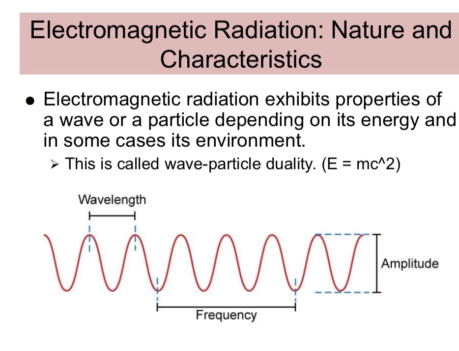
Electromagnetic Radiation: Nature and Characteristics
Electromagnetic radiation: electric and magnetic disturbance traveling at the speed of light
All spectrum components share the same velocity:
Vary in energy, wavelength, and frequency
EM radiation can exist apart from matter and travel through vacuum; originates from atoms
Electromagnetic Radiation: Course of Travel and Intensity
EM radiation travels as divergent rays from a source; intensity spread over a larger area
Intensity is energy flow per second (photon flux); greatest at the center
Intensity diminishes with distance; follows inverse relationships
Distance Formula and Inverse Square Law
Inverse square law for intensity:
I1: initial intensity, d1: initial distance, d2: final distance, I2: final intensity
Spectrum: Key Relationships
Electromagnetic spectrum from lowest to highest energy: radio waves, microwaves, infrared, visible, ultraviolet, X-rays, gamma rays
Wavelength range:
Frequency range:
Velocity and wavelength-frequency relation: ;
Velocity relation: (for EM radiation, )
Energy and Wave-Particle Duality
EM radiation exhibits wave-particle duality
Energy relates to frequency:
Planck’s constant (approximate):
Energy range of photons:
Rest of the Spectrum and Ionization.
Ionization status (as per summary):
Radio waves: No
Microwaves: No
Infrared: No
Visible light: No
Ultraviolet: No
X-rays: Yes
Gamma rays: Yes
X-Rays and Gamma Rays: Similarities and Differences
Similarities:
Exhibit wave-particle characteristics; high energy; can burn skin
Intensity follows inverse square law; can ionize matter
Differences:
Gamma rays originate from atomic nuclei (nuclear transitions)
X-rays originate from interactions between electrons and atoms
Particulate Radiation
Particulate radiation includes alpha and beta particles
Capable of ionizing matter; more common in nuclear medicine or radiation therapy
Alpha and Beta Particles
Alpha particles:
The nucleus: two protons and two neutrons
Positive charge; short range; cannot penetrate many materials
Beta particles:
Electrons emitted from unstable nuclei; originate in nucleus (not electron shell)
Lighter than alpha; may ionize along their path
Beta particles can be negative (beta minus) or positive (beta plus, a positron)
Radioactivity
Radioactivity: decay of unstable nuclei emitting gamma, alpha, or beta particles to reach stability
Decay transforms into new elements
Half-life: time for half of atoms to decay
Sources of Exposure
Natural/background and manmade sources
Subcategories: cosmic, terrestrial, internal, medical
Total dose varies by geographic location
Interaction with Matter: Reflection, Transmission, Absorption, Attenuation
Energy determines how EM radiation interacts with matter
Can be reflected, transmitted, absorbed, or attenuated by tissues
Radiopaque vs Radiolucent (Practical Imaging Concept)
Ra diopaque materials (bone) absorb X-rays (appear white)
Radiolucent materials (soft tissue) transmit more X-rays (appear darker)
Quick Reference: Core Takeaways
EM radiation properties: speed, wavelength, frequency, energy; wave-particle duality
Core equations:
Spectrum uses and ionization tendency: X-rays & Gamma rays ionize; others do not (per summary)
Alpha/beta particles: particulate radiation with distinct properties
Radioactivity and half-life: decay, stability, time scales
Exposure sources: natural vs manmade; medical contributions
Interactions with matter: reflection, transmission, absorption, attenuation
Imaging relevance: radiopaque vs radiolucent materials