The Mechanism of Muscle Contraction: Sarcomeres, Action Potential, and the Neuromuscular Junction
Structure of Skeletal Muscle
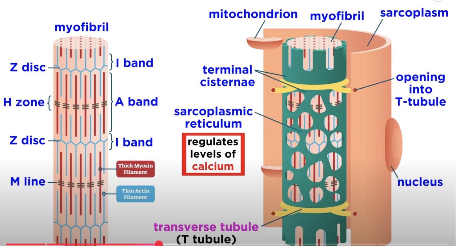
Skeletal Muscle Composition: Skeletal muscles are made up of fascicles, which are in turn made of muscle fibers (multinucleated muscle cells).
Myofibrils: Each muscle fiber contains myofibrils composed of myofilaments organized into contractile units called sarcomeres.
Contractile proteins: Actin and myosin
Regulatory proteins: Troponin (controls tropomyosin) and tropomyosin (blocks myosin binding to actin)
Accessory proteins: Nebulin (aligns actin) and Titin (elasticity and stabilizes)
Sarcomere Structure
A Bands and I Bands:
Dark A bands and light I bands contribute to the striated appearance of skeletal muscle.
The A band contains an H zone split by the M line, which is made of myomesin.
I bands are divided by Z discs, defining functional sarcomeres from one Z disc to the next.
Filament Arrangement:
Thick filaments contain myosin and span the A band, connected at the M line.
Thin filaments are composed of actin, extending across the I band into the A band.
Elastic filaments, composed of titin, extend from Z discs to the thick filament.
Myofilament Details
Myosin Structure:
Myosin consists of two globular heads and a long tail, with heads serving as the active sites.
Active sites contain ATP binding sites as well as actin binding sites.
Thick and thin filaments will interact at these sites by making cross bridges.
No myosin heads are present in the center of the sarcomere, which helps define the H zone.
Actin Structure:
Composed mainly of actin subunits that twist to form thin filaments.
G-actin —> F-Actin
Active sites on actin are blocked by tropomyosin in a relaxed muscle fiber.
Troponin complex binds to actin, tropomyosin, and calcium, regulating muscle contraction
When calcium ions are released from the sarcoplasmic reticulum, they bind to troponin, causing a conformational change that moves tropomyosin away from the active sites, allowing myosin heads to attach to actin and initiate the sliding filament mechanism.
Role of the Sarcoplasmic Reticulum
Sarcoplasmic Reticulum Function:
Surrounds each myofibril regulating calcium levels essential for muscle contraction.
Contains T tubules that help propagate signals throughout the muscle fiber.
T-tubules sit at each A-band and I-band junction
Sliding Filament Model of Contraction
Mechanism:
When stimulated by the nervous system, myosin heads form cross bridges with actin, pulling thin filaments towards the center of the sarcomere.
This process shortens the I bands, eliminates the H zone, and brings the A bands closer together, leading to muscle contraction.
Initiation of Muscle Contraction
Neuromuscular Junction:
Connection point between the nervous system and skeletal muscle, involving acetylcholine release from axon terminals into the synaptic cleft.
Pathway —> Excitation-contraction coupling:
Brain sends electrical signal through nerve to muscle (Start the race)
ACh releases by synaptic cleft, binds to nicotinic cholenergic receptors
Nerve releases a ACh (Pass the baton)
Binding of ACh opens the channels —> Na+/K+ moves across the membrane
ACh is removed by acetylcholinesterase
Na+ influx > K+ efflux = local depolarization occurs at synapse (EPP)
Ach tells muscle to create elec. sig. to muscle. (Spread the signal)
Action potential occurs at EPP down the T-tubules
DHP on Ca2+
DHP detects depolarization
DHP changes RyR = opening of Ca2+ at the sarcoplasmic reticulum
Signal reaches SR and calcium gates open (Open the gates)
Ca2+ to troponin on thin filament —> tropomyosin “on”
Calcium binds to troponin, moves tropomyosin out of the way, exposes “grab-sites” on actin
Calcium says “go”
Myosin to actin goes through cross bridge
muscle contracts
Acetylcholine binds to receptors causing sodium influx, resulting in depolarization and action potential generation.
Cross Bridge Cycle:
Active sit on actin is exposed when Ca2+ binds to troponin
calcium says go!
Myosin head binds to actin at the actin binding site and forms a weak crossbridge
Mysoin grabs actin
the inoragnaic phosphate releases from mysoin that causes the mysoin head to pivot toward the centre of the sarcomere
This pulls the thin filament toward the M line
Myosin pull actin (power stroke)
Release of ADP from myosin after the power stroke
Myosin lets go
myosin is now firmly bound to actin —> rigor state
a new molecule of ATP attaches to the myosin head, causing the crossbridge to detach
ATP resets myosin
The myosin head ATPase hydrolysis ATP—> ADP +Pi which returns myosin to the cocked position. Return to step 2 and continue the cycle if Ca2+ is bound to troponin.
Repeat as long as calcium is there
Action Potential Propagation:
The action potential moves along the sarcolemma and down T tubules, resulting in calcium ion release from the sarcoplasmic reticulum.
Repolarization occurs after action potential propagation when potassium channels open, restoring the negative charge inside the muscle fiber.
Excitation-Contraction Coupling
Calcium Role:
Rising levels of calcium ions in the cytosol displace tropomyosin from myosin binding sites on actin, facilitating contraction.
Myosin heads bind to actin, perform cross bridge cycling while hydrolyzing ATP, resulting in filament sliding that leads to muscle contraction.
Relaxation Phase:
As calcium levels decrease, troponin reverts to its original shape, tropomyosin re-blocks binding sites, and the muscle fiber relaxes.
Summary of Muscle Contraction Process
Muscle contraction begins at the neuromuscular junction with acetylcholine release.
Sodium and potassium ions create a voltage change, leading to action potential generation.
This action potential travels through T tubules, stimulating calcium release that enables myosin-actin interaction and muscle contraction.