L1: Bones & Thoracic Limb
Bone
Basic bone mass (bone matrix) is highly mineralized--hardness. The main mineral components of bone are:
- hydroxyapatite (Ca10 (PO4) 6 (OH) 2)
- calcium hydrogen phosphate (CaHPO4) and (MgHPO4)
- calcium carbonate (CaCO3)
Mineral constituent comprises up to 65% by weight of the bone. The presence collagen tissue gives skeletal tissue necessary flexibility.
In addition to the support function serves as a reservoir of bone minerals, which are issued as necessary into the bloodstream.
Calcium homeostasis:
union-- vitamin D3, calcitonin, parathyroid hormone - a constant level in the blood system by absorption from the small intestine -saving bone- Renal excretion, faeces
Osteocyte, Osteoblast, Osteoclast
- Bone cells - osteocytes are stored in tubes (lacunae, lat. Lacunae) which are interconnected by fine channels (lat. Canaliculi ossium). These cells release the mineral phase of bone tissue, therefore regulating blood calcium levels.
- Osteoblasts are involved in the creation of bone matrix, producing, in the form of precursors; after the formation of the extracellular matrix that surrounds them, and change into osteocytes.
- The osteocyte is the basic cell of the mature bone. They do not produce an extracellular matrix of bone. However, participates in the metabolism of releasing minerals from the base matrix of bone, it has a pivotal role in the regulation of calcium levels in the body.
- Osteocytes can be changed if necessary to osteoblast – (produces extracellular matrix), or reticular cell.
- The osteoclast is a type of bone cell that removes bone tissue (bone resorption). Osteoclasts and osteoblasts are instrumental in controlling the amount of bone tissue: osteoblasts form bone, and osteoclasts resorb bone. Osteoclasts are formed by the fusion of cells of the monocyte-macrophage cell.
Blood vessels and nerves
- Nutritive artery (lat. arteria nutricia)
- thicker; one or two; occurring at the site beginning ossification Nourishes its branches bone marrow and linked with the blood vessel in the Havers ducts associated with arteries of the periosteum.
- Periostial artery
- enter through Volkmann's canals and connects to the network in Havers tubules.
- Metaphysis artery (lat. arteriae metaphysariae)
- Branches deep in the spongiosa and nourish the area of bone and bone marrow between the diaphysis and epiphysis
- Epiphysis artery (lat. arteriae epiphysariae)
- majority arterial branches tangle of joint capsule.
- A flat bones - have greater nutritive artery and a large number of small periosteal artery.
Nerves
- The nerve fibres mainly penetrate from the richly innervated periosteum along the vascular channels through Havers to bone marrow. Probably only innervate the walls of the arteries. The impact of the loss of innervation of the bone is not exactly known, probably related to the complex innervation of muscle loss. Innervation of bone has not been demonstrated. Pain originating from bone thus periosteal pain - compact bone below the periosteum is insensitive!
Compacta Vs. Spongiosis
Substancia compacta
- Resists Pressure and tension
- The structure of the hollow fiber
- Osteon-Havers system-collagen fibers parallel or concentrically around the vascular channel
- Osteon apart delimited cement
- The transverse and oblique connections between osteon -Volkmann channels
Substancia spongiosa
- Moving the push and pull on compact
- Sponge-like structure
- It does not contain Havers system
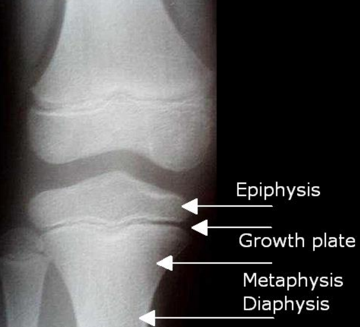
Intramembranous ossification Endochondral ossification cartilage model ( primary ossification center •secondary ossification center) •Bone Remodeling: PTH, Calcitonin,Thyroid hormones,Insulin,HGH,IGFs Bone Metabolism: Minerals, Vitamins, Exercise
Joints
- Synarthroses- form joints that are relatively rigid.
- Diarthroses- form joints that are freely movable. two bone ends covered with articular cartilage, synovial fluid
- Joint function is to absorb the force of impact, transfer the force via cartilage to bone and to allow a variable degree of movement.
- Synovial Joints
- Fibrous Joints
- Syndesmosis – connected by fibrous tissue
- Cartilaginous Joints
- Synchondrosis – connection with cartilage, changes to bone over time
- Symphysis – connection using cartilage
Type of joints :
- articulationes simplices – consists of two bones
- articulationes compositae - consists of 3 or more bones, or 2 bones and articular disc or meniscus
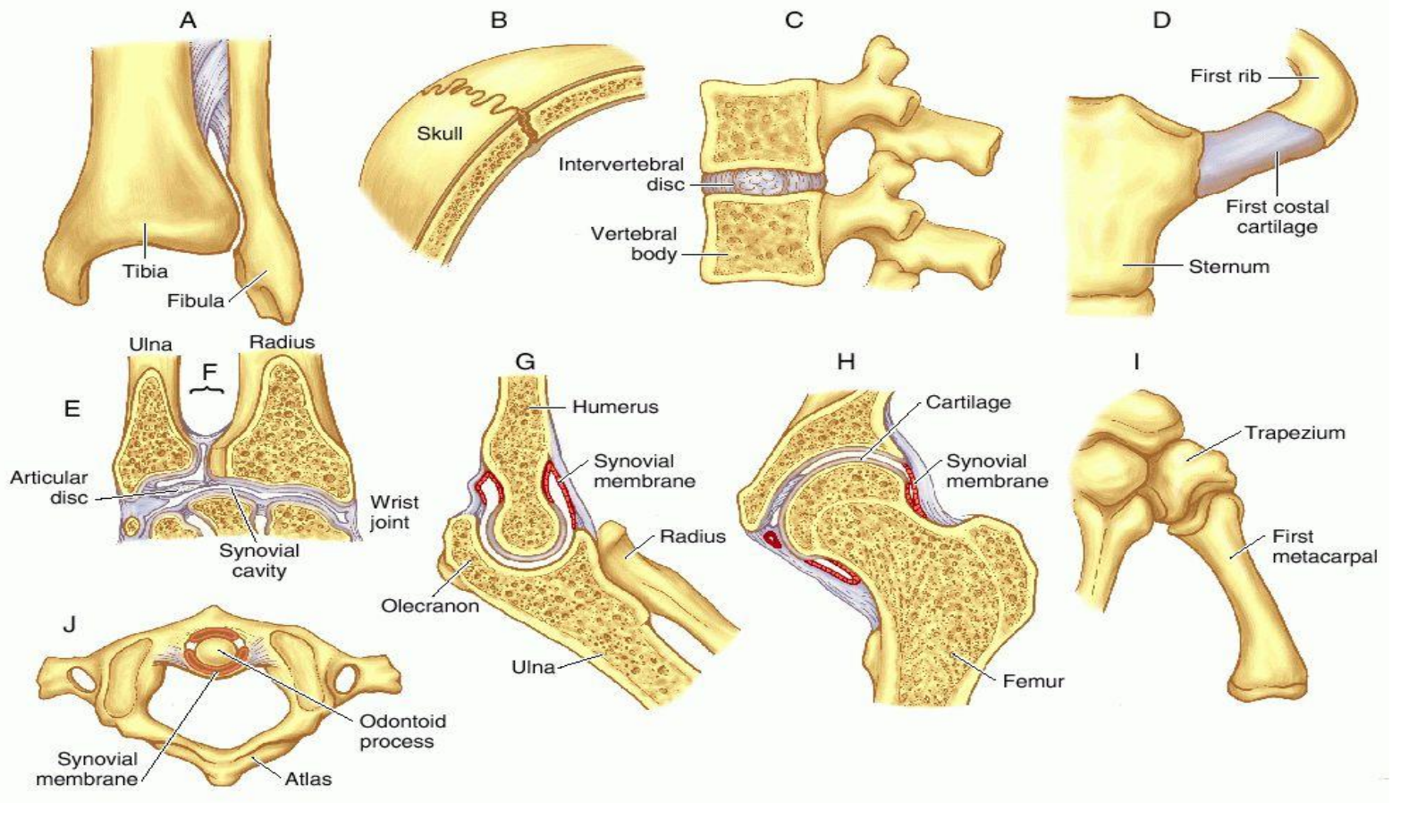
Various kinds of joints.
Fibrous: A, syndesmosis (tibiofibular); B, suture (skull). Cartilaginous: C, symphysis (vertebral bodies); D, synchondrosis (first rib and sternum).
Synovial: E, condyloid (wrist); F, gliding (radioulnar); G, hinge or ginglymus (elbow); H, ball and socket (hip); I, saddle (carpometacarpal of thumb); J, pivot (atlantoaxial).
Projections of the Thoracic Limb
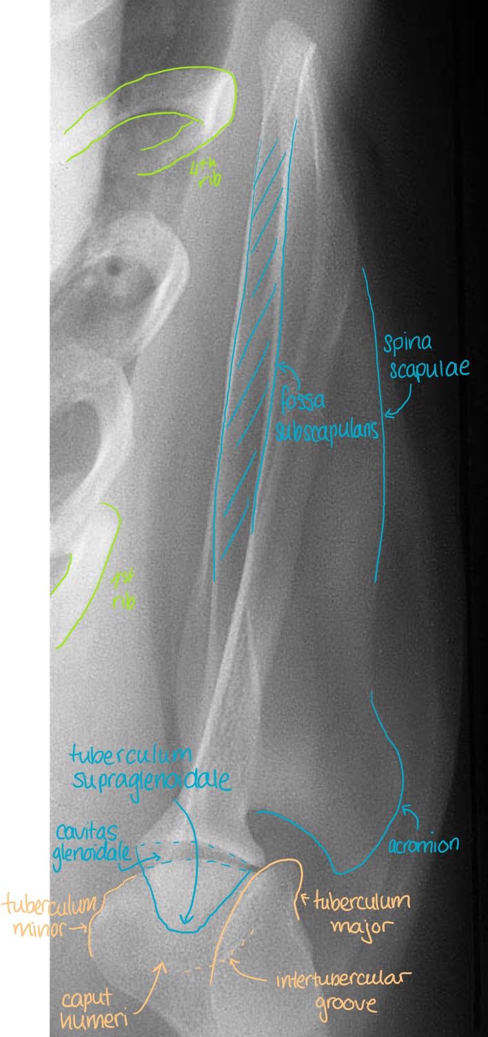
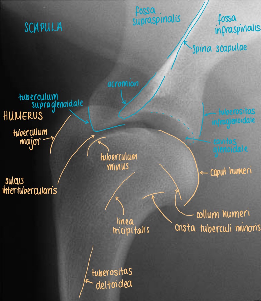
Articulatio Humeri
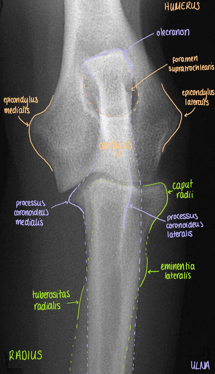
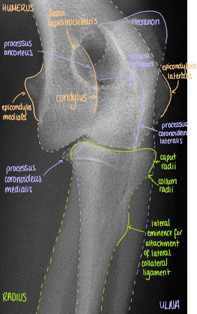
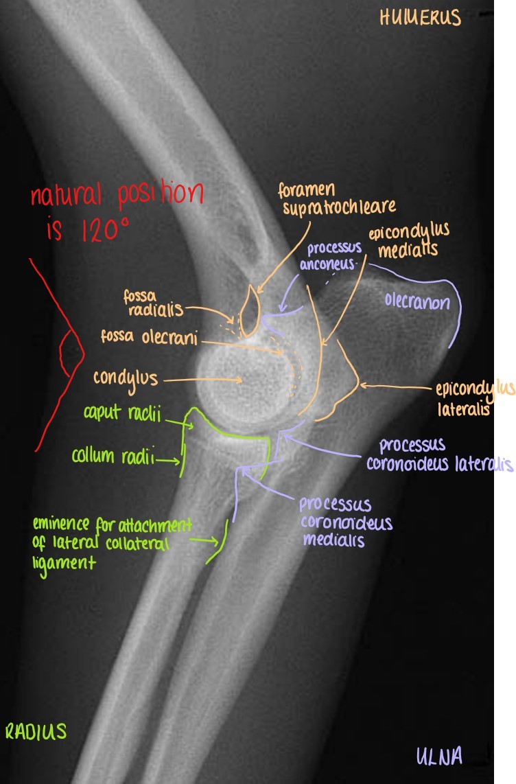
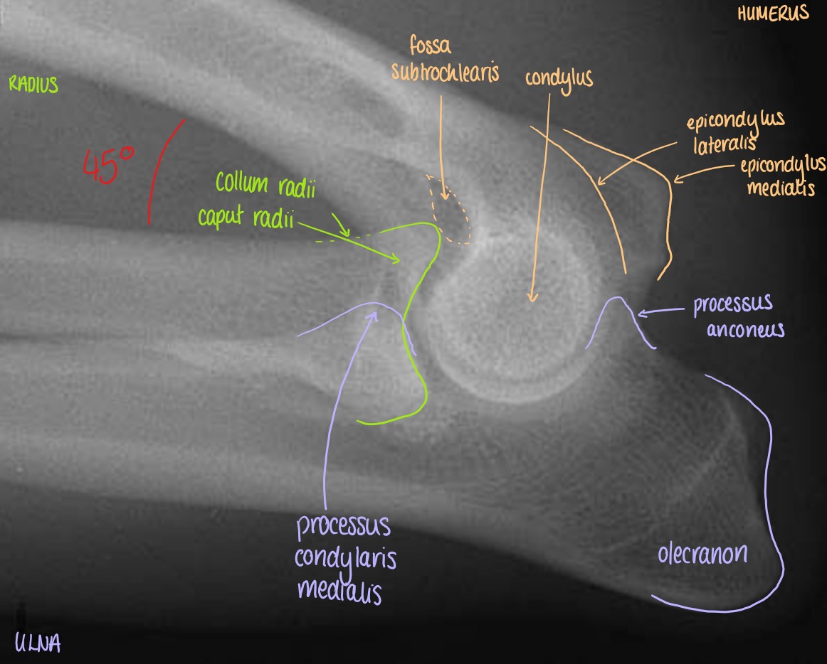
Articulatio Cubiti
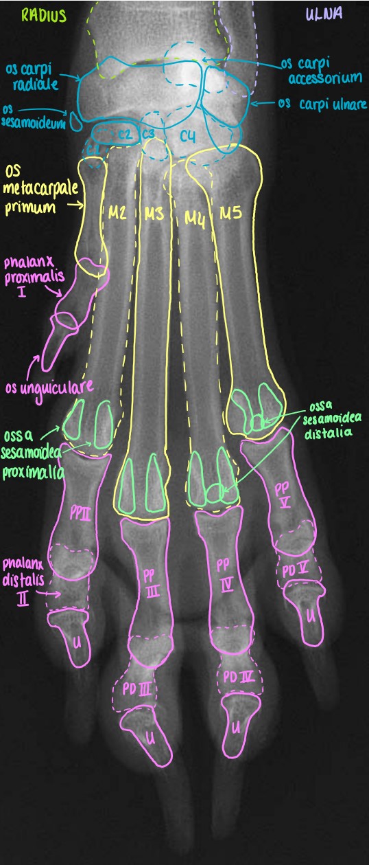
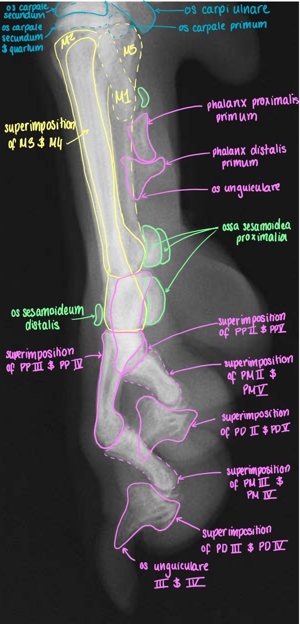
Articulatio Carpi
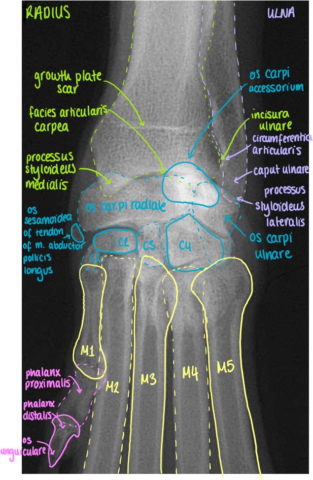
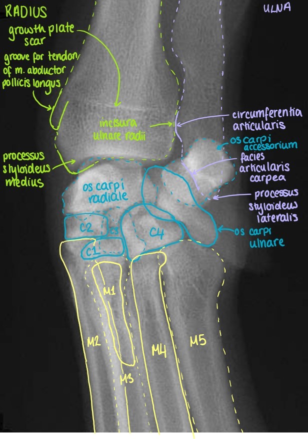
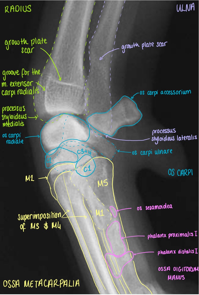
Manus