Wk4: eye, light and dark adaptation and receptive fields
4a: eye
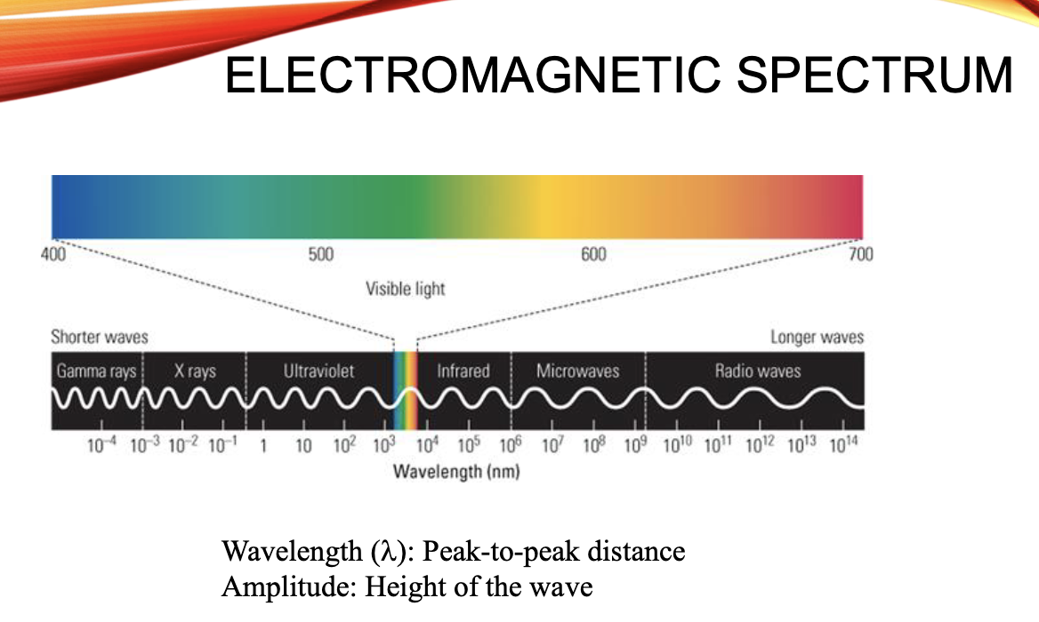
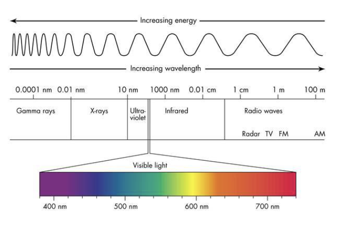
properties of light
the structure of the eye
image formation
properties of cones and rods
light intensity
wave
wavelength ( lambda)
amplitude (contrast)
particle
no. of photons
Cd/m² → candelas per m²
eye
cornea
clear covering over the lens
refracts incoming light
fixed refractive power
pupil
opening in the iris
partially regulates enetering light
lens
focuses incoming light
can change its refractive power
retina
neural tissue lining the back of the eye
location photoreceptors (rods and cones) that detect light (image)
rods and cones are part of the whole (retina)
optic disc and blind spot (nasal retina)
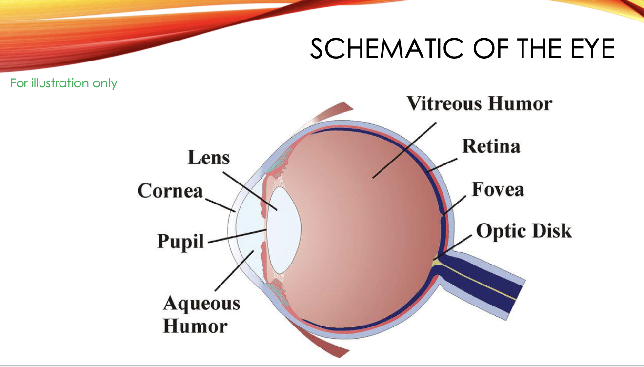
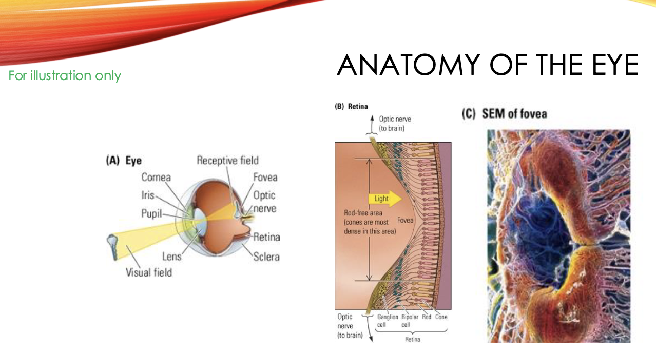
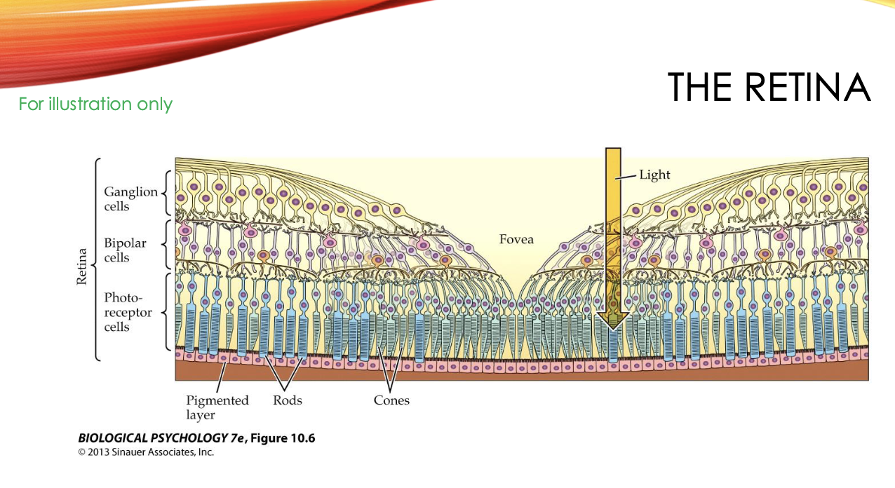
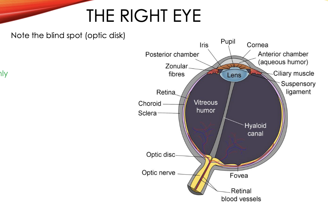
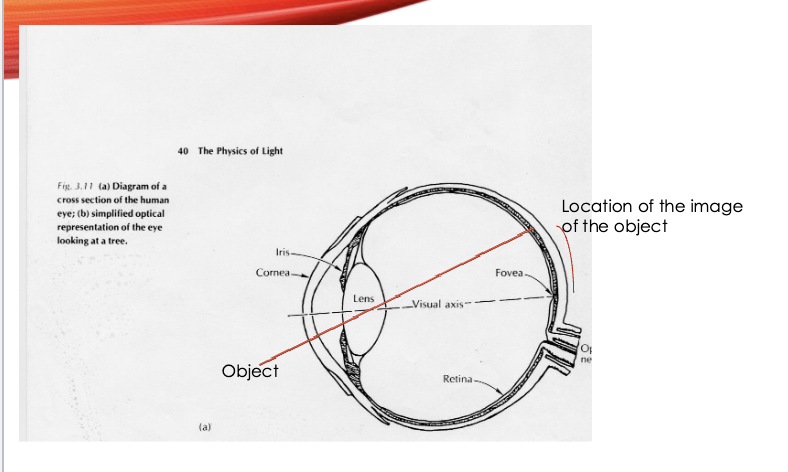
image formation
image focuse on the retina, where photoreceptors are (retina)
fovea: region of retina where photoreceptor density is high
where the image falls: fovea
the centre of where you’re looking
“central” vision not peripheral
distance and retina
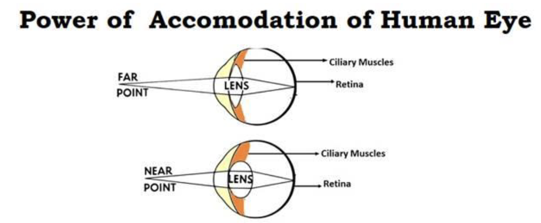
object close, image focused behind retina → blurry
hence, need more refractive power/light bending
lens more round (more curved)
object far, image focused in front of retina → still blurry
less refractive power/light bending
lens flatter (less curved)
near point: closest distance at which an object can be focused
limited by the maximum amount that a person’s lens can be curved
Accommodation conditions
emmetropia
normal vision
correctly focuses on objects at different distances
presbyopia
decrease in accommodation ability with aage
near point lengthens
myopia
too much refractive power
nearsightedness
far object blurred
near object focused
near point abnormally near
hypermetropia
not enough refractive power
farsightedness
near object blurred
far object focused
how children’s eyes start out
myopia: short-sightedness
environment induced myopia
high rate, makes up a lot of the population in many countries
cause:
highly correlated with educational level and degree of urbanisation
both lead to reduction in distances that people have to accommodate for
photoreceptors
rods
high luminance sensitivity
strong response at low light levels
rapid saturation
don’t operate in daylight conditions
cones
low luminance sensitivity
less rapid saturation
mediate colour vision and ability to see fine spatial detail
fovea only has cones, no rods
eccentricity
angle from line of sight (direction of fixation)
increasing the eccesntricity of an object:
object is more in your peripheral vision
results in image located further away from fovea.
spatial acuity
the ability to see small objects
cones provide best spatial acuity
decreases when increasing eccentricity
cone density decreases with eccentricity
neural convergence (no. of cells projecting to a single cell) increases with eccentricity
types of vision
scoptic: rods only
no colour
blind to images falling in the fovea
poor ability to see fine spatial detail
mesopic: rods and cones
photopic: cones only
colour vision
good ability to see spatial detail (especially fovea)
nighttime viewing
night characteristics:
low light → rods working not cones
no rods in fovea, image falls if you look directly at it
look slightly away from fovea → image falls away from fovea into peripheral retina → rods can process image
4b: Light and dark adaptations
three ways to achieve range of sensitivity
iris change is 8 fold
two types of photo detectors
rods low light
cones brighter condition
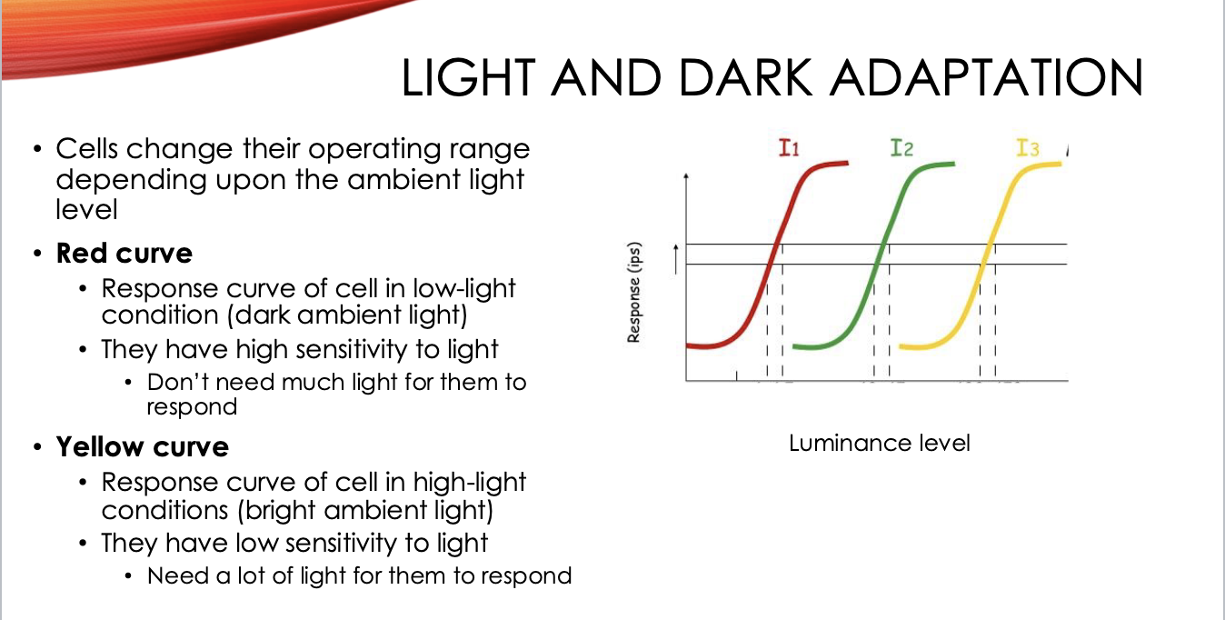
Light and dark adaptations
decrease/increase in the senitivity of cones and rods due to changes in light lvls → changes in the stimulus-response curve of cells
light adaptation → over time, cells become less sensitive to light lvls (gain is decreased)
dark adaptation → over time, cells become more sensitive to light levels (gain is increased)
GAIN: a control mech.
high gain: louder output for the same input (more sensitivity)
low gain: quieter output for the same input (less sensitivity)
example:
when going from bright sunshine to a dark room
initially can’t see anything but then after a while can
when going from a dark room into bright sunshine
initially just see total brightness but after a while can see variations
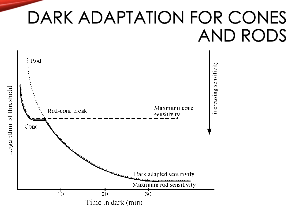
dark adaptations
light sensitivity imporovments increase longer for big dot compared to small dot.
large dot improvement intervals: 10 min→30min
small dot: 10min→stops
10 mins for both → cones
30 mins for large → rods
seeing at night without artificial lighting (dark)
both rods and cones dark adapt/improve sensitivity to light
rods adapt most (30mins)
cones adapt less but quicker and not as sensitive
example:
want to read a map at night but still maintain good night vision. what coloured light should you use?
what are the two competing funciton requirements?
which photoreceptors would mediate those functions and how
explanation:
the two requirements
read map
fine spatial detail (cone)
need light for cone
maintain good night vision
rods (low light)
want them to be fully dark adapted, so don’t want them to be exposed to light
want a light that
cones are sensitive to
rods are not/less sensitive to
red light (less than 630nm)
4c: eye and receptive fields (RFs)
receptive fields
receptive field of a cell
region in space in which stimulation leads to a response (change in firing rate) in the cell
for the cell to respond, the stimulation has to be of the correct type
that is the cell is tuned to particular aspects of the visual stimulus
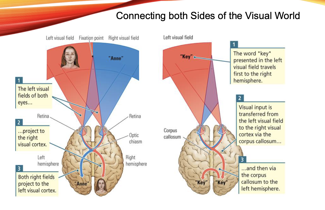
visual encoding/represenation
why can’t we ‘see’ the blind spot
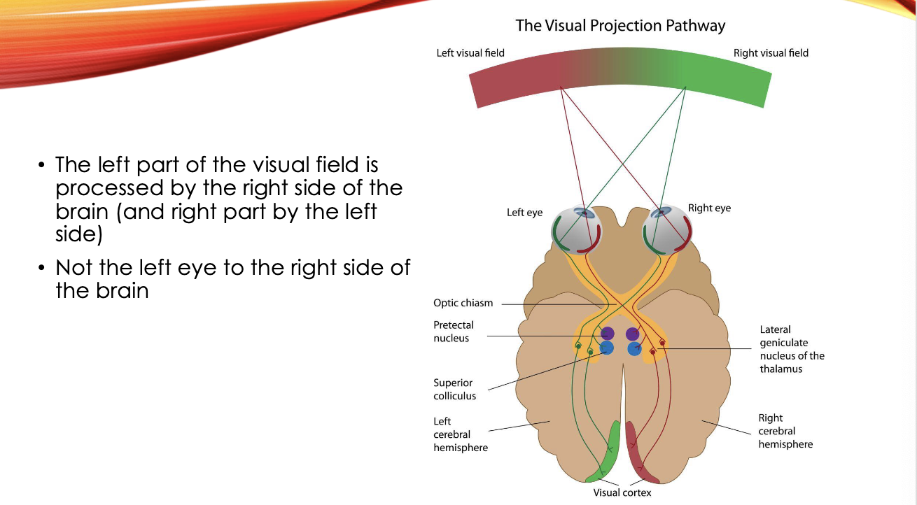
visual projection pathway: from the eye to the brain
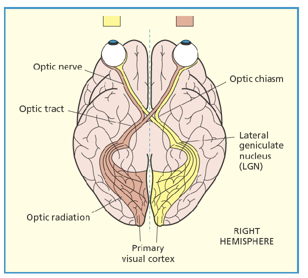
retinal ganglion - RG
lateral geniculate nucleus - LGN
primary visual cortex - V1
general properties of RFs
👁 What Are Receptive Fields (RFs)?
A receptive field is the specific region of the visual field where a stimulus will affect the firing of a particular neuron. In the visual system, RFs become increasingly complex as you move from the retina to the cortex.
🔍 Key Properties of RFs (up to V1: RF properties from retina to V1)
1. Spatially Localised
- Each RF corresponds to a small patch of the visual world.
- Neurons respond only to stimuli within their designated region.
- This allows for fine-grained spatial mapping of visual input.
2. Size Increases with Eccentricity
- RFs near the fovea (center of gaze) are small, supporting high acuity.
- RFs in the periphery are larger, sacrificing detail for broader coverage.
- This reflects the trade-off between resolution and coverage across the retina.
3. Spatially Opponent
- RFs are tuned to contrast, not uniform light.
- They respond to differences in luminance across space—edges, bars, patterns.
🧠 Types of Cells and Their RFs
🧱 Simple Cells
- Found in V1 (primary visual cortex).
- Have linear RFs with distinct regions of excitation and inhibition.
- Respond best to oriented edges or bars at specific locations.
- You can map their RFs using light/dark stimuli.
🌀 Complex Cells
- Also in V1, but with nonlinear RFs.
- Still sensitive to orientation and contrast, but:
- Less dependent on exact stimulus location within the RF.
- Respond to movement and patterns across their RF.
- More robust to spatial shifts, supporting motion detection.
eccentricity, spatial acuity as a function of eccentricity
angle from line of sight → direction of fixation
increasing eccentricity of an object
object more in your peripheral view
results in image being located further away from fovea
therefore, more blurry
spatial acuity as a function of eccentricity
spatial acuity decreases with increasing eccentricity
cone density decreases
neural convergence increases
retinal ganglion and LGN lateral geniculate nucleus
these levels have cells with similar RF properties
both RG and LGN have centre-surround RFs → response depends on whether light hits the centre or the surrounding area of their RF:
on centre type
detects when a bright dot is placed in its centre (increases firing rate)
no response to uniform light field
off centre type
detects when a dark dot is placed in its centre (increases firing rate)
centre and surround responses are balanced at the LGN levl
on centre: excited by light in the centre
off centre: excited by darkness in the surround
Type | Stimulus in Centre | Response | Stimulus in Surround | Response
-------------|--------------------|----------------|-----------------------|---------
On-centre | Bright spot | ↑ Firing rate | Bright spot | ↓ Firing rate
Off-centre | Dark spot | ↑ Firing rate | Dark spot | ↓ Firing rate
determining the response of spatially opponent cells
spatially opponent: description of centre-surround RF
calculation:
response = light on excitatory - light on inhibitory
both regions equally lit → no net response
on channels respond to increase in light intensity
off channels respond to decrease in light intensity
wiring up centre-surround cell
• Centre photoreceptors (cones/rods) send signals directly to the ganglion cell → Excitatory input.
• Surround photoreceptors send signals indirectly via horizontal cells, which inhibit the ganglion cell → Inhibitory input.
• This creates a spatially opponent structure: light in the centre excites the cell, light in the surround suppresses it.
centre-surround cells and visual processing
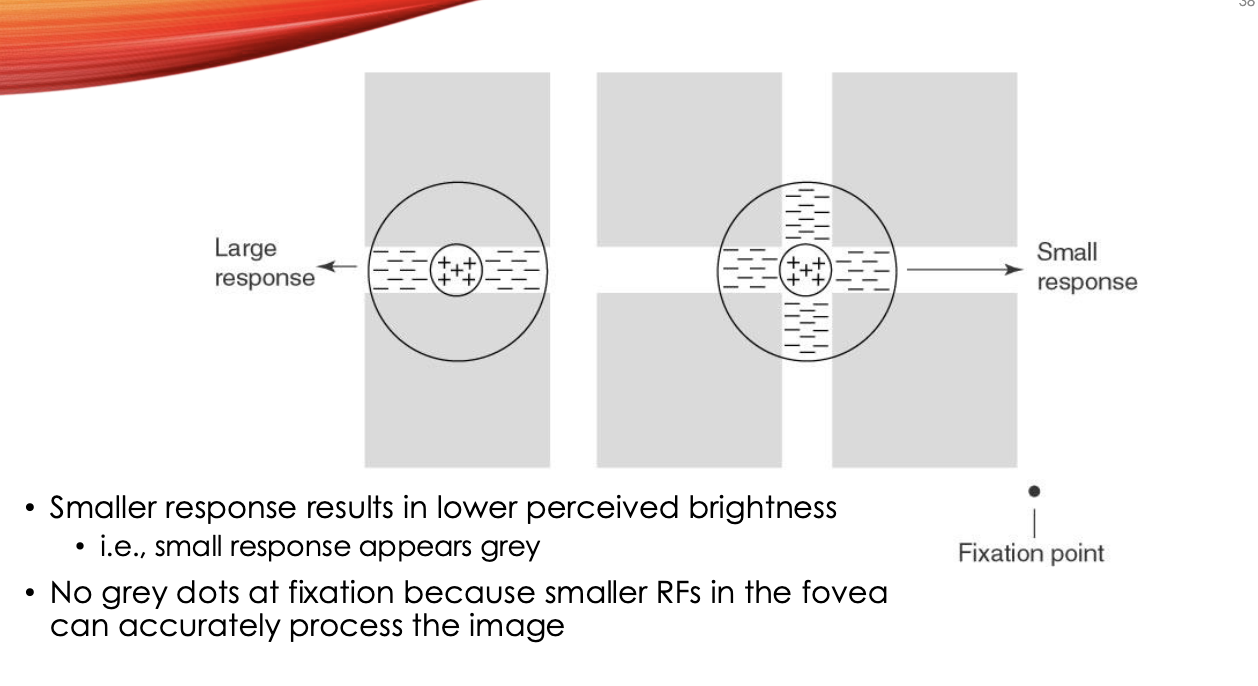
explaining hermann-hering grid illusion using properties of centre-surround cells
two aspects
get the grey dots
but not in central vision
peripheral vision, intersections have grey dots
at iintersections, surround regions of the RF is exposed to more white light than at line segments
this extra inhibition reduces the ganglion’s firing rate
your brain interprets this reduced signal as less brightness even though physical light is the same
only peripheral because fovea RF are small and precise, peripheral RF are larger, more surround light, stronger illusion
cortex V1
four types of cells:
simple
first cortical stage of visual feature detection
not circular in shape
sensitive to bars and edges (orientation and width)
linear
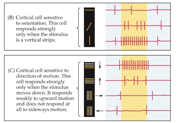
complex
also orientation and width
nonlinear
motion selective
pooling of simple cells
hyper complex (end stopped)
length selective
concentric
colour selective
tuning curve/bandwidth
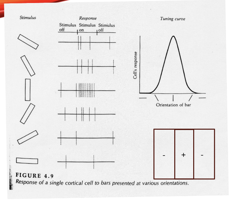
bandwidth:
how broadly tuned the cell is to that particular dimension
e.g. how many diff orientation a V1 simple cell is tuned to
additional property of V1 cells
receive binocular input
v1 cells the first to have this property
monocular RFs mostly overlap
monocular tuning properties mostly the same