Microscopy - Epithelial Tissue Histology
The goal of microscopy is to create a magnified image of objects too small to be seen with the eye alone.
Brightfield microscopes use a combination of glass lenses and light to view the specimen.
Microscopes are often used to study bacteria, cells, and tissues
Fields of study where a microscope is a necessary tool include:
Cytology - the study of cells
Histology - the study of tissues
Pathology - the study of disease
Microscopy allows for the visualization of cell morphology and their organization into a tissue.
There are four broad categories of tissues, epithelial, connective, muscle, and nervous.
Epithelial tissue covers our body surfaces and lines the inside of our hollow organs. They form sheets of cells that are exposed to an open space (lumen). Microscopic observation of epithelial tissue shows cells with distinct cell shapes and layers sitting on a basement membrane.
Epithelial cell shapes include:
Squamous - flat/scale-like
Cubodial - cube-shaped
Columnar - column-like
Transitional (changes shape from polyhedral to flat)
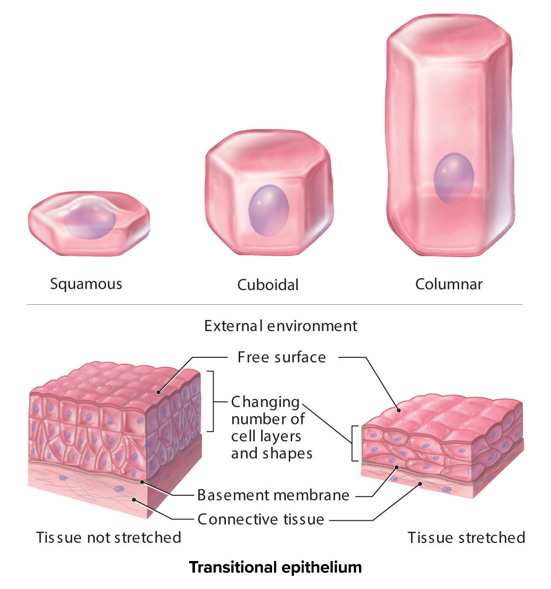
Epithelial tissue layers include:
Simple (single layer of cells)
Pseudostratified (single layer of cells that appears as more than one)
Stratified (multiple layers of cells)
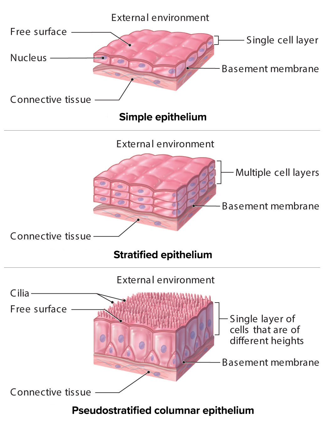
An epithelial tissue name typically consists of two words, combining the number of layers and cell shape, Stratified epithelial tissues are always named for the shape of cells at the apical surface.
Examples using these characteristics:
Tissue with a single layer of column-like cells would be identified as simple columnar epithelial tissue.
Tissue with multiple layers of cuboidal-shaped cells at the basal surface and flat cells at the apical surface would be identified as stratified squamous epithelial tissue.
Epithelial tissues are classified into the following groups:
Simple epithelial tissues
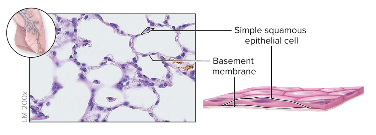
Simple Squamous Epithelium
Structure - Single layer of thin, flat cells resembling irregular floor tiles; the single nucleus of each cell bulges at its center
Function - Thinnest possible barrier to allow for rapid diffusion and filtration; secretion in serous membranes
Location - Air sacs in lungs (alveoli); lining of lumen of blood vessels and lymph vessels (endothelium): serous membranes of the body
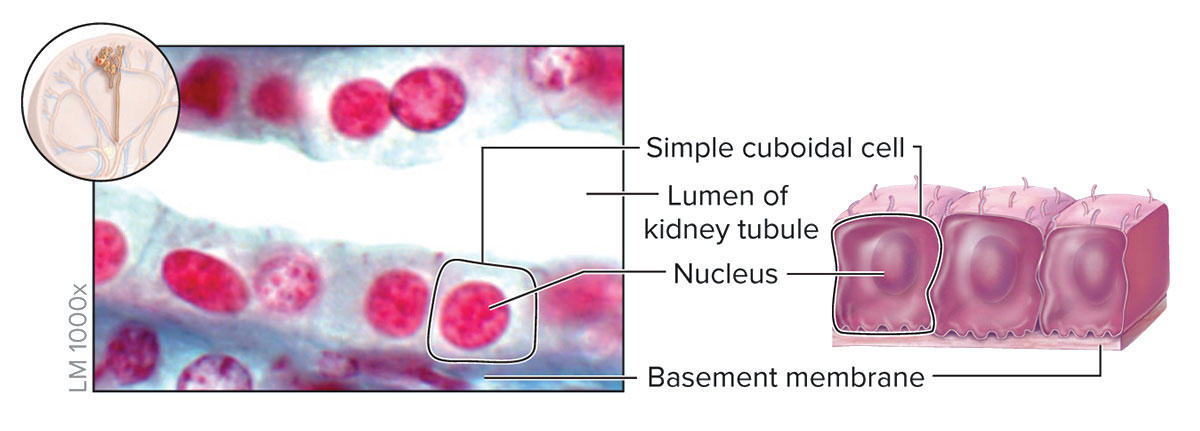
Simple Cuboidal Epithelium
Structure - Single layer of cells about as tall as they are wide; spherical and centrally located nucleus
Function - Absorption and secretion; forms secretory tissue of most glands and small ducts
Location - Kidney tubules, thyroid glance follicles; the surface of the ovary; secretory regions, and ducts of most glands
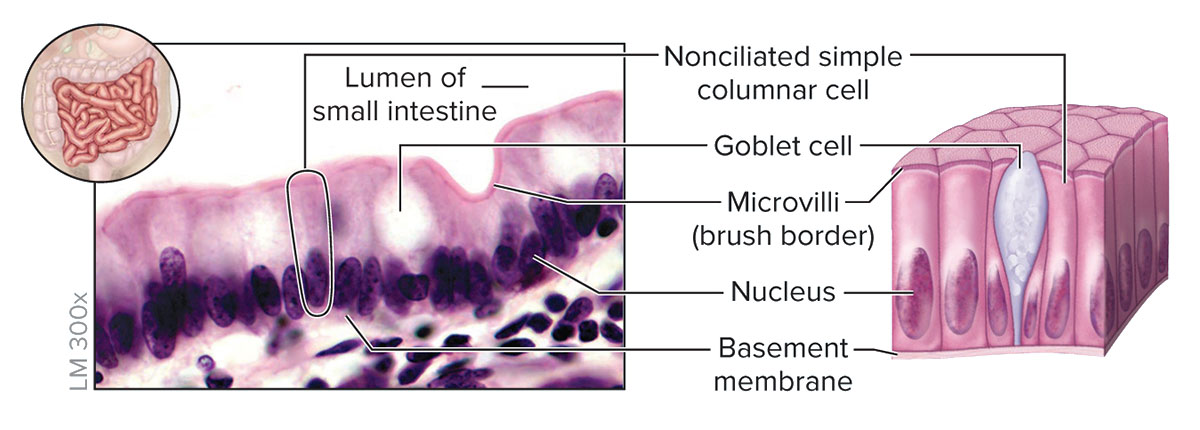
Simple Columnar Epithelium
Structure - Single layer of cells taller than they are wide; oval-shaped nucleus oriented lengthwise in basal region of cell; apical regions of the cell may have microvilli; may contain goblet cells that secret mucin
Function - Absorption and secretion; secretion of mucin
Location - Inner lining of most digestive tract (stomach, small intestine, and large intestine)
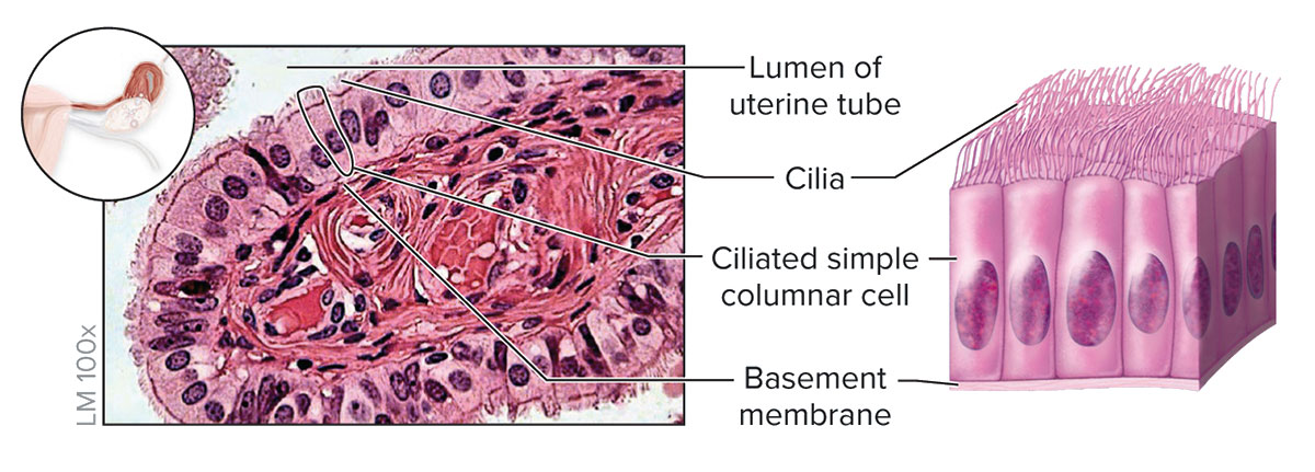
Ciliated Simple Columnar Epithelium
Structure - Single layer of ciliated cells taller than they are wide; oval-shaped nucleus oriented lengthwise in the basal region of cell; may contain goblet cells.
Function - Secretion of mucin and movement of mucus along the apical surface of epithelium by cilia; oocyte movement through the uterine tube
Location - Lining of the larger bronchioles (air passageways) of the lung and the uterine tubes
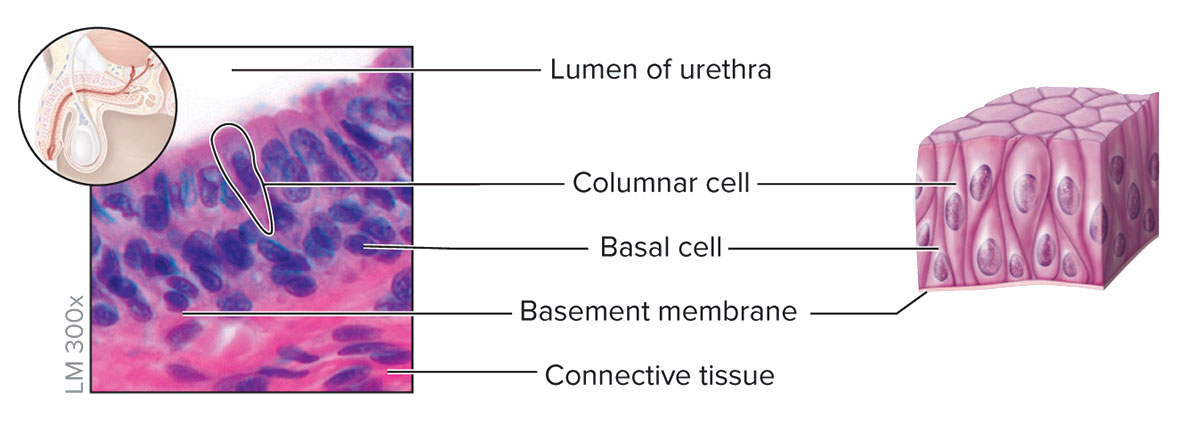
Nonciliated Pseudostratified Columnar Epithelium
Structure - Single layer of cells with varying heights; all cells connect to the basement membrane, but not all cells reach the apical surface; lacks goblet cells and cilia
Function - Protection
Location - rare lining of part of the male urethra and epididymis
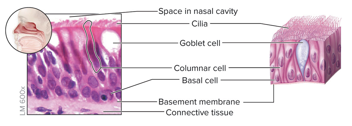
Ciliated Pseudostratified Columnar Epithelium
Structure - Single layer of cells with varying heights; all cells connect to the basement membrane, but not all cells reach the apical surface; has goblet calls and cilia
Function - Protection; secretion of mucin and movement of mucus along the apical surface of epithelium by cilia
Location - Lining of the larger airways of the respiratory tract, including the nasal cavity, part of the pharynx, parts of the larynx, trachea, and bronchi

Stratified Epithelial Tissues
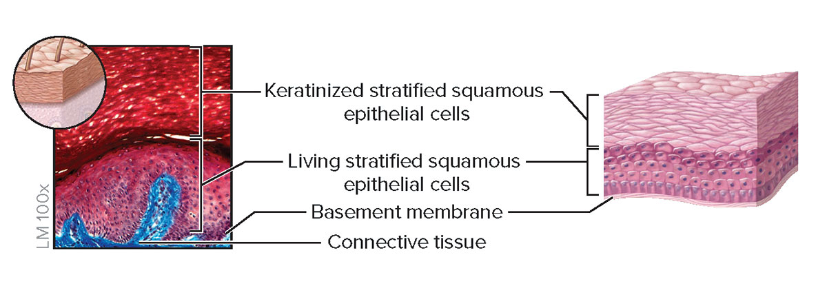
Keratinized Stratified Squamous Epithelium
Structure - Multiple cell layers; basal cells are cuboidal or polyhedral, whereas apical cells are squamous; apical cells are dead and filled with the protein keratin
Function - Protection of underlying tissue from abrasion
Location - Epidermis of skin
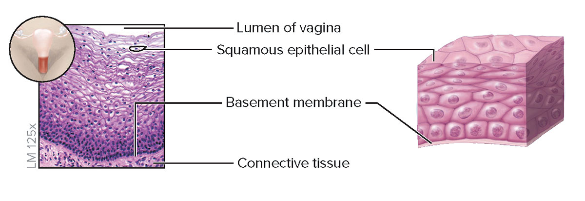
Nonkeratinized Stratified Squamous Epithelium
Structure - Multiple cell layers; basal cells are cuboidal or polyhedral, whereas apical (superficial) cells are squamous; cells lack keratin; superficial cells are alive and kept moist
Function - Protection of underlying tissue from abrasion
Location - Lining of the oral cavity, part of the pharynx, esophagus, the lining of the vagina, and anus
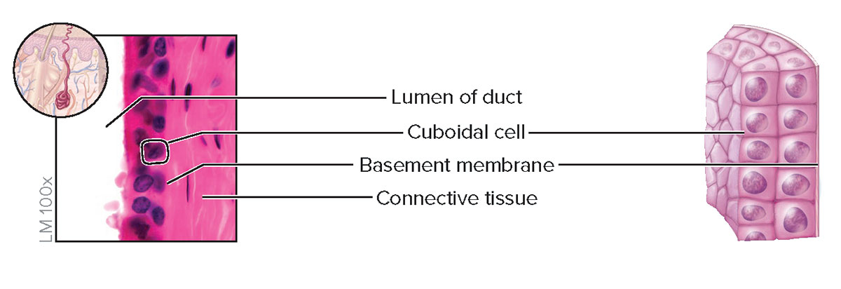
Stratified Cuboidal Epithelium
Structure - Two or more layers of cells; cells at the apical surface are about as tall as they are wide
Function - Protection and secretion
Location - Ducts of most exocrine glands and ovarian follicles
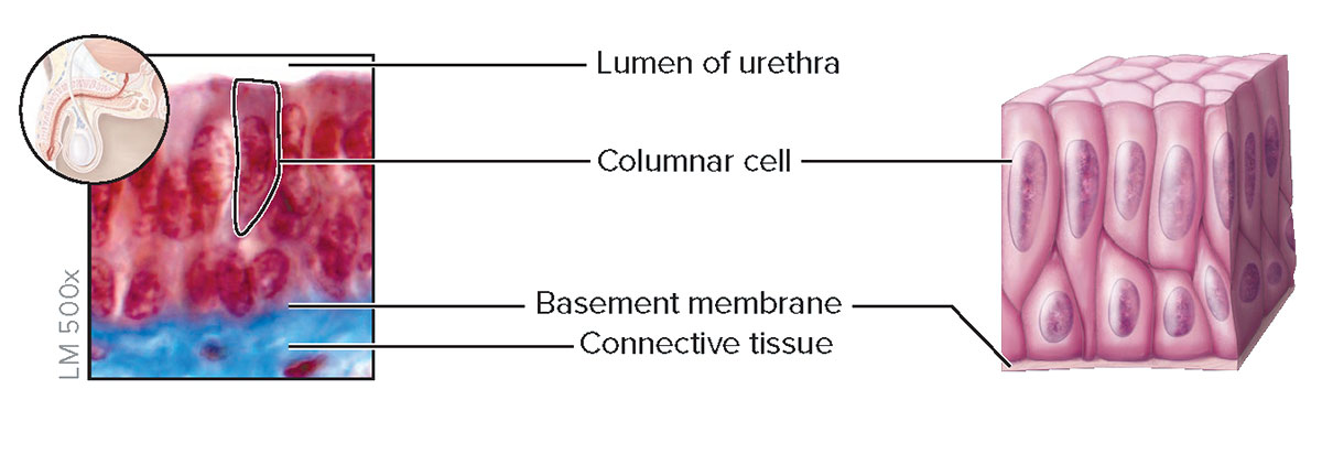
Stratified Columnar Epithelium
Structure - Two or more layers of cells; cells at the apical surface are taller than they are wide
Function - Protection and secretion
Location - Large ducts of salivary glands, conjunctiva covering the eye, and membrane part of the male urethra
Transitional Epithelial Tissues
Transitional Epithelium

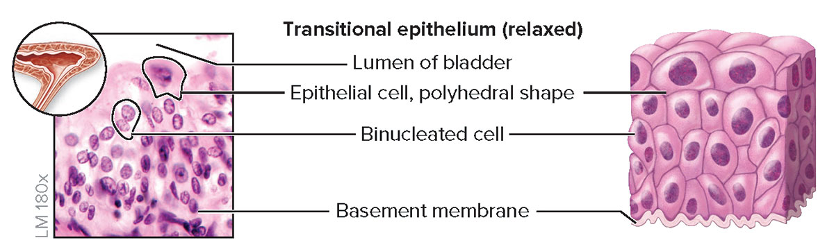
Structure - Epithelial appearance varies, depending upon whether the tissue is relaxed or distended (stretched); relaxed epithelium (top) has cuboidal or polyhedral cells and the apical cells are large and rounded, whereas distended epithelium (bottom has flattened cells at the apical surface; some cells are binucleated
Function - Accommodates urine volume changed (by distending or relaxing) in the urinary bladder, ureters, and part of urethra.
Location - Lining of urinary bladder, ureters, and part of urethra
Apical Surface - The surface of an epithelial cell facing the lumen or external environment.
Basal Surface - The surface of an epithelial cell facing the basement membrane.
Basement Membrane - The basement membrane is formed from the epithelial and underlying connective tissue layers. It is composed of noncellular layers of collagen, glycoproteins, and proteoglycans. The function of the basement membrane is to aid in attachment of the epithelial tissue and as a selective molecular barrier to underlying connective tissue.
Field of View - The field of view is the amount of the slide you are able to see through the eyepiece.
Lumen - Central cavity or open space within an organ
Total Magnification - Total magnification is the overall enlargement of the image of a specimen. To calculate total magnification. Multiply the magnification of the ocular lens (10X) with the magnification of the objective lens.