Bio Psychology
Foundations of Biological Psychology
Historical Overview
The foundation of biological psychology is rooted in historical discoveries and understandings of the brain and its functions.
A timeline of discoveries is utilized to orient the study of biological psychology.
Early Civilizations and the Brain
Egyptian Papyrus (~1700 BCE):
The Edwin Smith papyrus is notable for being one of the first recorded uses of the brain.
The importance of understanding early contributions to modern biological psychology is emphasized.
Egyptian hieroglyphs reflect early knowledge regarding the brain, including recognition of head trauma effects.
Recognition of the Cortex:
Ancient neuroscientists understood the existence of the cortex (the outer covering of the brain).
Awareness of conditions such as seizures and language impairments (aphasias) was present.
Knowledge regarding the laterality of movement was established, meaning movement is controlled by the opposite side of the brain.
Philosophical Developments
Hippocrates:
Considered the brain as the center of thoughts and emotions.
There was contention regarding whether the heart was the true seat of consciousness.
The debate on the brain's role persisted, with recognition that emotional experiences could be detected through heart rate.
Dualism:
The question of whether the brain and body are connected or separate arose, introducing the concept of dualism.
Rene Descartes:
Identified as the father of dualism, proposing that the mind and body are distinct entities.
Coined the term "reflex" and described the mind-body interaction as resembling two interactive machines.
Influenced by automatons, Descartes believed the mind (or soul) interacted with the body at the pineal gland, thought to be the seat of the soul.
Contributions to Biological Psychology
Interaction of Mind and Body:
Descartes theorized that actions arose when the soul influenced the pineal gland, subsequently pressuring muscles.
Modern science has evolved to understand that consciousness is a product of physiological biological processes, indicating that the mind and body are intertwined rather than completely separate.
Key Discoveries
Luigi Galvani:
Demonstrated that electrical stimulation could cause muscle contraction, advancing the understanding of nerve function.
Johannes Muller:
Proposed the concept of specific nerve energies, suggesting that different nerves have distinct functions corresponding to different brain regions.
Santiago Ramon y Cajal:
Utilized brain staining techniques to depict individual neurons, winning a Nobel Prize for his work.
Pioneered the understanding of the nervous system being composed of individual cells (neurons), similar to other body systems.
His detailed drawings mirrored what modern microscopy has confirmed about neural networks.
Anatomical Directions:
Neuraxis or CNS: Line drawn through the center of the brain and spinal cord, representing the axis of the nervous system.
PNS: Nerves that branch off CNS
Dorsal: Referring to the top or upper side of an organism (back of us humans) (Supieror)
Ventral: Referring to the beneath or lower side of an organism (Infeiror)
Caudal: Referring to the back or posterior part of an organism (Below of us humans) (Posterior)
Rostral: Referring to the front or anterior part of an organism (Anterior)
Ipsilateral: Referring to structures on the same side of the body (e.g., the right arm and right leg).
Contralateral: Referring to structures on opposite sides of the body (e.g., the right arm and the left leg).
Medial: Middle
Lateral: Referring to structures located towards the sides of the body
Anatomical Cuts:
Sagittal (Perpendicular to the ground)
Horizontal/Transverse (parallel to the ground)
Coronal (Like a deli)
What Is Neuroanatomy
Neuroanatomy = structure of the nervous system, especially the brain
This unit focuses on macro-level anatomy (whole brain structures)
Function at the cellular level comes later
Lab focuses on identifying structures on a sheep brain
How We Study the Brain
Lesion Studies
Lesion = damage or removal of a brain region to observe lost function
Helps establish structure–function relationships
Phineas Gage
Damage to frontal lobe
Result: poor impulse control, emotional dysregulation, personality change
Shows frontal lobe role in inhibition and executive control
Henry Molaison (H.M.)
Bilateral medial temporal lobectomy
Hippocampi removed
Could not form new declarative memories
Cerebellum was intact so was able to form new procedural memories
Old memories and procedural learning intact
Shows hippocampus is required for memory formation, not storage
Brain Recording & Imaging
EEG: excellent temporal resolution (when activity happens), poor spatial
MRI / fMRI: excellent spatial resolution (where activity happens), tracks blood flow
PET: shows neurotransmitter/metabolic activity
Phrenology (Historical Context)
Pseudoscience claiming skull bumps reflect traits
Used to justify racism and slavery
Incorrect, but introduced idea of regional specificity
Modern neuroscience keeps the idea, not the method
Overview of the Nervous System
Central Nervous System (CNS)
Brain
Spinal cord
Peripheral Nervous System (PNS)
Nerves
Ganglia
(Class focuses mainly on CNS)
Ventricular System
Ventricles (in order of flow)
Lateral ventricles (I & II) – supply cerebrum
Third ventricle – supplies diencephalon
Cerebral aqueduct – connects 3rd → 4th ventricle
Fourth ventricle – supplies hindbrain and spinal cord
Ventricles contain cerebrospinal fluid (CSF)
Cerebrospinal Fluid (CSF)
What CSF Is
Clear fluid in ventricles and subarachnoid space
Functions
Cushions the brain (shock absorption)
Allows brain to float in skull
Removes toxins (glymphatic system, especially during sleep)
Maintains stable neural environment
Production
Produced by choroid plexus in all ventricles
Constantly produced and reabsorbed
Clinical Relevance: Hydrocephalus
Excess CSF buildup
Causes brain compression and atrophy
Can result from blocked cerebral aqueduct or poor reabsorption
Treated by draining CSF or correcting cause
Cerebral Cortex
General Features
Outer layer of brain (“bark”)
Composed of gray matter (cell bodies)
Inside is white matter (myelinated axons)
Gyri and Sulci
Gyri = ridges
Sulci = grooves
Fissures = deep sulci
Purpose: increase surface area → more neurons
Gray vs White Matter
Gray matter: processing, develops earlier, declines with age
White matter: connections, develops into adulthood, increases efficiency
Major Brain Divisions (Evolutionary Order)
Forebrain
Telencephalon: cortex, basal ganglia, limbic system. Most recently evolved
Diencephalon: thalamus, hypothalamus
Midbrain
Mesencephalon
Hindbrain
Metencephalon: cerebellum, pons
Myelencephalon: medulla
Most recently evolved region:
Prefrontal cortex (part of telencephalon)
Four Lobes of the Cerebral Cortex
Frontal Lobe
Motor cortex (precentral gyrus)
Speech production
Planning and decision-making
Impulse control
Executive function
Damage → poor inhibition, personality changes
Parietal Lobe
Somatosensory cortex (postcentral gyrus)
Touch, pain, temperature
Proprioception
Spatial awareness
Occipital Lobe
Primary visual cortex
Processes and integrates visual information
Damage → cortical blindness even with intact eyes
Temporal Lobe
Auditory cortex
Language comprehension
Sound processing and meaning
Listening to music primarily uses the temporal lobe
Limbic System (Subcortical)
Hippocampus
Greek for “seahorse”
Learning and memory formation
Acts as a memory gateway, not storage
Essential for consolidation
Sleep is critical for hippocampal function
Damage → inability to form new memories (H.M.)
Amygdala
Greek for “almond”
Fear and anxiety
Strengthens emotional memories
Site of action for benzodiazepines (e.g., Xanax)
Fear = immediate threat
Anxiety = fear outside of immediate context
Diencephalon
Thalamus (“inner chamber”)
Central relay station
Gateway for all sensory systems except olfaction
Sensory info passes through before reaching cortex
Key nuclei:
Lateral geniculate nucleus (LGN) → vision
Medial geniculate nucleus (MGN) → hearing
Important exam point:
Smell bypasses the thalamus
Hypothalamus (“under thalamus”)
Master controller of the endocrine system
Controls pituitary gland
Regulates the 4 F’s: fight, flight, feeding, mating
Produces hormones like oxytocin, vasopressin
Regulates growth and sex hormones
Suprachiasmatic nucleus (SCN)
Controls circadian rhythm and sleep–wake cycle
Receives light information directly from retina
Midbrain (Mesencephalon)
Tectum (“roof”)
Superior colliculus: eye movements
Inferior colliculus: sound localization
Tegmentum
Substantia nigra
Dopamine + motor control
Degeneration → Parkinson’s disease
Ventral tegmental area (VTA)
Dopamine + reward
Projects to nucleus accumbens
Core pathway in addiction, motivation, depression
Hindbrain
Cerebellum (“little brain”)
Balance and coordination
Procedural/muscle memory
Automatic motor skills
Involved in fine-tuning movement
Pons
Means “bridge”
Contains reticular activating system
Involved in arousal and wakefulness
Locus coeruleus
Regulates alertness and stress
Too active → anxiety, poor focus
Optimal activity → peak performance
Medulla Oblongata
Controls vital functions:
Breathing
Heart rate
Blood pressure
Skeletal muscle tone
Damage → incompatible with life
Spinal Cord
Structure
Gray matter inside, white matter outside (opposite of brain)
Function
Dorsal horn: sensory input enters
Ventral horn: motor output exits
Capable of reflexes without brain involvement
Example: withdrawing hand from a hot surface before conscious awareness
High-Yield Review Facts
Smell is the only sense that bypasses the thalamus
Hippocampus = memory formation, not storage
Amygdala = fear and anxiety
Prefrontal cortex = most recently evolved
Cerebellum = automatic movement and procedural memory
Medulla = life-sustaining functions
Meninges:
Major source of protection for CNS
Dura mater (Durability)
Subdural Space
Acrachnoid membrane (Weblike)
Subarchnoid Space
Pia Matter (delicate)
Neurophysiology:
A nueron is one of the many cells in a brain
Two types of main cells:
Neurons: Synaptic Transmission. The process by which signals are transmitted from one neuron to another across a synapse.
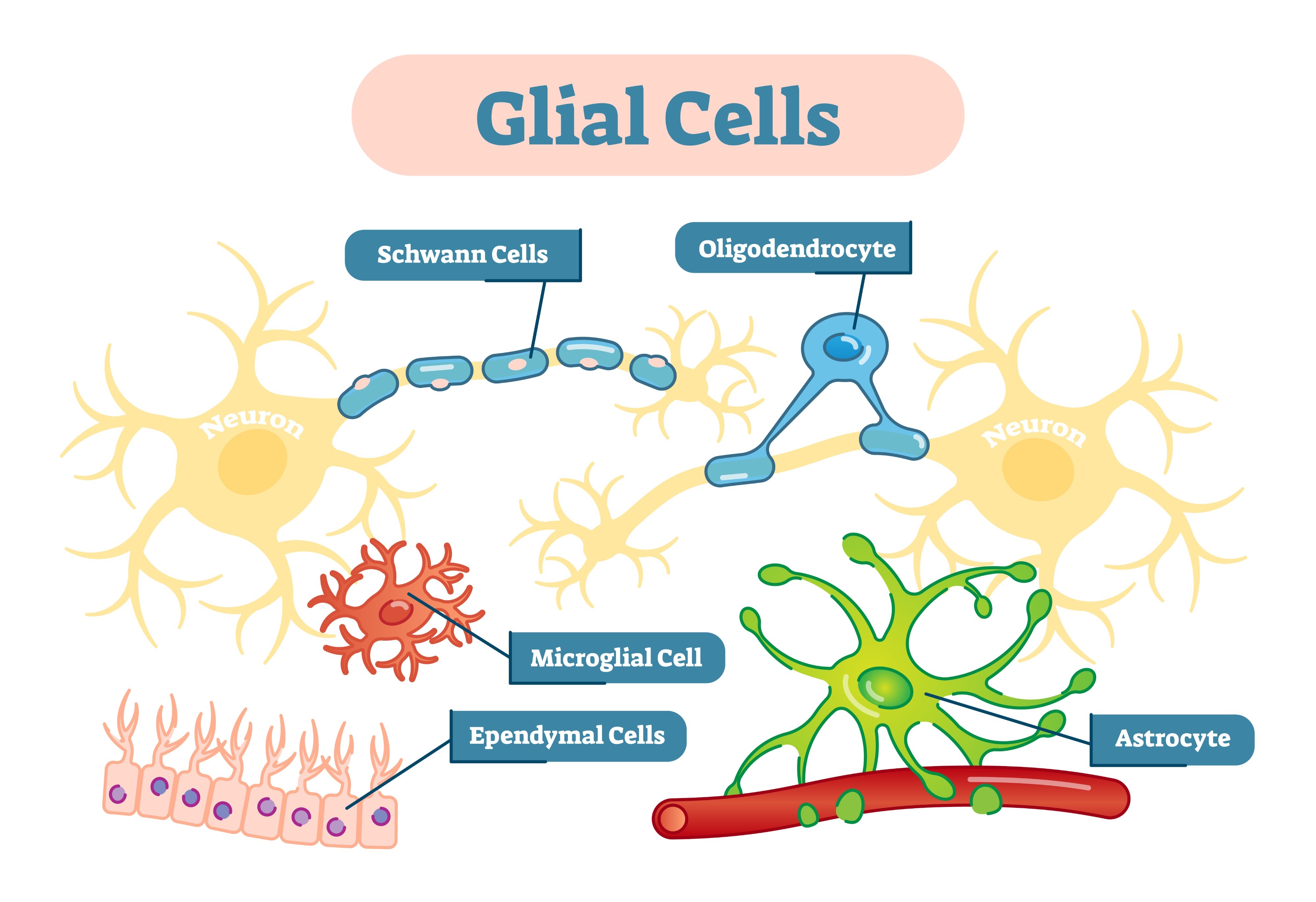
Glial Cells: Support and protect neurons by providing structural support, nutrition, and insulation, and by assisting in signal transmission.
Neurons of the central nervous system are provided nutrients, oxygen, and physical support by glial or neuroglial cells.
All connected to the Soma: the cell body of a neuron that contains the nucleus, DNA, mitochondria, and ribosomes and keeps it alive.
Dentrites: Finger-like projections of a neuron that receive signals from other neurons and transmit them towards the soma, playing a crucial role in how information is processed within the nervous system.
Axon: A long, thin projection of a neuron that has an action potential that transmits electrical impulses away from the soma to other neurons, muscles, or glands, facilitating communication throughout the nervous system.
The axon connects to the dendrites with a synapse.
Excited vs inhibit: Excited signals (like putting your hand on a hot pot) increase the likelihood that a neuron will fire an action potential, while inhibitory signals decrease that likelihood.
Neuronal Function & Synaptic Communication
1. Cells of the Nervous System
The nervous system contains billions of cells. Only about 10% are neurons, while the remaining 90% are supporting cells known as glia.
Three Types of Neurons
Sensory Neurons: Carry information from the peripheral nervous system (PNS) to the central nervous system (CNS).
Interneurons: Located entirely within the CNS. They relay information between sensory and motor neurons.
Motor Neurons: Carry instructions from the CNS to the muscles to produce movement.
Shutterstock
2. Anatomy of a Neuron
Neurons function like a complex electrical circuit. They are often compared to trees to help visualize their structure.
Soma (Cell Body): The heart of the neuron. It contains the nucleus and DNA. It provides the life processes of the cell through machinery like mitochondria (energy) and ribosomes (protein).
Dendrites: Branch-like structures that act as antennae. They receive messages from other neurons across the synapse. Dendritic branching (arborization) can grow or shrink based on learning or mental health conditions.
Axon: A long, slender tube that transmits electrical messages (action potentials) from the soma to the terminal buttons.
Myelin Sheath: A fatty insulation (lipids) that covers the axon. It speeds up the electrical signal and prevents it from leaking out.
Nodes of Ranvier: Gaps in the myelin sheath. The action potential regenerates at these points, a process called saltatory conduction.
Terminal Buttons: Located at the end of the axon branches. They secrete chemicals called neurotransmitters into the synapse.
3. Supporting Cells: Glia
Glia (meaning "glue") provide a scaffold for neurons and perform vital maintenance.
Glia Type | Location | Primary Functions |
Astrocytes | CNS | Provide physical support and nutrients (glucose to lactate). Maintain the blood-brain barrier. Clean up debris (phagocytosis). |
Microglia | CNS | Smallest glia. Act as the immune system of the brain. Trigger inflammation and clear debris. |
Oligodendrocytes | CNS | Produce myelin for multiple axons simultaneously. |
Schwann Cells | PNS | Produce myelin for a single axon segment. Support nerve regeneration if damaged. |
4. Resting Membrane Potential
The resting membrane potential is the electrical charge of a neuron when it is not active. The inside of the cell is typically -70 mV relative to the outside.
Key Ions
Sodium ($Na^+$): Highly concentrated outside the cell. It is positively charged.
Potassium ($K^+$): Highly concentrated inside the cell. It is positively charged. Potassium (K+) is the ion that predominantly in the intracellular fluid at rest.
Chloride ($Cl^-$): High concentration outside the cell. Negatively charged.
Organic Anions ($A^-$): Found only inside the cell. Negatively charged.
Physical Forces
Diffusion: The tendency of molecules to move from areas of high concentration to low concentration.
Electrostatic Pressure: The force that attracts opposite charges (+ and -) and repels like charges.
Both of these mediate resting membrane potential.Sodium-Potassium Pump: A protein that maintains the balance by pumping 3 sodium ions out and 2 potassium ions in.
5. The Action Potential
The action potential is an all-or-none electrical impulse. Once the cell reaches the threshold of excitation (roughly -65 to -60 mV), the process triggers automatically.
Steps of the Action Potential
Depolarization: Voltage-gated sodium channels open. Sodium ($Na^+$) rushes into the cell, making the inside positive (up to +40 mV).
Repolarization: Sodium channels close (refractory period). Potassium channels open and potassium ($K^+$) rushes out of the cell, making the inside negative again.
Hyperpolarization: The cell briefly becomes more negative than its resting state before the sodium-potassium pump restores the -70 mV balance.
6. Synaptic Transmission
Communication between neurons is an electrochemical event. An electrical signal (action potential) triggers the release of a chemical signal (neurotransmitter).
Synapse: The microscopic gap between the terminal button of the presynaptic neuron and the dendrite of the postsynaptic neuron.
Vesicles: Small orbs in the terminal button that store neurotransmitters.
Calcium ($Ca^{2+}$): When an action potential reaches the terminal, calcium enters the cell, causing vesicles to fuse with the membrane and release their contents.
Receptor Types
Ionotropic Receptors: Fast-acting. The neurotransmitter binds directly to a channel, causing it to open immediately.
Metabotropic Receptors: Slower. The neurotransmitter binds to a receptor that activates a G-protein, which then triggers a series of internal chemical steps to open a channel.
7. Postsynaptic Potentials (PSPs)
When a neurotransmitter binds to a receptor, it changes the charge of the receiving cell.
Excitatory PSP (EPSP): Makes the cell more positive (depolarization), increasing the chance it will fire. Usually involves sodium or calcium entering the cell.
Inhibitory PSP (IPSP): Makes the cell more negative (hyperpolarization), decreasing the chance it will fire. Usually involves potassium leaving or chloride entering the cell.
Neural Integration (Summation)
A single neuron may receive hundreds of messages at once. The cell body adds up the total number of EPSPs and IPSPs. If the net charge reaches the threshold of excitation, the neuron fires. If the inhibitory messages outweigh the excitatory ones, the neuron remains silent.
8. Termination of the Signal
To prevent a signal from continuing indefinitely, neurotransmitters must be removed from the synapse.
Reuptake: The presynaptic neuron uses transporters to pull the neurotransmitter back inside to be recycled.
Enzymatic Deactivation: Specific enzymes (like "Pac-Man") break the neurotransmitter apart so it can no longer bind to receptors.
Foundations of Behavioral Neuroscience
The goal of behavioral neuroscience is to explain behavior through two levels of explanation: generalization (deducing general laws from specific events) and reduction (explaining phenomena via basic elements).
The Mind-Body Question
Dualism: The belief that the mind and body are separate entities.
Monism: The belief that the mind is a product of the body; they are one. This is the modern scientific perspective.
René Descartes: A dualist who was the first to suggest a link between the mind and the brain. He believed the soul interacted with the body in the pineal body.
+2
Historical Milestones
Luigi Galvani: Used electrical stimulation on frog nerves to prove that muscle contraction did not require the brain, shifting the focus to the electrical nature of nerve messages.
Johannes Müller: Developed the Doctrine of Specific Nerve Energies, stating that while all nerves carry the same electrical signal, the perception depends on which part of the brain receives the signal.
Ramon Santiago y Cajal: Used Golgi staining to prove the nervous system is made of individual neurons rather than a continuous network.
Cellular Structure and Function
The Neuron
Neurons are the primary cells of the brain. They consist of:
Soma (Cell Body): Contains the nucleus and internal organelles.
Dendrites: Receive signals from other neurons.
Axon: Carries the action potential from the soma to the terminal buttons.
Terminal Buttons: Sites where neurotransmitters (NTs) are released into the synapse.
Internal Components
Mitochondria: Extract energy from nutrients to produce ATP.
Microtubules: Form the cytoskeleton and facilitate axoplasmic transport.
Anterograde: Movement from soma to buttons via the protein kinesin.
Retrograde: Movement from buttons back to soma via dynein.
Blood-Brain Barrier (BBB): A selectively permeable barrier that protects the CNS by controlling the composition of substances in the brain.
+1
Supporting Cells (Glia)
Glia provide physical and functional support to neurons.
Astrocytes: Provide nourishment by converting glucose to lactate and help isolate synapses.
+3
Oligodendrocytes: Produce the myelin sheath in the CNS.
Schwann Cells: Produce myelin in the PNS and help with nerve regeneration after injury.
Microglia: Act as part of the immune system by removing debris via phagocytosis.
Electrical Potentials and Communication
Resting and Action Potentials
The resting potential of a neuron is . This charge is maintained by the sodium-potassium pump, which pushes three $Na^{+}$ ions out for every two $K^{+}$ ions it brings in.
+2
The Action Potential Sequence:
The cell reaches the threshold of excitation.
Voltage-dependent $Na^{+}$ channels open; $Na^{+}$ rushes in, causing depolarization up to .
$K^{+}$ channels open; $K^{+}$ rushes out.
$Na^{+}$ channels close and become refractory.
$K^{+}$ continues to leave, causing the membrane to return toward its resting state and briefly hyperpolarize (overshoot).
Neuroanatomy
The Ventricular System and CSF
The brain floats in Cerebrospinal Fluid (CSF), which is produced by the choroid plexus. It circulates through the lateral ventricles, third ventricle, cerebral aqueduct, and fourth ventricle.
+3
Major Brain Regions
Division | Key Structures | Primary Functions |
Forebrain | Cerebral Cortex | Perception, movement, and higher reasoning. |
Limbic System | Emotion, learning, and memory (Hippocampus, Amygdala). | |
Basal Ganglia | Control of movement. | |
Thalamus | Relay station for sensory information to the cortex. | |
Hypothalamus | Regulates the ANS and endocrine system; the "Four F's". | |
Midbrain | Tectum | Superior colliculi (vision) and Inferior colliculi (audition). |
Tegmentum | Reticular formation (arousal) and Substantia Nigra (movement). | |
Hindbrain | Cerebellum | Balance and coordinated movement. |
Pons | Relay from cortex to cerebellum; sleep and arousal. | |
Medulla | Regulates cardiovascular system and respiration. |
Psychopharmacology
Pharmacokinetics (ADME)
Pharmacokinetics is the study of what the body does to a drug.
Absorption: Routes of administration determine speed. Intravenous (IV) is the fastest human route. Inhalation is also very quick as it goes from lungs to heart to brain.
Distribution: Lipid solubility is the most important factor in how fast a drug reaches the brain. Heroin reaches the brain faster than morphine because it is more lipid soluble, resulting in a "rush".
Metabolism: Primarily occurs in the liver via enzymes.
Excretion: Primarily occurs via the kidneys into urine.
Repeated Exposure
Tolerance: A decrease in drug effect after repeated use.
Sensitization: An increase in drug effect after repeated use.
Withdrawal: Compensatory symptoms that appear when drug use stops; signifies physical dependence.
Margin of Safety
The Therapeutic Index (TI) measures a drug's safety by comparing the toxic dose ($TD_{50}$) to the effective dose ($ED_{50}$).
Barbiturates: Have a low TI (2–3), making them dangerous.
Benzodiazepines: Have a high TI (100+), making them much safer for treating anxiety.
Neurotransmitter Systems
Neurotransmitter | Primary Function | Key Receptors/Details |
Glutamate | Main excitatory NT. | NMDA: Learning/memory; blocked by Magnesium. AMPA: Most common. |
GABA | Main inhibitory NT. | GABA-A: Ionotropic (Chloride channel). GABA-B: Metabotropic. |
Acetylcholine | Movement and memory. | Nicotinic: Brain/euphoria. Muscarinic: Peripheral movement. |
Dopamine | Reward and movement. | Nigrostriatal: Parkinson’s. Mesolimbic: Reward (VTA to N. Accumbens). |
Norepinephrine | Arousal/Alertness. | Produced in the Locus Coeruleus. |
Serotonin | Mood, appetite, sleep. | Produced in the Raphe Nuclei. Blocked by SSRIs. |
Opioids | Analgesia and reward. | Mu (): Euphoria. Kappa (): Dysphoria/opposite effects. |
Endocannabinoids | Appetite/movement. | CB1: Predominant in the brain. Not stored in vesicles; made on demand. |
More notes:
I. History and Evolution
Ancient Perspectives: While you noted Hippocrates, remember that Aristotle and other Greeks believed behavior was centered in the heart. Galen contributed by dissecting and studying non-human animal brains to understand behavior.
Functionalism: This principle states that an organism’s characteristics perform useful functions rather than having a predetermined "purpose". For example, the forelimbs of different species are adapted for specific functional needs.
Natural Selection: This is the mechanism where heritable variations that lead to greater reproductive success become more prevalent in a population over time.
Human Evolution: Homo erectus left Africa 1.7 million years ago. Homo neanderthalis lived in Western Europe between 120,000 and 30,000 years ago. Homo sapiens evolved in East Africa 100,000 years ago.
Why Large Brains? Human competition required large brains for tool use, color vision to spot food/danger, fire for cooking/heat, upright posture for long-distance travel, and language for group communication.
II. Internal Cell Structure and Transport
Organelles: The nucleolus is responsible for producing ribosomes, which synthesize proteins. Smooth Endoplasmic Reticulum produces lipid molecules. Lysosomes are sacs containing enzymes that break down waste products.
+3
Axoplasmic Transport: This process moves substances along microtubules within the axon.
Anterograde: Movement from the soma toward the terminal buttons using the protein kinesin.
Retrograde: Movement from the terminal buttons back to the soma using the protein dynein. It is approximately half as fast as anterograde transport.
III. The Synapse: Vesicles and Binding
Vesicle Release: When a vesicle releases neurotransmitters, it forms an omega structure. After release, the "Kiss and run" method allows the pore to close and the vesicle to refill without fully integrating into the membrane. Alternatively, the "Merge and recycle" method integrates the vesicle into the membrane.
Vesicle Pools:
Release-ready: Docked against the membrane; accounts for less than 1% of vesicles.
Recycling pool: Makes up 10 to 15%.
Reserve pool: Makes up 85 to 90% and only opens during high rates of axon firing.
Binding Types:
Competitive binding: Direct agonists or antagonists act directly on the neurotransmitter binding site.
Non-competitive binding: Indirect agonists or antagonists act on an alternative site to modify the effect of the neurotransmitter on the ion channel.
IV. Specific Neuroanatomy
Basal Ganglia: This system controls movement and consists of the caudate, putamen, and globus pallidus.
Thalamic Nuclei:
Lateral geniculate nucleus (LGN): Relays visual information.
Medial geniculate nucleus (MGN): Relays auditory information.
Ventrolateral nucleus: Relays information from the cerebellum to the primary motor cortex.
Autonomic Systems: The Sympathetic division is also known as the thoracolumbar system. The Parasympathetic division is known as the craniosacral system.
Vagus Nerve: The 10th cranial nerve that regulates the organs in the thoracic and abdominal cavities.
V. Pharmacology and Histamine
Injections: In lab animals, Intraperitoneal (IP) injection is the second fastest route of administration.
Histamine: This neurotransmitter is vital for wakefulness and arousal. It is synthesized from histidine and acts on H1 through H4 receptors.
Drugs that block histamine receptors produce drowsiness
Week 7:
Stages of Sleep and EEG Patterns
Sleep is categorized into distinct stages characterized by specific electrical activity in the brain.
Waking and NREM Sleep
Waking State: Characterized by alpha activity () and beta activity ().
Stage 1 (NREM): Consists of theta activity ().
Stage 2 (NREM): Contains sleep spindles ( occurring $2-5$ times during stages $1-3$) and K complexes, which are sudden, short waveforms.
Stage 3 (Slow-Wave Sleep): Defined by delta activity, which is characterized by frequencies <3.5\text{ Hz}.
REM Sleep
REM Characteristics: REM sleep consists of theta and beta activity.
Prefrontal Cortex: Shows low activity during REM, reflecting the lack of organization and planning found in dreams.
Extrastriate Cortex: Shows high activity during REM, reflecting visual hallucinations during dreaming.
Striate Cortex: Shows low activity during REM due to a lack of visual input.
Functions of Sleep and Learning
Sleep serves critical roles in physical recovery and memory consolidation.
Recovery and Development
Slow-Wave Sleep (SWS): Allows the brain to recover from physical activities of the day, though not necessarily cognitive activity.
Synaptic Erasure: Sleep assists in targeted synaptic erasure, which is essential for memory consolidation.
REM Sleep: Promotes brain development.
Age Differences: Young humans spend more time in REM ($9-10$ hours) compared to those with more developed brains (8 hours).
Memory Consolidation
Nondeclarative Memory: Consolidated primarily during REM sleep.
Declarative Memory: Consolidated primarily during slow-wave sleep.
Learning Tasks: Naps containing both SWS and REM show significantly higher discrimination improvement compared to no naps or SWS-only naps.
Neurotransmitters of Arousal and Sleep
Specific chemical systems modulate the transitions between waking and sleep states.
Neurotransmitter | Role and Characteristics |
Adenosine | Accumulates during wakefulness; serves as a sleep modulator. Glycogen increases adenosine, which is inhibitory. Levels are renewed during SWS. Caffeine works by blocking adenosine receptors. |
Acetylcholine (ACh) | High in the cortex and hippocampus during waking and REM sleep. Levels are low during SWS. |
Norepinephrine | Released from the locus coeruleus; involved in arousal and vigilance. Axons project widely throughout the brain. |
Serotonin (5-HT) | Found in the dorsal raphe nuclei; facilitates continuous, automatic movements like chewing or grooming. |
Orexin | Cell bodies located in the lateral hypothalamus; activation promotes wakefulness and tips the flip-flop circuit toward waking. |
Histamine | Located in the tuberomammillary nucleus; increases ACh release in the cortex to maintain arousal. Activity is high during waking but low during SWS and REM. |
Sleep/Wake Transitions: Flip-Flop Circuits
Transitions between states are managed by mutual inhibition between sleep-promoting and arousal-promoting regions.
Ventrolateral Preoptic Area (vlPOA): The primary sleep-promoting region containing the majority of sleep neurons.
Inhibitory Mechanism: These neurons secrete GABA and send axons to arousal regions.
Mutual Inhibition: The sleep neurons in the vlPOA receive inhibitory inputs from the locus coeruleus (LC), raphe nuclei (DR), and tuberomammillary nucleus (TN).
Flip-Flop "Off": When the vlPOA is activated, the brain enters slow-wave sleep.
Flip-Flop "On": When arousal systems are activated, they inhibit the vlPOA, resulting in an alert waking state.
Biological Clocks and Circadian Rhythms
Daily rhythmic changes in behavior and physiology are controlled by internal biological clocks.
Circadian Rhythm: Daily changes in physiological processes.
Zeitgeber: A stimulus, most commonly light, that resets the biological clock.
Suprachiasmatic Nucleus (SCN): Located in the hypothalamus, this region contains the biological clock for many circadian rhythms.
Melanopsin: A photopigment in retinal ganglion cells that provides light information to the SCN.
Pineal Gland: Secretes melatonin to control seasonal rhythms and rhythms influenced by day length.
Rhythm Disruption: Lesions to the SCN, paraventricular nucleus (PVN), or pineal gland disrupt rhythms controlled by day length.
Week 8:
Gemini said
These notes cover the core concepts of brain development from the PSYC 324 lecture, focusing on prenatal stages, anatomical structures, and the neurological shifts during adolescence.
Course Logistics
Instructor: Shawn Bates, PhD, Chico State.
Deadlines: Discussion board is due Wednesday; check-up is scheduled for Friday.
Early Brain & Prenatal Development
Brain development begins early in embryonic life as a hollow tube. This tube maintains its basic shape even as it matures, though it elongates, folds, and thickens to reach its final form.
+2
The Neural Tube (Days 20–24)
Day 20: The neural plate of the forebrain begins to form a neural groove and neural folds.
Day 21: The neural folds begin to fuse.
Day 24: The fused tube gives rise to the brain and spinal cord. The central canal of the spinal cord remains as the interior space.
+2
Early Sensory & Emotional States
Synesthesia: Infants experience a blending of senses.
Memory: Declarative memories (facts/events) do not form until approximately age 3.
Myelination: This process makes babies aware of the world. It happens in different regions at different times.
+1
Trauma: Early traumatic experiences can have long-lasting developmental effects.
Anatomical Subdivisions
The brain is categorized by major divisions, their associated ventricles, and the resulting principal structures.
+1
Major Division | Ventricle | Subdivision | Principal Structures |
|---|---|---|---|
Forebrain | Lateral / Third | Telencephalon / Diencephalon | Cerebral cortex, Basal ganglia, Limbic system, Thalamus, Hypothalamus |
Midbrain | Cerebral aqueduct | Mesencephalon | Tectum, Tegmentum |
Hindbrain | Fourth | Metencephalon / Myelencephalon | Cerebellum, Pons, Medulla oblongata |
Adolescent Brain Alterations
Adolescence is marked by significant reorganization at the cellular and behavioral levels.
+1
Neuronal Changes
Overproduction & Pruning: The brain overproduces neurons, followed by competitive pruning and apoptosis (programmed cell death).
Optimization: Myelination increases drastically, creating more efficient neural circuits.
Prefrontal Cortex (PFC): This is the last region to fully develop. A longer period of PFC alteration is associated with higher adult IQ.
+1
Reward & Emotion
Dopamine Signaling: Circuit density and dopamine signaling increase.
Reward Sensitivity: Strong rewards feel intense, while small rewards can feel aversive.
Regulation: Because the PFC is immature, the ventral striatum takes the lead in regulating emotions.
Reactivity: Adolescents are more reactive to emotional faces and regulate the amygdala differently than adults.
Mental Health: Most mental illnesses have their onset during adolescence.
Behavioral Patterns
Novelty Seeking: This behavior is common in primates and may be necessary for developing adult behaviors.
Risk Taking: High activation in reward areas leads to increased risk-taking and a higher likelihood of developing substance abuse issues.
These notes cover the biological foundations of sensation and perception, specifically focusing on the visual and auditory systems.
Fundamentals of Sensation and Perception
Perception is the process of the brain interpreting neural signals to represent the external world.
Action Potentials: The brain only receives input in the form of action potentials traveling along sensory pathways.
Transduction: This is the process where sensory receptors convert physical energy, like light or sound waves, into nerve impulses.
Sensory Encoding: The brain understands how energy turns into impulses and the pathways those impulses take, but it is less clear how these signals become a full representation of the world.
Perceptual Illusions: These demonstrate the gap between sensory input and our brain's interpretation.
The Visual System
The Stimulus and Anatomy of the Eye
Visual perception begins with light, which is the specific part of the electromagnetic spectrum humans can detect.
Dimension | Physical Property |
Hue | Dominant wavelength |
Brightness | Intensity of the light |
Retina: This is the back of the eye where transduction occurs.
Photoreceptors:
Rods: Approximately 120 million cells that are sensitive to low-intensity light, making them essential for night vision.
Cones: Approximately 6 million cells responsible for color vision and fine detail in bright light.
Fovea: The central region of the retina where cones are most dense and vision is sharpest.
Optic Disk: The location where ganglion cell axons exit the eye to form the optic nerve; it creates a blind spot because it lacks photoreceptors.
Visual Transduction and Retinal Circuitry
Transduction happens when light hits photopigments, such as rhodopsin, in the photoreceptors.
Hyperpolarization: Light causes the photoreceptor membrane to hyperpolarize.
Darkness: In the dark, photoreceptors actually depolarize bipolar cells.
Cell Layers: Information flows from photoreceptors to bipolar cells, and then to ganglion cells.
Horizontal and Amacrine Cells: These cells facilitate communication across the retinal layers.
Neural Pathways and the Brain
Information from the left visual field is processed in the right hemisphere, and information from the right visual field is processed in the left hemisphere.
Optic Nerve: Formed by ganglion cell axons.
Lateral Geniculate Nucleus (LGN): A part of the thalamus with six layers.
Striate Cortex (V1): The primary visual cortex in the occipital lobe where visual information is first combined.
LGN Layer Properties
The LGN segregates different types of visual information.
Layer Type | Characteristics | Function |
Magnocellular | Large cells; input from rods | Form, movement, depth, brightness |
Parvocellular | Small cells; input from cones | Color (red/green) and fine details |
Koniocellular | Found in gaps between layers | Input from blue cones |
Higher-Order Visual Processing
Beyond V1, the brain uses two distinct "streams" for processing.
Dorsal Stream (The "Where" Pathway):
Projects from V1 to the parietal lobe.
Processes spatial awareness and guides actions.
Receives primary input from the magnocellular system.
Ventral Stream (The "What" Pathway):
Projects from V1 to the inferior temporal lobe.
Processes object recognition and color.
Receives primary input from the parvocellular system.
Visual Pathologies (Agnosias)
Damage to the ventral stream can lead to agnosia, or "not knowing".
Visual Agnosia: Inability to recognize objects.
Achromatopsia: Inability to recognize colors.
Prosopagnosia: Inability to recognize faces.
The Auditory System
The Auditory Stimulus
Sound is produced by vibrating objects that cause air molecules to move in waves.
Perceptual Dimension | Physical Property |
Pitch | Frequency of vibration |
Loudness | Amplitude or intensity (measured in decibels) |
Timbre | Complexity of the sound wave |
Anatomy of the Ear
Outer Ear: The pinna directs sound waves into the ear canal.
Middle Ear:
Tympanic Membrane: The eardrum vibrates in response to sound.
Ossicles: Three tiny bones (malleus, incus, and stapes) that transmit vibrations to the inner ear.
Inner Ear (Cochlea): A snail-shaped structure containing the mechanisms for transduction.
Oval Window: Receives vibrations from the stapes to move fluid inside the cochlea.
Organ of Corti: The sensory organ on the basilar membrane.
Auditory Transduction
Hair Cells: These are the receptive cells of the auditory system.
Cilia and Tip Links: Movement of the basilar membrane causes the cilia (tiny hairs) to bend.
Ion Channels: When cilia bend toward the tallest one, tension on "tip links" opens ion channels (K+ and Ca2+), increasing the firing rate. Bending away closes the channels.
Neurotransmitter: Glutamate is the primary neurotransmitter used here.
Auditory Pathways and Perception
The auditory system is organized tonotopically, meaning different frequencies are processed in different locations.
Cochlear Mapping:
Basal End: Responds to high frequencies.
Apical End: Responds to low frequencies.
Neural Pathway: Information travels from the cochlear nerve to the medulla (cochlear nucleus and superior olivary complex), then to the inferior colliculus, the medial geniculate nucleus (MGN) of the thalamus, and finally the auditory cortex.
Auditory Cortex Streams:
Anterior Stream: Analyzes complex sounds (the "what").
Posterior Stream: Sound localization (the "where").
Coding of Pitch and Loudness
Place Coding: High and moderate frequencies are coded by which part of the basilar membrane is vibrating.
Rate Coding: Low frequencies are coded by the firing rate of neurons at the apical end.
Loudness Coding: Determined by the rate of action potentials and the shearing force on the cilia.
Week 10:
These notes cover the biological basis of emotion, aggression, and impulse control based on the PSYC 324 lecture material from Dr. Shawn Bates at Chico State.
I. Components of Emotional Response
An emotional response consists of three integrated components:
Behavioral: Muscular movements appropriate to the situation.
Autonomic: Physiological changes that facilitate the behavioral response.
Hormonal: Reinforcement of autonomic responses.
The adrenal medulla releases epinephrine and norepinephrine to increase blood flow to muscles.
The Amygdala: This structure controls the integration of these three components.
II. Fear and the Amygdala
The amygdala, located in the temporal lobes, is the primary region for processing fear and aversive stimuli.
Key Nuclei
Lateral Nucleus (LA): Receives information from all regions of the neocortex, thalamus, and hippocampal formation.
Central Nucleus (CeA): The region responsible for the expression of emotional responses.
Damage to CeA: Abolishes fear responses; animals become tamer, have lower stress hormones, and show no fear toward natural predators.
Activation of CeA: Heightens emotional reactivity; chronic activation can cause ulcers.
Conditioned Emotional Response
This is the most basic form of emotional learning, where a neutral stimulus is paired with an emotion-evoking one.
Neural Pathway: Information flows from the lateral nucleus to the central nucleus of the amygdala.
Social Learning: Humans and animals can also acquire fear by observing others (e.g., watching a dog attack someone else).
III. Aggression and Serotonin
Aggression involves threat behaviors (warning of attack), defensive behaviors, and submissive behaviors.
Neural Circuitry
Motor behaviors for aggression are organized in the brain stem, specifically the periaqueductal gray (PAG).
Dorsal PAG (dPAG): Involved in defensive behaviors.
Ventral PAG (vPAG): Involved in predation.
The hypothalamus (HT) and amygdala (AMY) influence the PAG to control these behaviors.
Role of Serotonin (5-HT)
Serotonin acts as an inhibitor of aggression in both animals and humans.
Decreased Serotonin: Associated with antisocial behavior and increased aggression.
Serotonin Agonists: Drugs that increase 5-HT activity (like SSRIs) decrease irritability and aggressive tendencies.
IV. Impulse Control and the vmPFC
The ventromedial prefrontal cortex (vmPFC) acts as an interface between emotional responses and the control of complex behaviors.
Damage/Lesions: Lead to emotional instability, impaired decision-making, and poor "real-world competencies".
Phineas Gage: A famous case where vmPFC damage resulted in a complete change in personality and loss of impulse control.
Iowa Gambling Task (IGT): Patients with vmPFC damage fail this task because they lack the normal Galvanic Skin Response (SCR) that typically precedes a risky choice.
Brain Development
Impulse control improves as the brain matures.
Amygdala: Matures early.
PFC: Matures late (into adulthood), which explains why adolescents often struggle with impulse control and cognitive processing speed.
V. Communication and Recognition of Emotion
Facial Expression
Innate Responses: Facial expressions of emotion appear to be innate and universal, rather than learned. This is evidenced by similar expressions across different cultures and species.
Facial Feedback Hypothesis: The contraction of facial muscles can intensify the emotion being felt (e.g., smiling can make you feel happier).
Neural Basis of Recognition
Laterality: The right hemisphere is more important for recognizing and expressing emotion.
Amygdala: Crucial for recognizing facial expressions of fear.
Insular Cortex: Specifically activated during the perception of disgust, likely for disease avoidance.
Superior Temporal Sulcus (STS): Important for recognizing the direction of gaze of others.
Empathy and Mirror Neurons
Mirror Neuron System: Located in the ventral premotor area of the frontal lobe, these neurons fire both when we perform an action and when we see someone else perform it.
Social Transfer of Pain: Empathy involves the Anterior Cingulate Cortex (ACC).
Transfer of pain is dependent on ACC-Nucleus Accumbens (NAc) projections.
Transfer of fear is dependent on ACC-Basolateral Amygdala (BLA) projections.