RPSGT Exam Secrets
Electroencephalogram (EEG) is a test that measures the electrical activity within the brain through scalp electrodes to rule out seizure disorders & to determine the characteristics of the sleep-wake state. Waves/cycles in 1 second intervals are measured in Hertz (Hz) to determine the stage of sleep:
Alpha: 8-13 Hz
Beta: 13-30 Hz
Delta: <0.5-4 Hz
Theta: > 4-7 Hz
The EEG equipment can be recorded digitally or analog. Digital EEG’s are preferred because they provide more accurate data collection, allow for easier analysis of brain wave patterns, & allows a variety of filters for different montages. Typically 6 leads are used (including 2 reference leads). The EEG is usually set to display in 10-second pages but can also be set to scroll continuously.
Face Electrodes
Face electrodes are used to ground & to record eye & chin activity & include:
Ground electrode: This electrode is usually placed on the forehead, but can be placed anywhere on the body, to help reduce electrical noise and provide a stable reference for the recordings. This electrode does not usually impact measurements with modern computerized equipment.
Chin electromyogram (EMG): The cEMG records muscle tone of the chin muscles & helps to identify REM sleep, during which REM activity decreases. This monitor can also provide information about teeth grinding, which can cause muscle movement, & snoring, as snoring causes artifacts.
Electrocardiogram
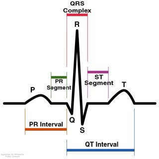
Electrocardiograms (ECGs) record & show a graphic display of the electrical activity of the heart through a number of different waveforms, complexes, & intervals:
P wave: The P wave represents the beginning of electrical impulses in the sinus node, which spread through the atria (muscle depolarization)
QRS complex: The QRS complex represents ventricular depolarization & atrial repolarization.
T wave: The T wave represents ventricular muscle repolarization (resting state) as cells regain negative charge
U wave: The U wave represents repolarization of the Purkinje fibers.
A modified lead II ECG is typically used for polysomnography to identify basic heart rhythms & dysrhythmias. Typical placement of leads for a 2-lead ECG is 3-5 cm inferior to the right clavicle & left lower ribcage. Typical placement for a 3-lead ECG is the right arm near the shoulder (RA), V5 position over the 5th intercostal space (LA), & the left upper leg near the groin (LL).
Anterior TIbialis Electromyogram
Anterior Tibialis Electromyograms (atEMGs) monitor the electrical activity in the leg muscles, allowing for monitoring of periodic leg movement during the polysomnogram (PSG) because electrical activity is absent when the muscle is relaxed & increases with movement. The atEMG may twitching & slight muscle activity & movement that may not be obvious from visual observation alone. Typically, muscle activity slows during sleep, especially during REM sleep. While intramuscular leads are used to diagnose neuromuscular disorders, surface leads are used for PSG because only a general overview of muscle activity is needed to assess sleep patterns and movements.
Sensors
Sensors may be used during the PSG to provide additional information about breathing during sleep:
Respiratory effort: Piezo-sensor hands or respiratory inductive plethysmography are used to indicate chest & abdominal movement during respiration as means of representing respiratory effort.
Snore: Microphones or piezo-sensors applied to the lateral-anterior neck superior to the larynx are used to indicate the degree & duration of snoring. Sensors are more accurate than microphones.
Airflow: Thermal sensors (thermistors or thermocouples) or pressure transducers (nasal) monitor both intake & outflow of air through the nostrils & the mouth. Typically, two prongs are inserted into the nose & a third prong is in front of the mouth.
Pulse oximetry
Pulse oximetry, continuous or intermittent, uses an external oximeter that attaches to the patient’s finger (or earlobe) to measure arterial oxygen saturation (SpO2), the percentage of hemoglobin that is saturated with oxygen. The oximeter also indicates the current heart rate. The oximeter uses light waves to determine SpO2. Normal SpO2 should be over 95% although some patients with chronic respiratory disorders, such as chronic obstructive pulmonary disease (COPD) may have lower SpO2 results. If SpO2 falls, the oximeter should be repositioned, as an incorrect position is a common cause of inaccurate readings. Oximeters do not provide information about carbon dioxide levels.
Inspection of equipment
Inspection of equipment should precede the study. Before beginning the PSG, the equipment is thoroughly inspected to ensure that the system is connected to electricity, that all cables/wires are secure, & that equipment is functioning properly. The computer is turned on & the patient information file is opened to ensure that information about the patient was entered correctly into the system. Any equipment issues should be resolved before beginning the test. All necessary supplies, such as leads, glue, & tape, are laid out & easily accessible to avoid unnecessary delays, thereby reducing patient stress. The physician’s orders should be checked to verify that the correct montage has been selected.
Selection of Montage
Selection of the montage depends on the presumptive diagnosis & the type of reading:
Nocturnal polysomnogram (PSG) | EEG, EOG, cEMG, atEMG, pulse oximetry, & sensors for respiratory effort, snore, & airflow | Used to diagnose obstructive sleep apnea syndrome & may be done before MSLT |
Multiple Sleep Latency Test (MSLT) | EEG (central & occipital), EOG, cEMG, & ECG: other channels are optional | Used to diagnose excessive daytime sleepiness (hypersomnia) & narcolepsy during waking hours; done after nocturnal PSG to ensure 6 hours of sleep preceding test |
Maintenance of wakefulness test | Same as MSLT | Used to evaluate success of treatment or ability to stay awake during the daytime; does not usually require a nocturnal PSG although it may be indicated for shift workers |
Applying Sensors
Electroencephalogram electrodes & sensors
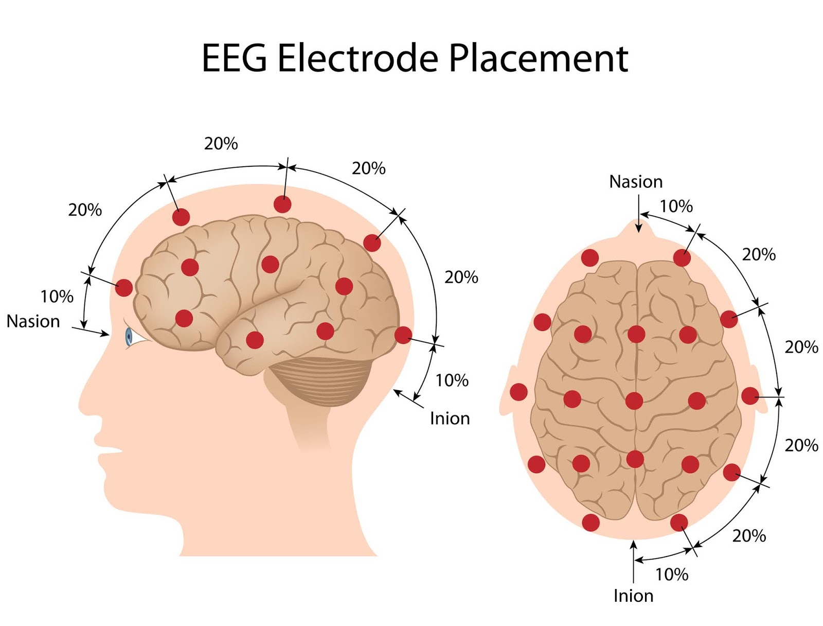
Electroencephalogram (EEG) electrodes & sensors must be applied properly in the correct position. EEG electrodes are placed using the International 10/20 measuring system which uses the nasion, inion, & preauricular points as landmarks while measuring the skull to determine lead placement at 10-20% distance from the landmarks. EEG leads are designated, according to the part of the skull to which they are applied, with even numbered subscripts on the right & odd numbered subscripts on the left. The system reference electrode is placed according to software requirements, often the central lead (Cz) at the vertex or top center of the head. Sleep technology often requires only a modified EEG with fewer electrodes, typically right & left central ( C4 & C3), right & left occipital (O1 & O2), & right & left reference mastoid leads (A2 & A1).
Scalp Electrode Site Measuring
Scalp electrode site measuring uses the international 10/20 measuring system based on landmarks. Sleep technicians must be proficient at performing these site measurements. Points are labeled, according to area of the brain & the exact site, so O1 is occipital area 1 with odd numbers indicating the left side:
Cz is measured vertically from the inion to the nasion & midline. The technician marks 50% (vertex) measure at the top of the head as well as 10% & two 20% measures on each side of the vertex, starting at the nasion & inion. The technician then measures from the left preauricular point to the right, intersecting at the 50% mark (vertex) & marking a 10% & two 20% measures on each side of the vertex, starting at the preauricular points. The point where the two vertical lines intersect at the vertex is Cz.
Other scalp electrode sites are measured once the vertical lines connecting the nasion & inion & the preauricular points are drawn & Cz is identified. The technologist measures the following:
Fpz & Oz: A vertical line is drawn 10% superior to the nasion, midline, to identify Fpz & 10% superior to the inion to identify Oz. A horizontal line is drawn 10% above the preauricular points to create a horizontal line that circles the head through Oz & Fpz.
C3: On the 30% mark (10% + 20%) above the left preauricular point
C4: On the 30% mark (10% + 20%) above the right preauricular point.
O1: On the 5% mark to the left of Oz on the horizontal line that is 10% above the left preauricular point
O2: On the 5% mark to the right of Oz on the horizontal line that is 10% above the left preauricular point
A1: Over the left mastoid process behind ear
A2: Over the right mastoid process behind ear
Face, electrooculogram, & chin electromyogram electrode placement
Face, electrooculogram, & chin electromyogram (cEMG) electrode placement during PSG includes the following:
Ground electrode | Place electrode on central forehead on line between nasion & hairline on a flat area of ski, avoiding deep wrinkles or creases. |
EOG | Right electrode: Place 1 cm lateral to & 1 cm superior to the outer canthus Left electrode: Place 1 cm lateral & 1 cm inferior to the outer canthus |
cEMG | Method 1: Place two electrodes on chin, 3 cm apart & 2 cm below lower lip Method 2: Place two electrodes on submentalis muscle, 3 cm apart (advised for thin but not obese patients) Method 3: Place one electrode on the chin (center) & the other on the submentalis muscle. |
Anterior tibialis electromyogram | Place two electrodes 3 cm apart along the anterior tibialis ridges on both legs, avoiding the tibias. |
Respiratory Effort, Snore, Airflow, & Oximeter
Respiratory effort | Bands (sized for the individual patient) containing sensors are placed securely (being careful not to restrict breathing) about the patient’s body with lead wires pointed upwards Thorax: secured just below axilla Abdomen: secured just about the waist |
Snore | Microphone: placed close to the patient Sensor: placed laterally on the anterior neck, superior to the larynx |
Airflow | Nasal prongs are inserted into the nostrils, & the third prong is positioned in front of the mouth |
Oximeter | The oximeter is clipped to the index finger closest to oximeter or earlobe. |
Head & Face Electrode Sites
Head & face electrodes are applied to areas that are clean & free of oil to ensure good quality signals. Before attachment, the skin must be cleaned thoroughly with an alcohol swab & then scrubbed for 5-10 seconds with an abrasive skin cleanser, such as NuPrep, using a cotton swab & being sure to scrub only the area of attachment, carefully separating the hair on the scalp.
Collodion Attachment
Collodion Attachment involves placing the electrode on the scrubbed site & covered with 2-3 cm size square of single-ply gauze. The air compressor stylus is inserted through the gauze & into the top center of the electrode & then the collodion glue is applied with an eyedropper to saturate the gauze. Smooth gauze is placed over the scalp or skin while drying with an air compressor, using care to avoid getting glue between electrode & skin. The stylus is removed. Using a blunt-tipped needle, the electrode is filled with electrolyte gel/cream.
Electrode Cream & Tape Attachment
Head & face electrodes are applied by different methods after the skin is scrubbed, including”
Electrode cream attachment: Electrode cream (EC2), which is an adhesive, is applied to one side of a 3 cm square of single-ply gauze. The electrode cup is filled with electrolyte paste/conductive gel, & the lead is placed in the middle of the hauze (gel side facing upward) to secure it to the gauze; the electrode & the gauze are then inverted onto the scrubbed skin, smoothing the gauze over the skin to secure it.
Electrode tape attachment (electrode collar): The electrode cup is filled with electrolyte paste/conductive gel & inverted onto the skin. The electrode is covered with tape, which is smoothed to secure the skin. The electrode is tucked under a corner of tape to facilitate later removal.
Body Electrode Sites
Body electrode sites include those for the electrocardiogram (ECG) & the anterior tibialis electromyogram (atEMG). Applying the electrodes includes:
Skin preparation: Electrode placement sites are thoroughly cleaned with alcohol, using a cotton applicator or premoistened alcohol swab. An abrasive cleaner, such as NuPrep, is indicated only if impedance is high. If the patient is hairy, the area may need to be shaved before cleaning the skin.
Application:
ECG: The lead wire is attached to the electrode before application. The adhesive backing is removed, & the gel side is placed against the skin, feeding the wires through the patient’s clothing, & then plugging them into the appropriate jack.
atEMG: Electrodes are attached with a double-sided electrode collar & taped (2-inches). The electrode wires are looped between the electrodes & secured to prevent dislodgement during the exam.
Verifying Impedance
Verifying impedances is done by the technologist after calibrating the machine to determine if the electrodes are properly applied & signal quality is appropriate. Electrodes pick up the electrical current generated by tissue & transmit this signal to the machine, which creates a wave pattern. If the connection between the electrode & the conducting gel is disrupted, the signal is distorted, causing motion artifacts. Silver/silver chloride electroes tend to have less motion artifacts than gold electrodes. Impedance refers to interference with the electrical signal form the point of contact to the recording device. Each type of electrode has an associated source impedance, but impedance levels for each individual electrode should remain low (< or equal to 5 kilohms). Each electrode should be individually tested, using the internal impedance meter or an external handheld meter. A difference in impedance between paired electrodes increases artifacts. If the impedance level of an electrode is high, then the electrode may need to be repositioned.
Educating Patients
Patient Orientation
Patient orientation begins with the patient arrival at the facility. Orientation should include:
Introduction: The patient should be introduced to the technologist & any other staff members who may be present. This is especially important if the patient is to be awakened.
Tour of location: The initial tour should include the patient’s individual sleeping area & storage space as well as the bathroom & shower facilities. The patient should observe the technologist’s monitoring area/any monitoring equipment.
Equipment: The technologist should identify & explain bedside equipment.
Alarms: Any alarms/call bells should be demonstrated so the patient knows how to use them & recognizes the sound
Patient’s rights: The technologist should apprise the patient of their rights, including the right to privacy & confidentiality & the right to refuse treatment, according to the rules of the Health Insurance Portability & Accountability Act (HIPAA)
Come back & add pages 21-25 if needed
Equipment Calibration
Equipment Calibration Before & After Exam
Equipment calibration (with standard settings for filters & sensitivity) of 30 seconds must be done & recorded before beginning the polysomnogram after the leads & sensors are applied & the patient is quiet. Calibration procedures vary somewhat, depending on the software & equipment used so following manufacturer instructions is important. Electroencephalograms (ECG), electrooculograms (EOG), & electromyograms (EMGS) generally require negative 50 microvolts per centimeter DC to all channels to obtain a deflection of the recording pen in the range of 5-10 mm. The technologist visually examines each calibration wave. As part of the calibration procedure, the technologist makes sure all equipment is properly plugged in, that all jacks are placed correctly, & that signal quality is adequate. Leads & sensors should be adjusted to control impedances & artifacts. Upon completion of the study, usually within 8 hours, the calibration procedure is repeated for 30 seconds & the results are compared with the initial calibration to ensure accuracy during the recording period.
Electrical Activity
Electrical activity is recorded by the PSG as tracings from three signal sources.
Bioelectrical signals: These are generated by the patient’s tissue & motion & recorded by surface electrodes to display of the ECG, EOG, & EMG
Transduced signals: These derive from sensors that convert action, such as chest wall movement into electrical signals with the electrical signal generated by the sensor instead of the body
Equipment signals: Sometimes ancillary equipment, such as a carbon dioxide analyzer, is used during the PSG. This equipment, which has separate signal displays, outputs, & processing units, may be stand-alone or interfaced with the digital PSG equipment
Physiological calibrations
Electrocardiogram (ECG), electroencephalogram (EEG), & electrooculogram (EOG)
Electrocardiogram (ECG): The technologist checks the polarity of the reading to ensure the ECG tracing is not inverted, which is a sign that the jacks are inserted in incorrect channels.
Electroencephalogram (EEG): The technologist asks the patient to relax with the eyes closed for 30 seconds or more, during which time the alpha waves on the EEG are typically prominent. Then the patient is asked to open eyes for 30 seconds.
Electrooculogram (EOG): When the patient is relaxed with the head still, the technologist asks he patient to look left & right a number of times, up & down a number of times; and finally blink 5 times. The technologist then examines the EOG tracings to ensure that the 3 different types of eye movements are distinct on the recording
Chin & Anterior Tibialis Electromyograms, Snore Sensor, Respiratory Sensors, & Airflow Sensors
Chin electromyogram (cEMG): The technologist asks the patient to relax & remain quiet while determining the baseline muscle tone remains 5 cm/amplitude or more. Then, the patient is asked to swallow, grit the teeth, & bite down to ensure that these actions show activity.
Anterior tibialis electromyogram (atEMG): The technologist asks the patient to extend the legs & then to flex & extend the great toe on each foot, which would show activiy.
Snore sensor: The technologist asks the patient to count to 5 out loud as this should cause deflection.
Respiratory effort sensors & airflow sensors: The technologist asks the patient to stop breathing & holds their breath in briefly to ensure that the respiratory tracing shows a flat line. The patient is then asked to mimic paradoxical breathing by tightening & relaxing the thorax.
Physiological calibrations should be repeated at the end of the PSG to ensure that leads & sensors remain in the correct position & that recordings were correct.
Frequency & Amplitude
Frequency & amplitude are waveforms that are recorded on PSG, using a standard time scale of 1 cm/sec. Frequency is the number of waves/cycles generated per second, & amplitude is the vertical height of a wave, determined by electrical voltage. Machines are calibrated with a known signal so that waveforms can be interrupted according to height & sensitivity settings. The setting of 50 microvolts/cm, typically used for electroencephalograms (EEG), electrooculograms (EOG), chin electromyograms (cEMG), & electromyograms (EEG), means that 50 microvolts of signal produce a standardized waveform that is 1 cm high. The wave height varies in relation to the sensitivity setting. While digital machines can record in a variety of ways, the data are displayed & recorded so that frequency & amplitude can be visually confirmed.
Filters
Filters are used to gain a more accurate recording by isolating bandwidths & reducing outside inference, such as from signals produced by the skin or muscle activity that causes artifacts. Most digital equipment records without the use of filters, but filters can later be applied to “clean up” the recording; however, filters (especially low frequency)can cause a phase shift that causes the wave to appear earlier or later. Filters are set in relation to the normal bandwidth of the test.
Low frequency filters eliminate signals below the normal bandwidth for a particular test
High frequency filters eliminate signals above the normal bandwidth for a particular test
60 Hz notch (band reject) filters remove signals (noise) produced in the 50-60 Hz range (power line interference) without affecting other frequencies, but this filter can interfere with recordings so it is rarely used except for anterior tibialis electromyograms (atEMG)
Band pass filters record frequencies only within a particular range
Frequency Settings
Frequency settings for filters vary according to the test
Electroencephalogram (EEG) (standard sensitivity of 50 microvolts/cm): Usually recorded in the range of 0.5-25 Hz, a low-frequency filter, therefore, would be set below the bottom range (about 0.3), effectively reducing output below that level & a high-frequency filter would be set above the top range (at 30-35 Hz), although when used to diagnose seizure activity, the high-frequency filter needs to be set higher (70-75 Hz) to allow for spiking during epileptic activity. Time constant is 0.25 seconds.
Electrooculogram (EOG): Settings are similar to those of an EEG.
Chin electromyogram (cEMG): The low-frequency filters are typically set at 10 Hz, & the high-frequency filters are set at 90-100 Hz. Time constant is 0.1 seconds.
Electrocardiogram (ECG) (sensitivity a 1 millivolt/cm): The low-frequency filter is set at 1 Hz, & the high-frequency filters are set at 30-35 Hz. Time constant is at 0.1 seconds.
Respiratory sensors: The low-frequency filters are set at 0.1 Hz & the high-frequency filters are set at 0.5 Hz. Time constant is around 1 second.
Oximetry (sensitivity at 1 volt/cm): The high-frequency filter is set at 15 Hz
Sampling Rate
Sampling rates must be selected before testing when converting analog recordings to digital as they cannot be changed afterward in the way that filters can be changed. The converter uses predetermined intervals to assign a numeric value to waveforms. This value determines the amplitude (height) of the waveform. The sampling rate is equal to the number of sampled intervals done in 1 second. According to sampling theory, the minimum sampling is equal to at least twice the highest frequency sampled, but this will not provide an accurate representation of the analog waveform, so a higher sampling rate is necessary to achieve an adequate waveform. For example, sampling rates must be 10 times higher for electroencephalograms (200-300 Hz with 30-35 Hz high-frequency settings) with adjustments if high-frequency filter settings are increased. Sampling rates should be selected for each channel.
Waveform Appearance
The appearance of the waveform is affected by filters. During calibration, when a 50-microvolt negative DC voltage is applied without filters, the waveform takes on a square appearance with an upward spike that is then sustained for the duration the voltage is applied. With low- & high-frequency filter settings, the shape & duration of waveforms alter as do the time constants, the difference between constant rise time & constant fall time:
Rise time: Interval of time required for calibration waves to fall to 60% of amplitude
Fall time: Interval of time required for calibration waves to fall to 37% of amplitude
Changing the setting of high- & low- frequency filters directly affects rise & fall time. Lowering the high-frequency filter setting increases rise time. Lowering the low-frequency filter setting increases fall time.
Documenting During Testing
EDS & Fatigue
Excessive Daytime Sleepiness (EDS), an increasing societal problem related to the lack of adequate sleep, causes the patient to feel sleepy during waking hours to the point at which the person may fall asleep or feel the need to nap.
Fatigue, on the other hand, is a general feeling or tiredness, weakness, or lack of energy & may be related to physical or emotional problems. A person who is fatigued generally does not feel sleepy or have difficulty staying awake. Patients often do not distinguish between sleepiness & fatigue so the technologist should question patients carefully to determine which they are experiencing.
Sleepiness & fatigue scale may also be administered to help with diagnosis. Patients should evaluate sleepiness & fatigue at different times of the day with 9 am & 9 pm when people are the most alert & 3 pm being the least.
Subjective Sleepiness Evaluations
Subjective sleepiness evaluations, using various scales, are sometimes provided by the patient. While these evaluations are easy to use, take little time, & are usually available at no cost in print or online, these results should not be considered definitive because a number of inherent weaknesses:
Patients do not always answer truthfully
Different scales measure different things & may not correlate
Some patients may receive a false positive & some a false negative
Scales are by their nature not objective so they are open to interpretation
The scales do not reflect comorbid conditions
People from different ethnic backgrounds may perceive sleep, sleepiness, & fatigue in different ways
Men & women may perceive sleep, sleepiness, & fatigue in different ways
Stanford Sleepiness Scale
The Stanford Sleepiness Scale is a brief assessment used to determine if people have Excessive Daytime Sleepiness (EDS). The scale is used to a number of different times during the day as people may report feeling sleepy at different times, especially in the late afternoon, a low period of alertness for most people. The scale lists seven different descriptors that rate increasing levels of sleepiness. People with an EDS score of 4-7 have a sleep debt that interferes with functioning:
1: Feeling alert & awake
2: Functioning & concentrating, slightly sluggish
3: Awake & functioning but not fully alert
4: Slightly foggy
5: Foggy, slightly drowsy
6: Feeling sleepy & drowsy & having difficulty staying awake
7: Nearing sleep onset & awake dreaming
8: Sleeping
Epworth Sleepiness Scale
The Epworth Sleepiness scale evaluates how likely a person is to fall asleep during a number of different activities. The person rates each situation on a scale of 0-3, corresponding to the chance of falling asleep. Scores 1-6 indicate adequate sleep. Scores 7-8 are average but scores 9+ indicate a high index for sleepiness & need for further testing. Descriptors include:
Sitting & reading
Watching TV
Sitting quietly in a public place
Sitting as a passenger in a car for an hour with no break
Lying down for an afternoon rest
Sitting & visiting with someone
Sitting quietly after eating lunch (no alcohol)
Sitting in a car while stopped in traffic for a few minutes
Sleep-Wake Activity Inventory
The Sleep-Wake Activity Inventory (SWAI) is a comprehensive instrument that includes subscales that measure a number of different aspects of sleep disorders: excessive daytime sleepiness (EDS), nocturnal sleep, relaxing ability, social desirability, & physiological distress. The EDS subscale asks the patient to score 9 different statements about sleepiness on a 1-9 scale (always present-never present). Scores are then added. 50+ is normal but 40-50 suggests sleepiness & a score of 40 or less indicates EDS.
I fall asleep while watching TV
I am able to nap anywhere
I fall asleep in the middle of a conversation
I fall drowsy within a few minutes of driving
I feel drowsy within 10 minutes of sitting quietly
I fall asleep during visits with friends
I feel sleepy after 15 minutes of reading
I fall asleep when I am relaxed
I fall asleep when I am riding in the car as a passenger
Fatigue Severity Scale
The Fatigue Severity Scale contains a list of 9 descriptions related to fatigue. The patient scores each statement on a 1-7 scale (strongly agree - strongly disagree). The scores are added together with scores 9-35 in the normal range & scores above 35 suggesting high fatigue.
I have less motivation when I am fatigued
I become fatigued when I exercise
I become fatigued easily
My fatigue interferes with my ability to function physically
I experience frequent problems because of fatigue
I cannot carry out sustained physical activity/functioning because of fatigue
I cannot adequately carry out all my duties/responsibilities
Fatigue is one of the three most disabling symptoms I experience
My work, social, & family life suffer because of my fatigue.
Recording
Documenting observations of the patient during the PSG (even if video monitoring is used) is important so that a accurate sleep study report can be generated at the end of the procedure as not all pertinent information is obvious from the data generated. The following must be documented:
Time lights go out & time lights go on
Total recording time
Patient’s emotional status, including anxiety or confusion that might impact results
Patient’s physical status, including unusual motor activity & nocturia
Patient’s position (supine, head elevated, sitting)
Descriptions of breathing/snoring or other audible sounds
Atypical findings such as REM sleep behavior disorder
The technologist should not rely on memory but should note unusual events as they occur, marking the time, duration, & frequency, so that the events can be correlated with the recordings.
Sleep Technologist’s Interventions
Sleep technologist’s interventions that occur during the PSG, including the application of electrodes & sensors, calibrations, & physiological calibrations, must be documented. Documentation should include a description of the event, the exact time of onset, duration of time, & time event ends. Important information to document includes:
Reattachment of dislodged electrodes & sensors
Assisting patient to change positions
Diaper changes & episodes of incontinence or nocturia
Patient requests
Patient or parent complains
Parasomnias
Parent or caregiver interventions
Arousals
Patient behaviors relevant to artifacts in recordings
Evidence of seizures
Identifying & Responding to Data Issues
Artifacts
Artifacts, extraneous signals, are common during PSG; some relate to normal activity, such a muscle movement or snoring. The technologist must monitor, identify, & correct artifacts as necessary by:
Checking other channels to determine if artifacts are occurring in only one channel or adjacent channels as well. If in only one channel, then the artifact may be related to a single lead.
Determining if the affected channels share a reference lead as this suggests the artifact relates to the reference.
Monitoring constantly & change derivations as needed through system referencing of the use of multiple channels.
Physiological Artifacts
Cause | Result | Correction |
Muscle (electromyogram EMG) | The background electroencephalogram (EEG) or electrooculogram (EOG) may be obscured & other signals distorted depending on the type of muscle activity | Encourage patient to relax, deep breathe, & slightly open the jaw to reduce tension. Report on visual observations when artifacts occur, noting signs of seizures or a sleep disorder. Ensure electrodes are correctly attached. |
Skin, irritation (rash) | Irritation, such as a skin rash, can alter the skin’s electrical signal, causing high impedance | Place electrode in a different area, avoiding irritated skin |
Vibration | Leg movement or snoring can cause high-frequency artifacts | Note & record all artifacts, correction is not usually possible |
Perspiration (EEG) | Excess perspiration may cause slow-frequency artifacts or can be similar to delta waves | Cool patient by changing room temperature or using fan |
Swallow (EEG) | Swallowing can result in slow waves in temporal areas, typical on arousal | Note & record, correction not usually possible |
Retinal disease (EOG) | The affected eye may interfere with electrical signal | Note activity of unaffected eye, or use other measures. |
Artificial eye (EOG) | The prosthetic eye does not generate electrical signals. | Note underlying frontal EEG activity. |
Blink (EOG) | Produces slow waves, depending on type & speed of activity | Note & record, correction not usually possible |
Eye muscle abnormality (weakness, hyperactivity, paralysis) (EEG, EOG) | Abnormalities may alter EOG readings, depending on the type of abnormality. Rectus movement can cause spike in EEG (frontotemporal) | Note activity of unaffected eye or use other measures. |
Mechanical Artifacts
Cause | Result | Correction |
ECG in EEG, EOG, or EMG channels | ECG tracing appears in other channels. | Re-reference (double reference) channels for EEG/EOG to reference leads A1 & A2. Ensure electrodes are correctly placed & attached. Avoid applying reference electrodes to fat, soft tissue. Use a common mode rejection for EMG artifacts, or reattach EMG electrodes. |
Electrode pop (sharp, spiking deflection) | Electrode pops are generally related to only one electrode but may be observed in multiple channels | Remove, reprep skin, & reattach electrode. Pops may result from pressure on electrodes, dirty electrodes, or loose wires |
Cardio-ballistic (sensors) | Sensors (respiratory effort, airflow, pick up pulse waves from ECG) and are crucial for accurately measuring physiological signals during sleep studies. | Note & record; correction not usually possible |
Condensation in CPAP tubing | Fluid in tubing can cause M-shaped waveforms in airflow channel | Remove fluid from tubing |
Loose belt (sensors) | Tracings are flat despite evidence of movement or respirations | Reapply belt correctly |
Misplacement of airflow sensor | Sensor does record changes in temperature/airflow | Reposition |
Electrodes, loose or improperly secured | High-frequency noise combined with high amplitude can slow activity | Remove, reprep skin, & reattach electrodes |
Oximetry channel | Inaccurate recordings of oxygen saturation can result from improperly attached oximeter. | Check placement & ensure fingernail is free of polish |
Recording-related Artifacts
Cause | Result | Correction |
50-60 Hz | Caused by poor grounding of EEG electrodes or interference from electrical leakage from other equipment; 50-60 Hz artifacts can occur in EMG channels especially in leg EMGs | Use common mode rejection. A 50-60 notch filter may remove artifacts at 50-60 Hz, but do NOT use in EEG or EOG channels as artifacts there usually indicate improper connection. In cases of excessive noise, consider re-evaluating electrode placement and ensuring that all connections are secure before proceeding with the recording. |
Multiple Channel Recordings | Multiple source artifacts can make a recording unreadable, & multiple channels cannot be shown at the same time so important data may be missed | Reconsider approach |
Excessive filtering | Filtering can distort data & mask problems that require correction | Avoid using filters to reduce artifacts, and do not use filters to remove artifacts unless underlying physiological signs are adequate. |
Multiple Sleep Latency Tests
The Multiple Sleep Latency Test (MSLT) measures sleepiness during waking hours & the tendency of a person to fall asleep. The MSLT may diagnose narcolepsy & idiopathic hypersomnia & determine the effectiveness of therapy. Elements include:
Patient keeps a 2 week sleep diary before testing, sometimes with actigraphy, to identify sleeping patterns
Medications are assessed & withheld when possible if they affect sleep. Stimulants should be discontinued 2 weeks before testing.
The MSLT must be preceded by nocturnal PSG & the montage must include EOG, EEG, cEMG, ECG.
MSLT includes 5 nap periods with the first within 3 hours of a nocturnal PSG & then spaced two hours after start of preceding nap
Patients report their subjective evaluations of sleepiness 45 minutes before 5 designated nap periods, & physiological calibrations are done within 5 minutes before onset of nap period
No smoking is allowed within 30 minutes of starting a nap & no strenuous activity within 15 minutes
Mean sleep latency is evaluated.
Maintenance of Wakefulness Test
The maintenance of wakefulness test (MWT) is done to assess sleepiness & effectiveness of treatment, determines the patient’s ability to stay awake in the daytime. The MWT may be done with the Multiple Sleep Latency Test (MSLT). Elements include:
A 2 week sleep diary & nocturnal PSG may be done before the MWT, depending on the patient
Montage is similar to the MSLT: EEG, EOG, cEMG, ECG
Patient is placed at rest, sitting in bed with low lights for four 40 minute periods, spaced at 2 hours, & advised to remain awake but not to engage in activities
Sleep latency (onset of sleep) is measured with fewer than 8 minutes considered abnormal. About half or normal sleepers remain awake during all 40 minute nap periods.
Patient is awakened 90 seconds after falling asleep as duration of sleep is not important for the MWT alone.
If the patient does not fall asleep during the 40 minute resting period, the test is terminated.
Wrist Actigraphy
Wrist actigraphy uses a portable device worn on the wrist that records & analyzes movement. Information stored in the device is downloaded into a computer. A number of different devices are available, & they evaluate movement in different ways, using a single channel, so determining the validity of the reports or comparing it to standard to standard PSG is problematic, especially if patients have movement disorders or periods of quiet (without moving) during waking hours. Because of these limitations, wrist actigraphy should be used for multiple days (at least three 24 hour periods with up to 7 days optimal) to help identify patterns of sleep/waking. Wrist actigraphy alone is not usually adequate for diagnosis of sleep disordered breathing or periodic limb movement but can be used to evaluate other sleep disorders. The patient should keep a diary of activities during wrist actigraphy to identify artifacts & to aid in interpretation of the results. Scoring varies according to manufacturer guidelines.
Infant Polysomnograms
Infant PSGs may be done for short (between feedings) or extended (overnight) periods, but short examinations may not render adequate information so longer testing is recommended. Some modifications are necessary for infant PSGs. Usually a parent/caregiver remains in the room although the person should be advised not to disturb the child unless necessary. The child should be in a crib or secure bed with side rails. Using play & dolls to show placement of electrodes may be helpful for small children. Safety measures should be in place to secure all electrical outlets, supplies, & equipment.
Placing Electrodes & Sensors for Infant/Child PSGs
Infant/child PSGs may require some modification when placing electrodes & sensors. Parents may hold or soothe the child while leads are applied. There are a number of factors to consider:
Collodion may cause eye irritation so paste or other adhesive may be used. Pasted electrodes increase slow-frequency electroencephalogram (ECG) artifacts, reduced by increasing a low-frequency filter to 1 Hz (but this will interfere with slow-wave EEG activity.
Toddlers may require a gauze or turban cap to keep scalp electrodes in place.
Infants may need to hands covered with socks & toddlers may need parents to prevent the child from touching leads until the child is asleep.
Bundling & taping of leads can prevent dislodgement.
Re-referencing/re-montaging may be necessary if children are active
Higher sampling rates are needed to detect seizure activity (EEG > or equal to 500 Hz & EOG/EMG > or equal to 200 Hz.
cEMG should be placed where it will not come in contact with drool.
Respiratory effort belts & sensors may need to be secured with tape.
PH Sensor
A pH sensor may be placed in the esophagus to diagnose gastric acid reflux in infants & children. In infants, a thin wire with a sensor at the end is passed nasally but older children/adults may be able to swallow the sensor with fluids. There are two types of sensors used:
Antimony/antimony oxide electrode: The small pellet-like sensor is antimony coated with antimony oxide. A skin reference electrode is needed for this type.
Glass electrode: The glass electrode is a combined sensor & reference electrode (2-4 mm diameter). It is inserted into the esophagus through the nasal passage or swallowed. The glass electrode has a high level of electrical impedance that can interfere with readings & can be broken if not handled properly, although it is unlikely to break when positioned in the esophagus
Infant Polysomnogram, Sensors, & Oximeter
Respiratory effort sensors: It is important that the sensors (inductance plethysmography or piezo crystal bands) are the correct size & secured with tape if necessary. The thoracic band is placed immediately above the nipple & the abdominal band, about the umbilicus.
Position sensor: This sensor is placed according to the manufacturer’s directions, usually on the lower back (over the diaper) with the infant in supine position
Motion sensor: This sensor is placed on a limb, which is moved to ensure that the signal is adequate.
Oximeter: This sensor is placed on the hand or foot in the infant & secured with wrapping as necessary avoiding excess padding hat might increase heat or affect readings.
Interventions During Infant Polysomnogram
Interventions during an infant PSG are made as indicated by what the technologist observes. Emergency action may be needed if an infant has apneic periods for over 20 seconds:
The oxygen saturation & heart rate are noted to determinte if in infant is showing a decrease, but no intervention is necessary until the oxygen saturation level is less than 85% or the heart rate is less than 60 for 10 seconds. When oxygen saturation & heart rate have fallen to the critical points, intervention is indicated:
The infant is stimulated by flickering the thumb against the heels or the bottom of the feet
If there is no improvement, the airway & position are checked, using suction to clear airways if necessary. Oxygen per beg is provided, a few puffs are adminstered & continued until oxygen saturation & heart rate return to normal
The EEG is checked for indications of seizure activity.
American Academy of Pediatrics Clinical Guidelines for OSAs
The American Academy of Pediatrics has issued clinical guidelines for the diagnosis & treatment of OSAs. Recommendations include:
All children are screened for snoring, observed apnea, restlessness during sleep, daytime sleepiness, or neurobehavioral abnormalities
Physical exam notes abnormalities that may relate to OSAs
If indications of OSAs are present:
High-risk children with co-morbid conditions should be immediately referred to a specialist
Children who are not high risk but show evidence of cardiac or respiratory failure should have further evaluation in consultation with a specialist.
Children who are not high risk & do not show evidence of cardiac or respiratory failure should be referred for PSG to diagnose OSAs
Tonsillectomy/adenoidectomy is the first-line treatment for OSAs with CPAP as an option for those unable to have surgery or for those who do not respond to surgical treatment.
High-risk children should be monitored as inpatients after surgery
Re-evaluation is needed after surgery to determine effectiveness.
American Thoracic Society, Standards/Indications for Cardiopulmonary Sleep Studies in Children
Differentiating benign snoring or primary snoring from pathological snoring that involves periods of apnea
Evaluating children who experience disturbances in patterns of sleep, including waking, sleepiness, who fail to thrive, or who exhibit cor pulmonale or polycythemia especially in children who snore
Clarifying clinical observations & diagnosis
Evaluating children with laryngomalacia & stridor (worsening at night)
Evaluating the effects of obesity if other symptoms such as snoring, sleep disturbance, or hypercapnia, are present
Evaluating the condition of the child with sickle cell disease with evidence of OSAs or veno-occlusive disease
Noting progress after treatment (4 week postsurgical) or weight loss (if indicated to control OSAs)
Assisting with titrating continuous positive airway pressure
Childhood Disorders: Bronchopulmonary Dysplasia, Cystic Fibrosis, Asthma, Neuromuscular Disorders, Alveolar Hypoventilation, & Infantile Apnea/Bradycardia
Bronchopulmonary Dysplasia | Children may receive supplemental oxygen to maintain oxygen saturation at more than 92%. Assess oxygen saturation levels during both waking & sleeping hours to determine if hypoxemia occurs. Assess oxygen saturation levels after oxygen is discontinued if unexplained symptoms occur such as cor pulmonale, polycythemia, & failure to thrive. Evaluate bradycardia occurring without apnea & snoring or suspected upper airway obstruction. Evaluate gastric reflux disorder (pH sensor) |
Cystic fibrosis | Oxygen desaturation may occur without apnea. Provide continuous nocturnal oximetry if daytime oxygen saturation level is less than 95%. Use nocturnal oximetry for at least 8 hours if child has headaches in the morning, cor pulmonale, polycythemia, or daytime sleepiness. Diagnose OSAs in a child receiving supplemental oxygen with symptoms of cor pulmonale, polycythemia, & decrease in nocturnal oxygen saturation. Determine adverse effects of supplemental oxygen in the presence of severe lung disease. |
Asthma | Do a PSG with pH sensor if nocturnal symptoms may be related to gastroesophageal reflux disease. Use nocturnal oximetry for children who experience asthma attacks during the night, complain of headaches on waking, or have other types of disturbed sleep or cor pulmonale |
Neuro-muscular disorders (muscular dystrophy, cerebral palsy) | Do a PSG with end-tidal or transcutaneous carbon dioxide monitoring: If respiratory muscles are weak & forced vital capacity is less than 40%, PIP is less than 15 cm H2O, snoring is evident, or child has difficulty swallowing. If impairment is beyond that expected by diagnosis occurs, including snoring, cor pulmonale, headache on waking, failure to thrive, & delay in development. As part of planning for nocturnal mechanical ventilation. For evaluation of respiratory treatment & care. As preoperative or postoperative assessment. |
Alveolar Hypoventilation Syndrome | PSG with carbon dioxide monitoring is indicated; To determine severity of disorder. To evaluate condition/treatment (periodically). To evaluate clinically unstable children with symptoms that indicate cor pulmonale, polycythemia, failure to thrive, developmental delay, headaches upon waking, or altered mental status |
Infantile apnea/bradycardia | While PSG is not recommended for routine evaluations of infants with apnea/bradycardia experiencing an apparent life-threatening event, it may be indicated: To clarify the frequency of apnea & type & alternations in the EEG, ECG, & to other parameters. Especially if OSAs or ineffective control of respiration is suspected or bradycardia occurs without central apnea. |
Identifying & Responding to Patient Needs
Nonrespiratory conditions that impact sleep in infants & small children.
Neonates & infants | Colic is a circadian disorder in which the child develops abdominal cramping & pain during the evening & night. Gastric reflux or milk intolerance also can result in crying & discomfort but usually is also evident during the daytime. CSA & OSA may result in crying at night. |
Ages 1-5 | Non-REM parasomnias may occur. The child may have reduced sleeping needs for both nighttime sleeping & napping, & this can lead to frustration & resistance to sleeping that results in sleeping disorders such as conditioned insomnia. The child may experience sleep terrors, nightmares, or nocturnal seizures that interfere with sleep. |
Nonrespiratory Conditions Impacting Sleep in School-age Children & Adolescents
Ages 5-11 | Non-REM parasomnias may occur. The child may be sensitive to noise & arouse easily. Sleep hygiene may be inadequate The child may experience exaggerated fears that interfere with sleep |
Adolescents | Medications used to treat behavioral/psychiatric disorders may interfere with sleep. Busy schedules often preclude adequate sleep. Delayed sleep phase is common, so the adolescent goes to sleep alter & has difficulty awakening because of inadequate sleep time Onset of narcolepsy may occur during adolescence |
Sleep Requirements for Infants
0-1 months | The newborn sleeps about 16.5 hr/d, evenly spaced through both day & night |
2-4 months | The infant continues to sleep a lot, about 15 hr/d but is often awake for periods in the morning, afternoon, evening, so sleeping time during the night exceeds sleep time during the daytime by about an hour |
4-6 months | The child sleeps about 10-11 hours at night with 2-3 daytime naps & total sleep time of about 14-14.25 hours |
6-8 months | The child begins to have more waking hours, sleeping 10-11 hours with two naps & total sleep time of about 14 hours during the day, which may consist of one longer nap and one shorter nap. |
10-12 months | The child continues to sleep 10-11 hours at night with two naps in the daytime with total sleep time of about 13.75 hours. |
Sleep Requirements for Children
Sleep requirements for children slowly decrease as they eliminate daytime naps & become more engaged in activities
1-2 years | As the infant becomes more active, the nighttime requirements remain at 10.75 hours, but daytime sleeping of two naps decreases in duration of 2-3 hours for a total of 13 hours of sleep by age 2. |
3-4 years | During this transitional stage, the child’s nighttime sleeping time increases slightly as the daytime nap is eliminated, so those napping sleep a total of 10.25 hours during the night, increasing to 11.5 hours when naps cease |
5-8 years | The child is more active, & the schedule is more regimented, precluding naps, as the child attends school with overall sleeping time decreasing from 11 hours total at 5 years to 10.25 hours at age 8 |
9-11 years | As the child becomes more engaged in activities, total sleep time slowly decreases from 10 hours at age 9 to 9.5 hours at age 11. |
Sleep Requirements for Adolescents
Sleep requirements during adolescence remain relatively high during early adolescence but decrease to adult levels by age 18. Because children mature at different rates, not all children will have the same requirements at the same age. Girls tend to mature earlier than boys, & this can impact their sleep requirements.
11-14 years | This is a transitional time for children as their hormones & their bodies go through changes that may increase anxiety & impact sleep time. Children mature at varying rates, so there are wide differences with total sleeping hours usually ranging from 9.5-9 hours. |
15-18 years | As the child begins to mature into an adult, they may begin to engage in activities that impact sleep, but sleeping requirements range from 8.75 hours at age 15 to 8.25 hours at age 18 |
Infant/Child PSG
During infant/child PSG, close observation of children is necessary to relate activities to recorded data, as this information may be critical in developing a diagnosis for sleep-related disorders.
Newborn to 5 years | The lab tries to emulate the sleep environment & activities of the child’s home as much as possible, & the technologist observes the behaviors of both the child & parents, including interactions & bedtime rituals |
5-12 years | The technologist reviews the child’s sleep log or parental report of child’s sleeping habits & carefully notes behaviors, such as stalling or tantrums that may delay bedtime. The child’s ability to initiate sleep should be observed as well as episodes of fidgeting, restlessness, or crying. Bedtime habits or rituals are followed & documented as they make impact sleep onset. |
12-18 years | While observations are similar to those of adults, adolescents are often concerned about privacy & may feel uncomfortable sleeping under the observation of a technologist of the opposite gender; thus, a frank conversation that allows the adolescent to express opinions is important |
Neuromuscular Diseases
Neuromuscular diseases can impair respiratory muscles, including pharyngeal, intercostal, & diaphragm muscles, resulting in the increased risk for OSA as well as aspiration during sleep & hypoventilation. The PSG aids in determining respiratory impairment & the need for assisted ventilation.
Spinal Cord Injury
Impairment relates to the level of injury:
C4 & higher: Complete paralysis of muscles of respiration (i.e., intercostal, diaphragmatic, abdominal) so the patient requires a ventilator
C4-T6: Varying degrees of muscle weakness & paralysis so even though the person does not require a ventilator, respirations may not be adequate
T6-T12: Allows for normal breathing, but the muscles that control cough are impaired
Below T12: Does not affect muscles related to respirations or coughing. The PSG assesses the patient for hypoventilation & hypoxia.
Spina Bifida & Myelomeningocele
Spina Bifida is a neural tube defect with an incomplete spinal cord & often missing vertebrae that allow the meninges & spinal cord to protrude through the opening.
Myelomeningocele is a spina bifida cystica with the meningeal sac containing spinal fluid & part of the spinal cord & nerves, resulting in varying degrees of muscle paralysis & loss of sensation below the area of involvement as well as hydrocephalus. Children are at an increased risk for apnea (especially OSA, CSA) hypoventilation, & aspiration. The nocturnal PSG is important as patterns of hypoventilation & sleep-disordered breathing may not be obvious during waking hours. Infants with myelomeningocele are less likely to arouse in response to hypercapnia than other infants.
Pseudohypertrophic Duchenne Muscular Dystrophy
Pseudohypertrophic Duchenne muscular dystrophy is the most common form of muscular dystrophy. Pseudohypertrophic refers to the enlargement of the muscles by fatty infiltration associated with muscular atrophy, which causes contractures & deformities of joints & abnormal skeletal development such as scoliosis that can impair breathing. As the disease progresses, it involves the muscles of the diaphragm & other muscles, such as the oropharyngeal, which are needed for respiration. Sleep-disordered breathing may be obvious during the PSG even though pulmonary function tests are normal during waking hours. Typical sleep related problems can include the following:
Increasing sleep disruption
Decrease in vital capacity to less than 2 L
OSA
Hypercapnia & oxygen desaturation during REM sleep, progressing to non-REM sleep as the condition worsens
Children whose disease has progressed may require ventilatory support
Spinal Muscular Atrophy
Spinal muscular atrophy (SMA) compromises a number of different neuromuscular diseases with types 1 (Werdnig-Hoffman disease or “floppy infant syndrome”) the most severe with progressive weakness & wasting of skeletal muscles caused by degeneration if anterior horn cells of the spinal cord & the motor nuclei of the brainstem. Children with type 1 are typically hypotonic at birth & are prone to aspiration because of weakness of the intercostal muscles, although the diaphragm is usually unaffected. These children may have frequent aspirations & pneumonia. PSG may show hypoventilation, sleep apnea, & hypoxemia. Types II & III are characterized by weakness of peripheral muscles & scoliosis. Respiratory muscles may also have some degree of weakness, leading to respiratory failure. With SMA, PSG may indicate hypoventilation & hypoxemia, suggesting the need for noninvasive ventilation to prevent or delay progression of respiratory failure.
Congenital Myotonic Dystrophy
Congenital myotonic dystrophy causes damage during the fetal period that results in hypoplasia of the lungs & diaphragm ; thus, the infant requires ventilatory support at birth. Both apnea & sleep disordered breathing may occur in the neonatal period, and nocturnal hypoventilation may persist. Symptoms vary, depending on the severity of the disease, with some children exhibiting only slight hypotonia & impaired sucking & swallowing reflexes (increasing the chance for aspiration), while others present with severe respiratory failure. Older children & adolescents may have hypersomnolence, OSA, & disruption of sleep as well as gastroesophageal reflux, so a PSG may require a pH sensor. Cardiac arrythmias are common & may be associated with hypercapnia, hypoxemia, & hypoventilation (resulting in acidosis) so careful observation is critical
Infant & Pediatric PSG & Neuromuscular Diseases
Infant/pediatric PSG is an essential component of evaluation of children with neuromuscular diseases to determine cardiorespiratory impairment & to establish the need for assisted ventilation during sleep because impaired sleeping/ventilation increases the risk of respiratory failure. PSG should be done as soon after diagnosis as possible to establish baseline readings, followed by periodic PSG to note the progress of the disease. Because of the child’s impaired sensation or ability to move, care must be taken during the PSG to prevent injury irritation.
Montage requirements for children with neuromuscular diseases:
EEG, EOG, & cEMG | Sleep staging shows progression of disease. Mild: Frequent arousal & decreased REM & stage 1 non-REM sleep Moderate: Frequency arousals & awakenings & decreased REM Advanced: Short arousal & awakening after prolonged periods of desaturation & absent REM sleep |
Oximetry | Mild: < or equal to 96% Moderate: < or equal to 94% with desaturation during REM sleep Advanced: < or equal to 92% with desaturation during REM & non-REM |
Respiratory effort | Respiratory effort varies with the type of disorder & the degree of muscle impairment; thus, it may be difficult to judge respiratory effort. Inductance plethysmography is most accurate. |
Airflow | Mild: Respiratory rate is increased Moderate: Respiratory rate is normal to increased Advanced: Respiratory rate is normal. Snoring may indicate obstruction. |
etCO2 Continue transition monitoring | Mild: < 45 torr Moderate: > or equal to 45 torr Advanced: > or equal to 50 torr Increases may be evident before oxygen desaturation with onset of hypoventilation |
ECG | Cardiac arrythmias are common with neuromuscular diseases |
Video | Correlating activity with recordings is essential |
Noninvasive Ventilation
Noninvasive ventilation (NIV) is used with patients with neuromuscular disease to prevent or delay respiratory failure & the use of more invasive ventilatory measures; however, in some cases, such as Duchenne’s muscular dystrophy, too early use of NIV may worsen respiratory failure. NIV may be implemented during PSG. Factors to consider include:
Gas exchange goals: This includes acceptable oxygen saturation & carbon dioxide levels & parameters for use. The physician’s orders should explicitly state the levels at which NIV is to be implemented
Types of ventilation, device & settings
Accessibility of a physician in case of emergencies
Interface: The oronasal mask must be used with care for neuromuscular patients because they can not easily remove the mask. Nasal prongs may not be an appropriate fit for small children & may not provide adequate ventilation.
Physiological Effects of Sleep
The physiological effects of sleep must be considered with children with pulmonary disorders because sleep can exacerbate respiratory problems
Reduced tidal volume resulting in reduced lung volume & minute ventilation
Reduced functional residual volume (& reduced store of oxygen), resulting from hypotonia of respiratory muscles (primarily during REM sleep), displacement of the diaphragm (cephalad), central pooling of blood, & increased elasticity of lungs
Increased chest wall compliance from muscle hypotonia
Increased airway resistance
Decreased basal metabolic rate, resulting in a decreased production of carbon dioxide counterbalanced by a simultaneous decrease in alveolar ventilation that results in an overall increase in carbon dioxide by 5-6 torr above normal value
Decreased central nervous system response to chemical changes or mechanical changes, resulting in reduced respiratory drive especially during REM sleep
Altered arousal threshold
Proper Body Mechanics
Proper body mechanics must be used by sleep technicians to prevent back injury when assisting patients to change position during the night. If a patient falls, the technician should not attempt to lift the patient back into bed but should instead call for assistance; the patient must be examined for injuries & lifted safely. Lifting techniques should include the following:
Avoid bending at the waist or reach for items. Stoop down with the knees bent
Avoid stretching overhead to reach for items on high shelves or out of reach. Use a step stool or grip tool with extension
Avoid reaching, bending, or twisting to lift. Stand close to the person or item to be lifted, bend knees & hips, & use muscles in the legs to support weight rather than the back or arms
Thermoregulation
Thermoregulation involves systems that are controlled automatically with the exception of engaging in physical activity & sleeping. The normal body temperature is about 37 degrees Celsius but this varies according to the circadian cycle & activities, with low points about 3 am & high points about 6 am. People who suffer sleep deprivation tend to maintain a higher temperature in the morning with less overall variation. Body heat dissipates through conduction, convection, radiation, & evaporation. The body retrains or increases heat through shivering, muscle activity, changes in hormone, changes in posture, vasoconstriction, & environment changes. Thermoregulation is less stable in older adults. Production of heat decreases & heat loss increases. The temperature-regulating mechanism of the hypothalamus may reset internal temperature control at a lower level. Infants & children have larger body surface-to-weight ratios than adults, thinner skin, & a lower fat content. Infants can only produce heat by activity, shivering (> 3 months of age), & nonshivering thermogenesis.
Sleep Deprivation
Sleep deprivation is becoming more common as many adults as well as teenagers & some children sleep less than 7 hours a night. Short term sleep deprivation (1-2 days) seems to have no long lasting effects, but long term sleep deprivation can cause some of the following effects: changes in thermoregulation, emotional lability, increased stress & increased response to stress, increased blood pressure, impaired functioning, increased risk of accidents, chronic sleepiness & fatigue. When sleep deprivation is chronic, people are often unable to judge their degree of sleepiness, using common tests, so subjective reports may not indicate the actual degree of sleepiness. Sleep deprivation may be related to poor health, medications, & lifestyle choices.
Stress & Anxiety & the Sympathetic Nervous System
The sympathetic nervous system turn on the physiologic response to stress & anxiety & readies the body to react. The hypothalamus stimulates the pituitary gland to secret a hormone, leading to increased cortisol levels. The sympathetic nervous system usually leads to a decrease in blood flow in the gastrointestinal tract. This decreases appetite & movement of the intestinal tract. The neuromuscular system is charged up & ready to respond. Reflex time is increased, & there can be some twitching or shaking of muscles. The need for sleep is greatly reduced, leadings to periods of insomnia. The facial expression may be tense & anxious, & the individual may actually pace about. The individual may have uncontrolled muscle movements, restlessness, & fast speech & may startle easily. The skin may become easily flushed or itchy with increased sweat gland production.