Health Science I
Tab 1
Unit 1- Body Tissues
Tissues- A group of cells similar in structure and perform common or related functions
4 Types of Tissues: Epithelial, Connective, Muscle, and Nervous tissues
Epithelial Tissue
Epithelial tissue- Composed of epithelium attached to a layer of connective tissue. The lining gastrointestinal tract and other hollow organs like the skin surface in the epidermis, cover the inside and outside body surfaces.
Usually, epithelial tissue is tightly packed as a way to form a protective barrier (skin, organ lining).
Since epithelial don’t have a vascular supply they are attached to a layer of connective tissue, epithelial get their nutrients from connective tissues which are connected underneath them, and have blood vessels that provide nutrients.
Functions
Protection- the skin protects is from physical and chemical injuries and microbial infections
Absorption- stomach and intestinal lining absorb nutrients during digestion.
Filtration and Extraction- Kidneys get rid of excess waste
Secretion- Make us the glands that synthesize fluid and hormones for the body and secrete them
Sensory Perception- From stimuli if it’s innervated by nerves.
Classification
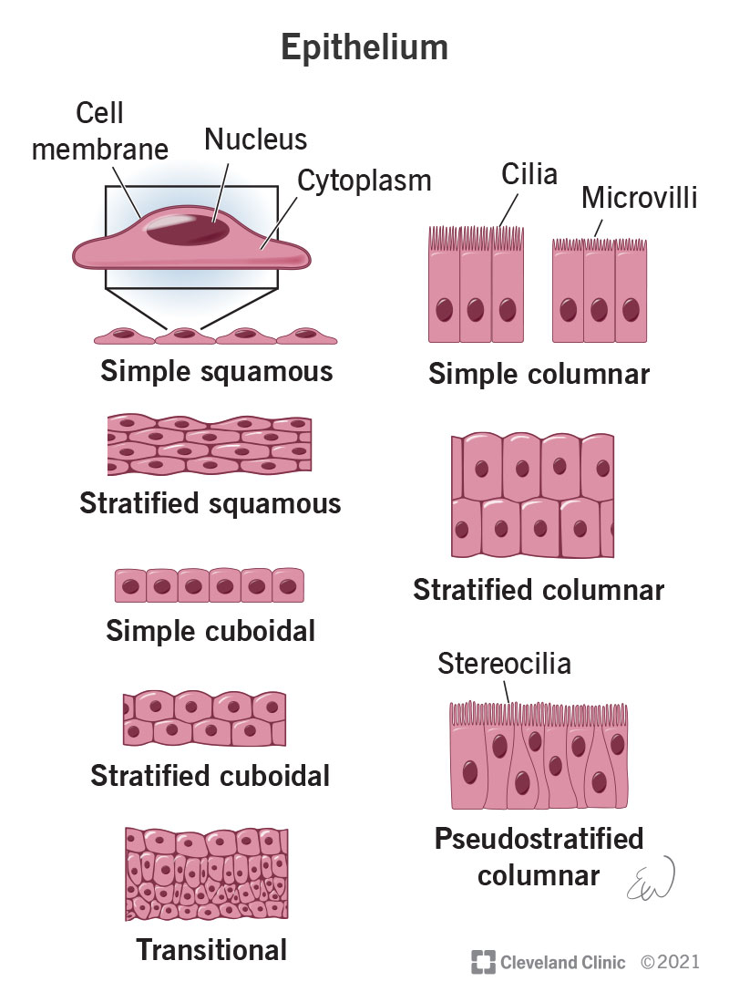
Epithelial Tissue can be classified by their Cell Shape and Cell Arrangement.
Cell Shape:
Squamous- Flat and scale-like
Cuboidal- Cube- Shaped
Columnar- Tall, and elongated cells
Cell Arrangement:
Simple- A single layer of cells thick for absorption and filtration, forms a solid layer of cells lining blood vessels and organs and functions with filtration and diffusion.
Stratified- Stacked cell layers that work best for protection, from the epidermis
Types:
simple columnar- one cell thick, found in the lining of the digestive tract, functions with reabsorption and secretion
stratified columnar- multiple layers thick, rare, and found in few locations in portions of the pharynx and larynx,
Mucous Membrane- a composite of connective and epithelial tissues. Sometimes called mucosae, these epithelial membranes line the body cavities and hollow passageways that open to the external environment and include the digestive, respiratory, excretory, and reproductive tracts.
Mucous- Mucus, produced by the epithelial exocrine glands, covers the epithelial layer.
Serous Membrane- The outer lining of organs and body cavities of the abdomen and chest, including the stomach. Made up of two layers
The parietal layer- lines the walls of the body cavity. (Outer layer)
“Pariet-” refers to a cavity wall.
Visceral layer- covers the organ (the viscera). (Inner layer)
“Viscera-” refers to organ.
What Does It Make?
Epithelial Tissue makes up the Endocrine Glands and Exocrine Glands
Exocrine Glands- release their substances to the surface of the body (sweat glands located deep in the skin but what they produce is released to the surface)
Endocrine Glands- ductless glands, as ducts are lost during development, produce hormones and then released to the interstitial space around the cells, they will diffuse into nearby capillaries and then be carried around the body until they reach their target organ (thyroid glands, islet cells of pancreas)
Muscle Tissue
Muscle tissue that allows movement (voluntary control). They respond to a stimulus. As Cardiac muscle doesn’t allow for voluntary movement.
Skeletal Muscle- Voluntary movement, muscled bundles that attach to bones for movement. Usually found in large body muscles (biceps, Hamstrings)
Smooth Muscle- Involuntary movement, function is to move blood, food, and waste through the body's organs. Forms the contractile component of the digestive, urinary, and reproductive systems as well as the airways and arteries
Cardiac Muscle- Involuntary movement, controls synchronized contraction of the heart to produce the heartbeat and is found ONLY in the heart walls
Nervous Tissue
Found in the brain, spinal cord, and nerves (the main component of the nervous system)
Function
Function as being excitable and capable of sending and receiving electrochemical signals that provide the body with information. As Neurons are the main component of nervous tissue along with neuroligins, sending and receiving action potentials as signals across the body
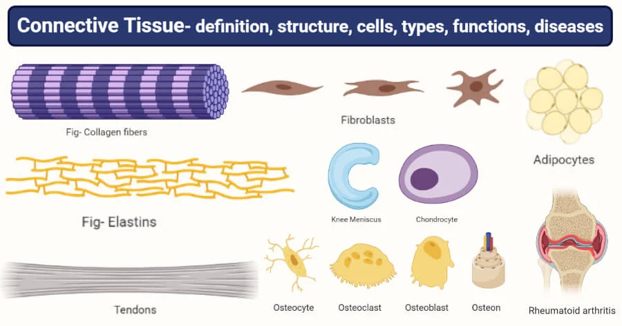
Connective Tissue
composed of cells closely packed with little or no extracellular space in between, connective tissue cells are dispersed in a matrix. Connective tissues come in a vast variety of forms, they typically have in common three characteristic components: cells, large amounts of amorphous ground substance, and protein fibers.
Extracellular Matrix- consists of living and nonliving substances called the “ground substance”. It can have various consistencies such as fluids, solids, gel-like, or very hard. It is mainly found in the connective tissues.
Functions
Connects- binds and supports structures
Protects- cushions organs and tissues
Extracellular matrix- can bear weight, stretch, and take abuse
Adipose- Insulates and stores fats, fills up space
Blood- Transport nutrients and substances
Bone- Provides framework and protection of our organs
Module 2 - Anatomical Structural Organization
Anatomists and healthcare providers use terminology that can be bewildering. However, the purpose of this language is not to confuse, but rather to increase precision and reduce medical errors. Using precise anatomical terminology, we eliminate ambiguity.
Anatomical Positions
To further increase precision, anatomists standardize the way in which they view the body.
There are 5 basic Anatomical Positions: Basic Anatomical Position (BAP), Prone, Supine, Trendelenburg, Fowler's
Basic Anatomical Position (BAP)- the body standing upright, with the feet at shoulder width and parallel, toes forward. The upper limbs are held out to each side, and the palms of the hands face forward. →
Reduces confusion
Creates a common point of reference for all who study the human body
Directional Terms (anterior, posterior) are used when the body is in an anatomical position
Prone- A face-down orientation (lying on your stomach) →
often used for neck and spinal surgeries.
Supine- A face-up orientation “you’re on your spine” (lying on your back) →
Trendelenburg- table or bed is tilted so the head is lower than the feet. →
Usually used as an emergency treatment for hypotension or shock
Fowler’s- Sitting Position. 3 different positions each different angle: 30, 45, 90 degrees. →
Fowler’s- 45 degree angle
Low Fowler’s- 30 degree angle
High Fowler’s- 90 degree angle
Directional Terms
Essential for describing the relative locations of different body structures. (one band of tissue as “inferior to” another).
Anterior (or ventral)- The front or direction toward the front of the body. (The toes are anterior to the foot.)
Posterior (or dorsal)- The back or direction toward the back of the body. (The popliteus is posterior to the patella.)
Superior (or cranial)- A position above or higher than another part of the body proper. (The arm is superior to the leg).
Inferior (or caudal)- A position below or lower than another part of the body proper; near or toward the tail. (The pelvis is inferior to the abdomen.)
Lateral- The side or direction toward the side of the body. (little toe is on the lateral side of the foot)
Medial- The middle or direction toward the middle of the body. (big toe is on the medial side of the foot)
Proximal- A position in a limb that is nearer to the point of attachment or the trunk of the body. (proximal end of femur joins with pelvic bone)
Distal- A position in a limb that is farther from the point of attachment or the trunk of the body. (hand is on the distal end of forearm)
Superficial- describes a position closer to the surface of the body. (The skin is superficial to the bones.)
Deep- describes a position farther from the surface of the body. (The brain is deep into the skull.)
Cranial- Describes a position closer/ upper to the head of the body. (toward the head)
Caudal- Describes a position closer/ lower to the end of the tail. (toward the tail)
Craniocaudal- From head to tail
Superior---Inferior, Anterior----Posterior, Medial---Lateral, Proximal---Distal, Superficial--Deep,
Cranial--- Caudal
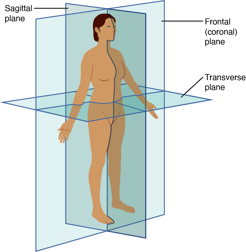
Body Planes
Imaginary two-dimensional surface that passes through the body. Consists of 3 planes:
Sagittal (median), Coronal (frontal), Transverse (horizontal)
Sagittal (Median)- The median plane is a vertical plane that divides the body into Right and Left halves. (If the body is divided into EQUAL right & left sides, then it would be known as the midsagittal plane).
Coronal (Frontal)- The frontal plane is at a right angle to the sagittal plane that divides the body into Anterior and Posterior halves
Transverse (Horizontal)- The horizontal plane is perpendicular to the sagittal and coronal planes that divide the body into Superior and Inferior halves
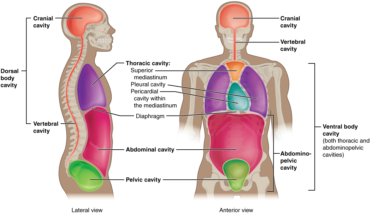
Body Cavities
The body maintains its internal organization utilizing membranes, sheaths, and other structures that separate compartments. The Dorsal (posterior) cavity and the Ventral (anterior) cavity are the largest body compartments. These cavities contain and protect delicate internal organs, and the ventral cavity allows for significant changes in the size and shape of the organs as they perform their functions.
Dorsal (Posterior/ Back) Cavity
Consists of the Cranial and Spinal Cavities.
Cranial Cavity- Which consists of the Brain
Spinal Cavity- Which contains the Spinal Cord
The brain and spinal cord are protected by the bones of the skull and vertebral column and by cerebrospinal fluid, a colorless fluid produced by the brain, which cushions the brain and spinal cord within the posterior (dorsal) cavity.
Ventral (Anterior/ Front) Cavity
Consists of two main subdivisions: the Thoracic cavity and the Abdominopelvic cavity.
The Ventral Cavity is protected by the Serous Membrane.
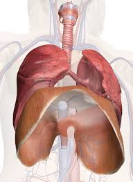
Thoracic Cavity- A superior subdivision of the anterior cavity, and it is enclosed by the rib cage.
Contains the Lungs and Heart, located in the Mediastinum.
The lungs contain the major of the respiratory system (bronchi, bronchioles, alveoli, trachea)
and the esophagus and thymus
The diaphragm forms the floor of the thoracic cavity and separates it from the inferior Abdominopelvic Cavity.
Abdominopelvic Cavity- The largest cavity in the body. No physical membrane to divide the cavity but consists of the Abdominal and Pelvic Cavities
Abdominal Cavity- The division that houses the digestive organs. (stomach, liver, gallbladder, pancreas, spleen, large and small intestine, appendix)
Pelvic Cavity- The division that houses the organs of reproduction. (urinary bladder, reproductive organs, rectum)
Abdominal Regions and Quadrants
To promote clear communication, healthcare providers typically divide up the cavity into either nine regions or four quadrants.
Abdominopelvic Quadrants
4 subdivisions of the abdomen, the navel (belly button) is the landmark.
The terms “left/right” and “upper/lower” are always from the perspective of BAP, not your perspective, the patient’s.
Right Upper Quadrant- Liver, stomach, gallbladder, duodenum, right kidney, pancreas, and the right adrenal gland.
Right Lower Quadrant- appendix, reproductive organs, right ureter.
Left Upper Quadrant- Liver, stomach, pancreas, left kidney, spleen, and the left adrenal gland.
Left Lower Quadrant- left ureter, reproductive organs
*All four quadrants contain portions of the small and large intestines.
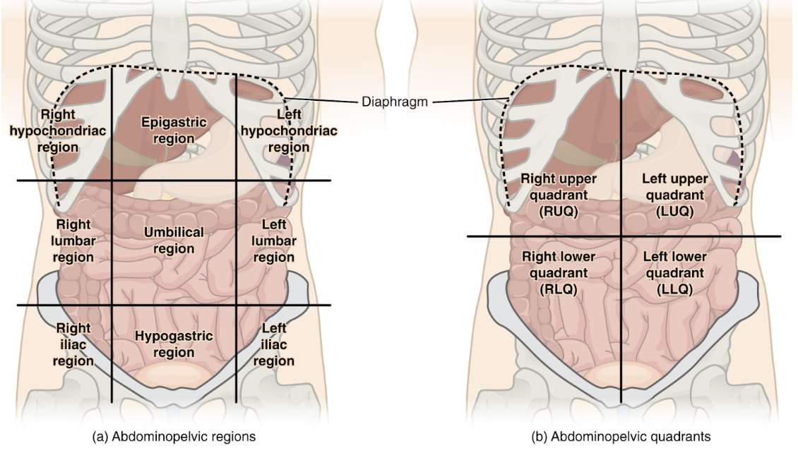
Abdominopelvic Regions
The more detailed regional approach
Consists of 9 regions
Hypochondriac Regions (Left and Right)- the prefix “hypo” means below or under. The word “chondriac” means cartilage, which refers to the cartilage of the ribs.
Lumbar Regions (Left and Right)- The word “lumbar” refers to the vertebrae in your lower back.
Iliac Region (Left and Right)- The top of the hip bone has what is called the iliac crest.
The middle column-named after their location relative to the stomach.
Epigastric Region- The prefix “epi” means above, or over, and “gastric” means stomach or belly.
Umbilical Region- This is easy to remember because the umbilical region contains your navel, which is also called the umbilicus.
Hypogastric Region- Hypo = “below,” and “gastric” refers to the stomach or belly.
Module 4 - Skeletal System
The skeletal system is the body system composed of bones and cartilage and performs the following critical functions for the human body
Skeletal System
The skeleton is subdivided into 2 major divisions: Axial and Appendicular
Functions:
Provides Support- Without the skeletal system you would be a limp mass of organs, muscle, and skin
Protection for the Organs- From internal injury by covering or surrounding them
Muscle Attachments- Some support the muscles, others transmit the forces produced when your muscles contract
Blood cell Formation- In the middle, there is Bone marrow. 2 types of bone marrow: Yellow and Red. Red marrow is where Hematopoiesis (production of blood cells) takes place.
Fat and Mineral Storage- Acts as a reservoir for a number of minerals important to the functioning of the body (calcium, and phosphorus)
Human Skeletal System Consists of:
Joints- Where 2 bones come together
Cartilage- Hyaline, Elastic, Fibro
Ligaments- Connects bone to bone
Bones: Anatomy
Babies are born with (270-300) bones at birth. These bones eventually fuse to form the 206 Bones Adults have.
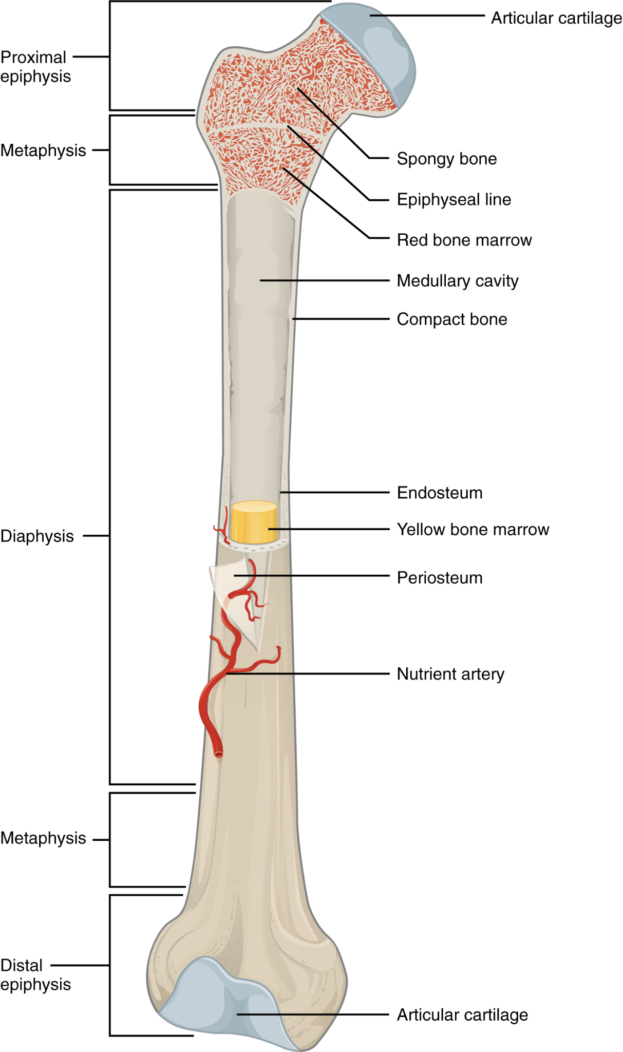
Anatomy of Long Bones:
Anatomy of a bone as 2 parts: Diaphysis and Epiphysis
Diaphysis (shaft)- Hollow cylinder of hard compact bone (defense bone)
In the hollow region of the Diaphysis (or the inside) is where the Medullary Cavity is.
Medullary Cavity (Canal)- filled with yellow marrow
Endosteum- delicate membranous lining in the Medullary Cavity in which bone growth, repair and remodeling occur
Epiphysis (end of the bones)- the wider section at each end of the bone. where less strength is needed in the bone.
In the inside red bone marrow fills the inside in which is where Erythrocytes (red blood cells) are made
Periosteum- fibrous membrane covering the surface of the bone. Contains blood vessels, nerves, and lymphatic vessels that nourish compact bone. (only covers till the end of the Diaphysis not the Epiphysis)
Articular Cartilage- thin layer of cartilage that reduces friction and acts as a shock absorber (only to the ends of the Epiphysis)
Bones: Formation
3 types of cells are found within the bone tissue: Osteoblasts, Osteocytes, Osteoclasts, (and Osteogenic cells. They develop into osteoblasts).
Ossification- Occurring by the 7-8th week of embryonic life, the process of Bone Development, from cartilage to bone. 2 pathways: Intramembranous, Endochondral but bone is the same regardless of pathway.
Fontanel (Soft Spot)- space between bones of the skull from birth to 1 year of age. eventually hardens through ossification
Throughout fetal development and into childhood growth and development bone forms on the cartilaginous matrix. By the time a fetus is born most of the cartilage has been replaced with bone.
Beginning, specialized cells with become osteogenic cells and then to Osteoblasts, they will cluster then spread out by the formation of bone tissue
Osteoblasts- bone cells responsible for forming new bones. Synthesize and secrete the collagen matrix and calcium salts.
As the secreted matrix surrounding the osteoblast hardens, the osteoblast becomes trapped, changing in structure and becomes an Osteocyte
Bones increase in circumference by addition of more bone to the outside surface of the diaphysis (shaft).
As diameter increases, bone material is dissolved from the central part of the diaphysis (shaft).
This creates an internal cavity called the marrow cavity (medullary canal).
Osteocytes- the primary cell of mature bone and the most common type of bone cell. Maintain the mineral concentration of the matrix via the secretion of enzymes
Formed when osteoblasts become embedded in the matrix it has secreted.
Osteoclasts- cells responsible for bone resorption, or breakdown.
They continually breaking down old bone while osteoblasts are continually forming new bone
Usually found on bone surfaces and at sites of old, injured or unneeded bone
Types of Bones
The 206 bones that compose the adult skeleton are divided into 5 categories based on their Shapes. Each shape of the bone has a distinct function. This includes: Long, Short, Flat, Irregular, Sesamoid
Long Bones- Longer than they are wide, most of the bones of the limb are this type (not including the wrist or ankle),
even if the bone is short if it is longer than its wide it’s considered a “long bone”
Found in the arms (humerus, ulna, radius) and legs (femur, tibia, fibula) as well as in the fingers and toes
Long bones Function as “Levers” they move when muscles contract
Short Bones- Cube-shaped, including tarsals, carpals, cuneiforms
Approximately equal in length, width, and thickness
Function to provide stability and support as well as some limited motion
Flat Bones- Flat, thin bones, including sternum, ribs, scapula, and some cranial bones
Typically thin, it is also often curved
Function is to serve as points of attachment for muscles and often protect internal organs
Irregular Bones- Weird shaped, that doesn't have an easily characterized shape (does not fit any other classification). Including vertebrae, facial bones, talus
Vertebrae that support the spinal cord and protect it from compressive forces
These bones tend to have more complex shapes
Sesamoid Bones- Small round bones, embedded in a tendon, including patella, pisiform, or small bones within the first and second metacarpal and the first metatarsal
These bones form in tendons (the sheaths of tissue that connect bones to muscles) where a great deal of pressure is generated in a joint
Function to protect tendons by helping them overcome compressive forces
Axial Skeleton
Forms the Vertical, central axis of the body and includes all bones of the Skull, Spinal column, Ribs, Sternum (Breastbone), Hyoid bone
Consists of 80 out of the 206 bones (Skull, Vertebral Column, and the Thoracic Cage)
Functions:
Serves to protect the brain, spinal cord, heart, and lungs
Serves as the attachment site for muscles that move the head, neck, back, and muscles that act across the shoulder and hip joints to move
The Skull (Cranium)
Formed by 22 Bones (8 Cranial Bones and 14 Facial Bones).
Suture- an imobile joint between adjacent bones of the skull (what holds the bones together)
Functions:
To protect the brain from accidents or fights
Protect the major sense organs (eyes, nose, ears)
Provide attachment sites for the muscle
Bones of the Cranium:
Frontal- the single bone that forms the forehead.
Parietal- the base of the skull (Top), roof and sides of skull
Temporal- Side of our face (barley above the ears), houses the ears
Occipital- base of the skull (backend of the skull)
Nasal- Bridge of nose
Vomer- Lower part of nasal septum
Maxilla- Upper jaw
Mandible- Lower jaw, The only Moveable joint in the face
Zygomatic arch- Cheekbone area
Hyoid- U shaped bone in neck
Facial Bones- guard and support eyes, ears, nose, mouth
The Vertebral Column (Spinal Column/ Spine)
Consists of a sequence of vertebrae each of which is separated and united by an Intervertebral Disc
Intervertebral Disc.- separate and unite each vertebrae, together, vertebrae and intervertebral discs form the vertebral column.
Act as a shock absorber/cushion.
Throughout our lifetime the discs become thinner - which is why you get shorter with age.
Foramen- Holes in vertebrae that allow for the passage of spinal cord
Encloses spinal cord
Functions:
Strong, Flexible column that supports the head, neck, and body and allows for their movement
Protects the spinal cord, which passes down the back through openings in the vertebrae
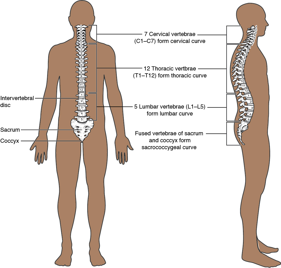
Regions:
Sub divided into 3 regions. Cervical, Thoracic, Lumbar
Cervical Vertebrae- (7) has a small body, which carry the least amount of body weight
1st Cervical (Atlas)- supports the skull on top of the vertebral column. Does Not have a body or spinous process. Ring shaped. Allows us to nod our heads and articulates with the occipital bone of the skull
2nd Cervical (Axis)- serves as the axis for rotation when turning the head toward the right or left.
Thoracic Vertebrae- (12) Located in the chest area. Articulate with the ribs
Lumbar Vertebrae- (5) located in the low back. Large vertebral bodies that bear most of the body's weight
Others:
Sacral vertebrae- Wedge shaped bone formed by 5 fused bones
Coccyx- “Tailbone” formed by 4 fused bones
Ribs and Sternum
The rib cage forms the chest portion of the body. The ribs consist of 12 Ribs along with their costal cartilages and Sternum
Function- The Rib Cage protects the heart and lungs
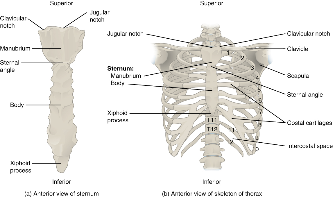
Sternum
The elongated bony structure that anchors the anterior rib cage. Also known as the Breastbone.
Consists of 3 parts: Manubrium, Body, Xiphoid Process
Manubrium- the wider, upper portion of the sternum, has a shallow U shaped border
Can be felt at the anterior base of the neck between the medial ends of the clavicles
Body- Central (middle) portion of the sternum. Ribs 3-7 attach to the body
Xiphoid Process- small inferior (lowest) tip of the sternum.
Ribs
Curved, flattened bone that contributes to the wall of the rib cage. There are 12 pairs of ribs
True ribs- (1-7) pairs of costal cartilages join 7 pairs of ribs directly to the sternum
False ribs- (8-10) their costal cartilages are attached to the 7th rib instead of directly to the sternum
Floating ribs- (11-12) connected either costal cartilage nor the sternum
Hyoid Bone- does not attach to any other bone
Appendicular Skeleton
Consists of the Shoulder girdle,
Module 5 - Muscular System
The Muscular system comprises nearly half of our weight, with over 650 muscles
Types of Muscles
There are types of muscles found in the body: Skeletal, Smooth, Cardiac (Sphincter)
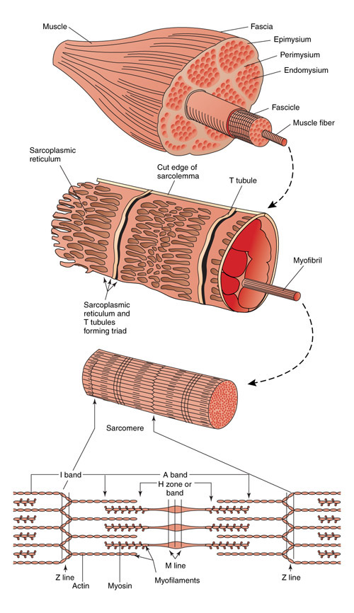
Each muscle is made of hundreds or thousands of Muscle fibers (Muscle cells)
Skeletal Muscle- the major muscle group of the body (biceps, quadriceps). Skeletal muscle allows us to perform motor activities, protects internal organs by serving as an extra barrier layer, protects the skeleton by stabilizing it and preventing structural damage to the skeleton
Skeletal muscle is VOLUNTARY, it can be moved by will
Skeletal muscle is STRIATED, which means it contains a Striped look. This allows force and contraction
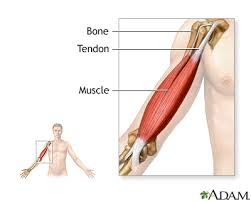
Skeletal muscle often runs across a joint and Connects to bones via TENDONS
Smooth Muscle- Found in the walls of organs (intestines, stomach, bladder, esophagus). Cells of the Smooth muscle are spindle-shaped and are much shorter than Skeletal muscles.
Smooth muscle is INVOLUNTARY, it moves unconsciously, contractions are rhythmic and slow
Smooth muscle is NOT STRIATED
Smooth muscle Contracts the uterus during labor, Controls diameter of blood vessels as blood circulates through the body
Cardiac Muscle- Only found in the heart. Cardiac muscle fibers are extensively branched and connected with each other at their ends with Intercalated Discs. Conductivity of this allows for transmissions of electrical signals in a wave like pattern so the heart works as a pump when it contracts (when one contracts, all contracts)
Cardiac muscle is INVOLUNTARY
Cardiac muscle is STRIATED, BRANCHED
Cardiac muscle is meant to Contract the heart
Intercalated Disks- connect the ends of the cardiac muscle
Allows the transmission of electrical signals (like a wave pattern) so the heart works as a pump when it contracts
Sphincter Muscle- (Dilator Muscle) special circular muscles located in the Digestive system (anus, opening between esophagus and stomach, small intestines) and Urinary system (bladder)
Open and close to control the passage of substances (Both Voluntary and Involuntary)
Sphincter muscle is STRIATED
Connective Tissues
Tendons- Dense bands that connect skeletal muscle to the bone.
Fascia- Fibrous connective tissue (primarily collagen) sheets that Wrap around muscle bundles to hold them in place.
attaches, stabilizes, encloses, and separates muscles and other internal organs
Characteristics of Muscles
These include Contractibility, Excitability, Extensibility, Elasticity
Contractility: Quality that no other tissue possesses
Skeletal: Contraction of skeletal muscle causes movement of the bones. Muscles become SHORTER and THICKER
Cardiac: Contraction of cardiac muscle makes the heart pump blood to the entire body
Smooth: Contraction of smooth muscle it surrounds blood vessels, intestines, and numerous other organs which causes the diameter of these tubs to increase and decrease
Excitability or Irritability- Ability to respond to certain stimuli (the production of action potentials)
Characteristic of both muscle and nervous cells (neurons)
Extensibility- Ability to be Stretched. Muscles on the back are either extended or stretched
Elasticity- Ability to return to its original state when relaxing (like a rubber band)
Muscle Tone- Partial contraction. Muscles should always be slightly contracted and ready to pull. Maintained with proper nutrition and regular exercise
Atrophy- Muscles become weak and flaccid, from lack of movement. Waiting away of muscle
Hypertrophy- Muscles become enlarged, from over exercise of muscles. Overgrowth
Muscle Fatigue- inability to generate force. Caused by Accumulation of Lactic Acid in the muscles, can lead to fatigue and cramps
Accumulation of lactic Acid, if from long periods of exercise where blood is unable to transport enough oxygen for the complete oxidation of glucose in the muscles, which causes muscles to contract Anaerobically (without oxygen)
Symptoms: Muscle weakness, soreness, localized pain, shortness of breath, muscle twitching, trembling, weak grip.
Treatment: Stop, Rest, and Take enough oxygen. This changes lactic acid back into glucose for muscle cells
Naming Muscles
Muscles can be classified and named by: Shape, Size compared to other muscles in the area, location in the body or location of its attachments to the skeleton, how many origins it has, its action
*Study Quizlet Terms
Muscles of the Body
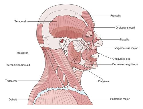
Head and Neck Muscles
Frontalis- Moves eyebrows up and wrinkles forehead
Masseter- chewing muscle of the jaw
Orbicularis Oculi- closes the eyelids
Orbicularis Oris- encircles the mouth
Temporalis- chewing muscle of the upper jaw that covers the temporal bone
Zygomatic Major- smiling muscle
Sternocledidomastoid- rotates the head and flexes the neck
Torso/Trunk (Anterior/ Front)
External Intercostals- Between the ribs. Assists the lungs by raising the ribcage
Diaphragm- thin, dome shaped muscle that separates the thoracic and abdominal cavity. Contracts/relaxes when you inhale
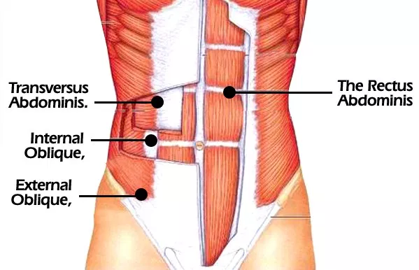
Rectus Abdominis- the “six pack”, help bend your abdomen
External Oblique- located on each side of the rectus abdominis. Twists the trunk
Internal Oblique- located on each side of the rectus abdominis below the external obliques. Assists with twisting the trunk and respiration
Torso/Trunk (Posterior/ Back)
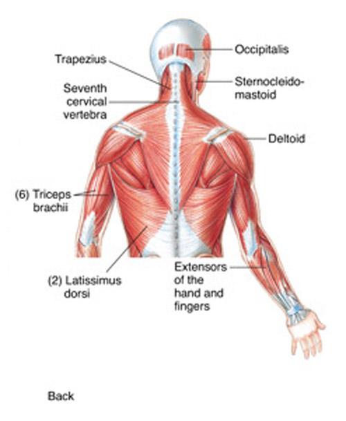
Latissimus Dorsi- “butterfly wings” lower back muscle, known as the largest muscle in the body
Upper Extremities:
Trapezius- upper back, supports the arm and helps with scapula
Deltoid- helps lift the upper arm, one of the sites for injections
Pectoralis Major- chest muscles, assist with arm movements and motion
Biceps Brachii- lifts the forearm. On the anterior of the upper arm
Triceps Brachii- posterior arm. Straightens the arm
Brachioradialis- flexes the forearm. Pronation and supination
Lower Extremities
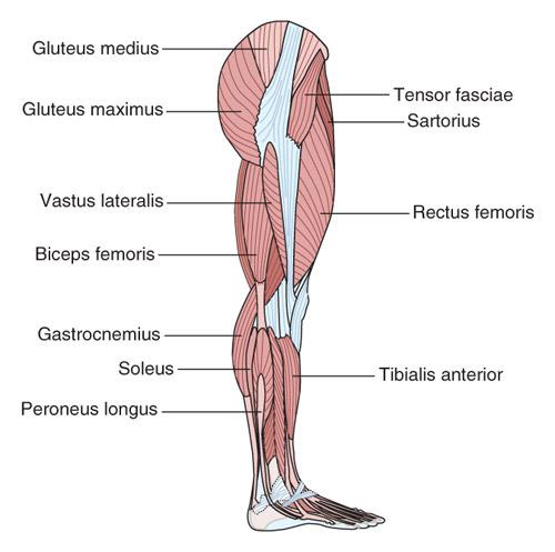
Gluteus Medius- sits on the outer surface of the ilium, helps with hip movement
Gluteus Maximus- butt, helps with several hip movements
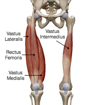
Rectus Femoris- long, thin muscles on the femur, part of the quads
Vastus Lateralis- side of the thigh, helps extend the knee and move leg forward
Sartorius- longest muscle in the body, runs from hip to to the medial knee
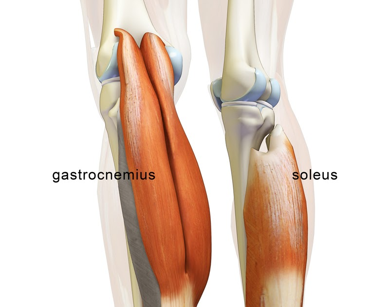
Gastrocnemius- calf muscle, works with the soleus
Soleus- under the gastrocnemius. Runs from the knee to the heel. Helps with standing and walking
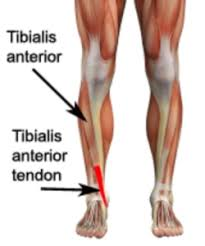
Tibialis Anterior- on the front of the tibia. Dorsiflexes and inverts the foot
Functions of the Muscular System
Movement, Posture, Body Heat, Protection of internal organs
Movement: of the digestive system by contractions, Circulatory system and its regulation of the heart beats
Body Heat: Homeostasis (shiver, sweat, exercise. Basically the body adjusting to the environment automatically)
Disorders
Fibromyalgia- Disorder characterized by widespread musculoskeletal pain accompanied by fatigue, sleep, memory and mood issues.
Causes- Unknown, a variety of causes: genetics, infections, psychological or emotional trauma
Symptoms- Widespread pain, fatigue, cognitive difficulties (memory loss), mood swings
Diagnosis- diagnosis can be made if a patient has widespread pain for more than 3 months with no underlying medical condition that would cause pain.
Treatment- No cure. Medication and self carem improve sleep, reduce stress to lessen symptoms
Hernia- Occurs when an organ protrudes through an opening in a weak muscle.
Causes- muscle weakness and strain
Locations: 3 locations of a hernia
Abdominal hernia- Occurs when organs protrude through the abdominal wall.
Inguinal hernia- Occurs in the inguinal area.
Hiatal hernia- When stomach pushes through the diaphragm
Symptoms- pain or discomfort in affected area, especially when bending over coughing or lifting
Diagnosis- physical exam, endoscopy, barium x-ray
Treatment- surgery, lifestyle changes, medication
Muscle Spasms- A “cramp” or sustained contraction of a muscle
Causes- Overuse/overuse, Compression of nerves, Dehydration, Low levels of electrolytes (i.e. magnesium, potassium, calcium), Not enough blood to muscles, Pregnancy, Certain medications
Locations- can happen anywhere but most commonly: thighs, feet, hands, arms, abdomen, and along rib cage
Module 7 - Digestive System
Break down the foods we eat, release their nutrients, and absorb those nutrients into the body. Divided into 2 groups that contain different organs systems: Alimentary canal, Accessory digestive organs.
The Digestive System
Uses both Mechanical and Chemical activities to break down food into absorbable substances.
Digestion Process:
This process of digestion includes 6 Steps: Ingestion, Propulsion, Mechanical/Physical digestion, Chemical digestion, Absorption, Defecation
Ingestion- 1st step. Entry of food into the Alimentary Canal through the mouth, the food is chewed and mixed with saliva (contains Enzymes that break down carbohydrates plus some lipid [fat] digestion via lingual lipase), chewing increases the surface area of the food into what is now called a Bolus.
Bolus- A mixture of food and saliva, once its soft and pliable
Propulsion- 2nd step. Refers to the movement of food through the digestive tract. Includes both voluntary (swallowing) and involuntary (process of peristalsis) processes
Peristalsis- consists of sequential, alternating waves of contraction and relaxation of alimentary wall smooth muscles that help propel food through the digestive tract.
These waves mix food with digestive juices.
Very powerful that you can swallow, even if your standing on your head
Mechanical Digestion- (part of steps) a physical process that does not change the chemical nature of the food. Instead makes the food smaller to increase both surface area and mobility
Mastication- “Chewing” as well as tongue movements that help break down food, mixing saliva with the food
Churning- stomach breaking down food to expose it to more digestive juices
Segmentation- occurs in the small intestine, contractions of circular muscle of the muscularis layer to continuously divide, break up and mixing the contents further with digestive juices
Chemical Digestion- digestive secretions break down complex food molecules into their chemical building blocks (proteins to amino acids). Secretions contain water, various enzymes, acids, and salts. Completed in the small intestine
Absorption- 3rd step. Primarily in the small intestine, food that has been broken down is of no value to the body unless it enters the bloodstream and its nutrients are absorbed.
Defecation- 4th step. Undigested materials are removed from the body as Feces.
*Steps in the ACTUAL NOTES: Physical, Chemical, Absorption, Elimination.
Alimentary Canal
Also called the gastrointestinal (GI) tract or Gut. A One Way tube, from Mouth to Anus
Function- the organs of the alimentary canal is to nourish the body
Contains: Mouth (buccal cavity), Pharynx (throat), Esophagus, Stomach, Small intestines, Large intestines
Mouth (Buccal Cavity)
The Mechanical Portion of digestion that occurs in the mouth, involving the Teeth, tearing and crushing the food into smaller pieces and the Tongue smashing it against the hard Palate of the mouth.
Reduces the size of the food particles and increase the surface area available to interact with the mouths chemical agents (saliva)
Consists: Tongue, Teeth, Salivary Glands, Palate, Uvula
Tongue
Accessory Structures
The accessory organs aid in the breakdown of food.
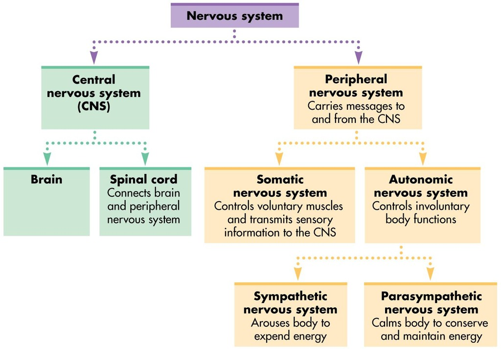
Module 9 - Nervous System
The Nervous system is Divided into 2 divisions: Central nervous system (CNS), Peripheral nervous system (PNS)
The nervous system communicates with various nerve cells (neurons) activated by action potentials (stimulus) either my motion, or senses that go through the body.
Central Nervous System (CNS) Anatomy
Includes the Brain and the Spinal cord. A person's conscious experiences are based on neural activity in the brain aswell as the regulation of homeostasis (by a specific region in the brain) in the Brain. While, coordination of reflexes depend on the integration of sensory and motor pathways in the Spinal cord.
The Brain
The adult brain is described in terms of 4 Major Regions: Cerebrum, Diencephalon, Brain stem, Cerebellum.
Weighs 3 lbs; contains 100+ billion neurons, brain tissue dies in 4-6 min without adequate oxygen
The Cerebrum
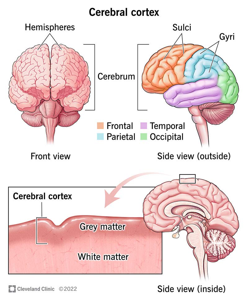
Takes up most mass of the brain. (largest part of the brain).
Comprises of the Outer Grey matter (Cerebral Cortex) and White matter
The cerebrum is divided into 2 Hemispheres: Lef and Right Hemispheres. This is separated by the Longitudinal Fissure.
Longitudinal Fissure- separates the cerebrum into 2 hemispheres (Cerebral hemispheres)
Corpus Callosum- provides the major pathway for communication between the 2 hemispheres of the cerebral cortex (not mentioned in the notes)
Separated into 4 Major regions: Temporal lobe, Parietal lobe, Frontal lobe, Occipital lobe
Temporal lobe- primary auditory sensation (part of the limbic system), memory function is associated with this lobe (face recognition)
Responsible for establishing long term memory
Can recall sensations of smell, movement, sensory feedback
Parietal lobe- All of the tactile senses are processed in this area: touch, pressure, tickle, pain, itch, vibration (general senses of the body) senses are processed here.
Frontal Lobe- primarily associated with motor functions. Aswell as thinking of a movement to be made, production of language or controlling movements responsible for speech, cognitive function of the basis of personality, short term memory, and consciousness.
motor neurons that instruct cells of the spinal cord to move skeletal muscles
Occipital Lobe- responsible for primary visual perception. Visual info is processed in the temporal and parietal lobes.
The Cerebral Cortex
A continuous outer layer of gray matter, thin extensive region of wrinkled matter that covers the cerebrum
Gyrus- the ridge of one of the wrinkles (plural= gyri)
Sulcus- the groove between 2 gyri
The Diencephalon
The connection between the cerebrum and the rest of the nervous system. The rest of the brian, spinal cord, PNS all send info to the cerebrum through the diencephalon.
Located between the cerebrum and the midbrain
The diencephalon contains 2 Major regions: Thalamus, Hypothalamus
Thalamus
Relay station for incoming & outgoing nerve impulses from the various sense organs of the body (with exception of olfactory sensations).
All sensory info (except smell) passes through the thalamus before processing by the cortex for interpretation
Takes in information (sensory, synapse, motor neurons) processes, filters, and relays that information to the appropriate regions of the cerebral cortex.
Like a “double checker” before passing it to the cerebral cortex for actual processing.
Hypothalamus
Collection of nuclei that is largely involved in Regulating homeostasis.
The executive region in charge of the Autonomic nervous system and the endocrine system through regulation of the anterior pituitary gland.
Other parts of the hypothalamus are involved with memory and emotion as part of the Limbic system
Below the thalamus
Limbic System- collection of structure involved in emotion, memory, and behavior
Consists of 9 Vital functions:
Autonomic nervous control (sympathetic & parasympathetic)
Cardiovascular control (controls BP, heart beat, blood vessel dilation & constriction….)
Temperature control (98.6 degrees Fahrenheit)
Appetite control (hunger pangs & satiety center)
Water balance (thirsty = body needs more water)
Manufacture of oxytocin (uterus contraction during labor)
Gastrointestinal control (increases peristalsis & secretion from intestinal glands)
Emotional state (controls display of emotions)
Sleep control (sleep-wake cycle)
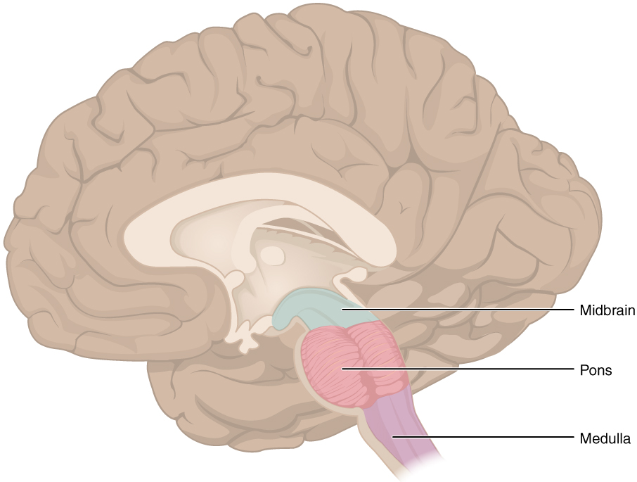
The Brain Stem
Controls the flow of messages between the brain and the body.
Consists of 3 regions: Midbrain, Pons, Medulla
Midbrain- coordinates sensory representations of the visual, auditory, and somatosensory perceptual spaces
Pons- main connection with the cerebellum. Assist with the Medulla to regulate functions
Medulla- regulate crucial functions: cardiovascular and respiratory systems and rates (works with the Pons)
* Damage to the Brain Stem inflicts a Coma.
The Cerebellum
“Little brain” Responsible for comparing information from the cerebrum with sensory feedback from the periphery through the spinal cord
Composed of 2 hemispheres.
Controls all body functions related to skeletal muscles, including: Balance, Muscle tone, Muscle movement
Protective Coverings of the Brain and Spinal Cord
The outer surface of the CNS is covered by a series of membranes composed of connective tissues called Meaninges
Meninges- protect the brain and line the skull and vertebral canal and protect the brain and spinal cord
Consists of 3 Membranes: Dura mater, Arachnoid mater, Pia mater
Dura Mater- Outermost thick fibrous layer and a strong protective sheath over the entire brain and spinal cord. Accorded to the inner surface of the cranium and vertebral cavity
Arachnoid Mater- Middle membrane of thin fibrous tissue that forms a loose sac around the CNS. Resembles a spider web
Pia Mater- Delicate innermost thin fibrous membrane that follows the convolutions of gyri and sulci in the cerebral cortex and fits into other grooves and indentations.
Subarachnoid space- between arachnoid and pia mater, filled with cerebrospinal fluid
Cerebrospinal Fluid- acts as a liquid shock absorber and source of nutrients for the brain
Neurons
Considered to be the basis of nervous tissue. Can send messages (action potentials).
Are responsible for the electrical signals that communicate information about sensations and that produce movements in response to those stimuli, along with inducing thought processes within the brain.
Parts of the Neuron:
Main Parts:
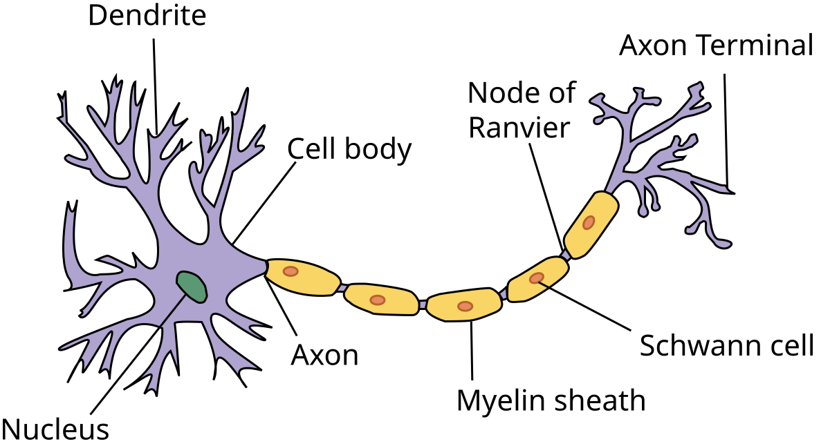
Soma (cell body)- contains the nucleus and most of the major organelles
Axon- A fiber that merges from the cell body and projects to target cells. Cane branch repeatedly to communicate with many target cells. Propagates the nerve impulse.
Dendrites- small finger-like branches (from the hand). They receive information from other neurons at specialized areas of contact called synapses
Synapses- Space separating two neurons where the impulse jumps to the next neuron.
Other parts that connect to other parts of the neuron:
Myelin- Made of glial cells, acts as an insulator that wraps around the axon that has gaps along the fiber, which speeds up the process of communication of each neuron. Those gaps are called “Node of ranvier”
Node of Ranvier- an important way that electrical signals travel down the axon
Schwann Cell- 2nd type of glial cell. Insulates axons with myelin and periphery. Wraps around a portion of only 1 axon segment.
Process of Synapse Exchange: (simple terms: its a chemical change)
Action potential (message from neurons) flows down, calcium flows into nerve, neurotransmitters (GABA, hormones) get released and move across the synapse, match with an ion channel and gets shape change, the action potential moves in reverse
*The neurotransmitter between muscle cells & the nervous system is Acetylcholine.
*In order for a nerve impulse to begin, it must be initiated by a stimulus.
Types of Neurons:
Sensory Neuron- send information from sensory receptors (i.e. skin, eyes, ears, nose, tongue) to the spinal cord and brain.
Motor Neuron- send information from the central nervous system back to the body. Responsible for causing muscle fibers to contract
Associative (interneurons)- send information between sensory and motor neurons.
The Spinal Cord
Maintains the tube structure and is only specialized into certain regions.
Begins at foramen magnum.
Continues down to 2nd lumbar vertebrae.
White and soft in the spinal canal.
Surrounded by cerebrospinal fluid.
Functions as a reflex center and a conduction pathway to and from the brain.
Divisions of the Nervous System
Peripheral Nervous System (PNS)
Defined as everything that is not the CNS. Basically the spinal nerves and other nerves of the body.
Provides a complete network of motor and sensory fibers connecting the CNS with the rest of the body.
Includes 12 pairs of Cranial nerves and 31 pairs of Spinal nerves.
Cranial nerves: Spinal nerves:
Olfactory = smell
Optic = vision, eyesight
Oculomotor = movement of eye muscle
Trochlear = movement of eye muscle
Trigeminal = face & teeth muscles, chewing
Abducens = movement of eye muscle
Facial = facial expressions, taste
Vestibulocochlear = hearing & balance
Glossopharyngeal = movement of throat muscle, taste
Vagus = movement of throat, affects heart, digestive system
Accessory = movement of neck muscles
Hypoglossal = movement of tongue
Divided into 2 Divisions: Somatic and Autonomic Nervous Systems
Somatic Nervous System
Module 10 - Sensory System
The Central Nervous System (CNS), which consists of the brain and spinal cord collect information from the senses and spread the information throughout the body. We have senses to collect information from our surroundings and send the info to our brains to react.
The body has 5 senses: Vision, Hearing, Smell, Touch, Taste
Eyes (Vision)
Vision is the special sense of sight that is based on the transduction of light stimuli received through the eyes.
External Structure of the Eyes:
The Orbital Cavity- cone-shaped cavity formed by the skull that houses & protects the eyeball. (contains all listed below)
Eyelids (with Lashes)- help to protect the eye from abrasions by blocking particles that may land on the surface of the eye.
Conjunctiva- Thin membrane that extends over the white areas of the eye (the sclera), connecting the eyelids to the eyeball
Lacrimal Apparatus- the entire tear system which includes: Lacrimal duct, Lacrimal gland
Lacrimal Duct- Tears produced by the gland flow through the lacrimal duct to the medial corner of the eye, where the tears flow over the conjunctiva, washing away foreign particles. Why water also comes out of nose
Lacrimal Gland- what produces tears. located just inside the orbit, superior and lateral to the eyeball
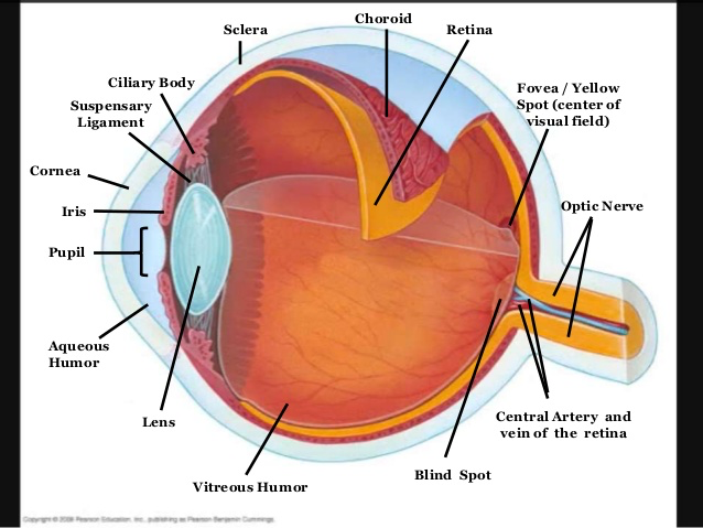
Layers of The Eyes
There are 3 layers of the eye: Sclera, Choroid, Retina
Sclera- Tough, outer layer; “white of the eye;” helps maintain shape of the eye. Extrinsic muscles are attached to the sclera and help move the eyeball
Choroid- The middle layer of highly vascularized connective tissue that provides a blood supply to the eyeball (nourishes outer layers of the retina).
Retina (neural tunic)- the innermost layer, which contains the nervous tissue responsible for photoreception (light rays reflect here to form an image). contains rods and cones. And Optic Disc.
Rods- sensitive to dim light.
Cones- sensitive to bright light and color
Optic Disc - on retina (back end); known as blind spot, nerve fibers form optic nerve here.
Internal Structure of The Eye
Cornea → Pupil → Lens → Retina → Rods & Cones → Optic Nerve → Brain (Passageway of light to brain)
Cornea- “window of the eye” transparent and permits the passage of light rays.
Pupil- the hole at the center of the eye that allows light to enter.
Iris- the colored part of the eye. is a smooth muscle that opens or closes the pupil. Contains intrinsic muscles within the iris that control the size of the pupil and amount of light entering the eye
Lens- Crystalline structure located behind the iris and pupil; separates anterior and posterior chambers; focuses images on the retina.
Optic Nerve- carries the impulses formed by the retina to the brain for interpretation.
Brain- The optic lobe of the brain and then process the signals and construct the images we see.
The eye is also divided into two cavities: the Anterior cavity and the Posterior cavity.
The anterior cavity is the space between the cornea and lens, including the iris and ciliary body. It is filled with a watery fluid called the aqueous humor
The posterior cavity is the space behind the lens that extends to the posterior side of the interior eyeball, where the retina is located. The posterior cavity is filled with a more viscous fluid called the vitreous humor.
Aqueous humor- Anterior chamber filled with watery fluid called aqueous humor that nourishes the lens and maintains pressure in the eye..
Vitreous humor- jelly-like fluid that maintains eye shape and refracts light rays.
These 2 help:
Maintain the eyeball’s spherical shape
Refraction (bending) light rays as they pass through the eye.
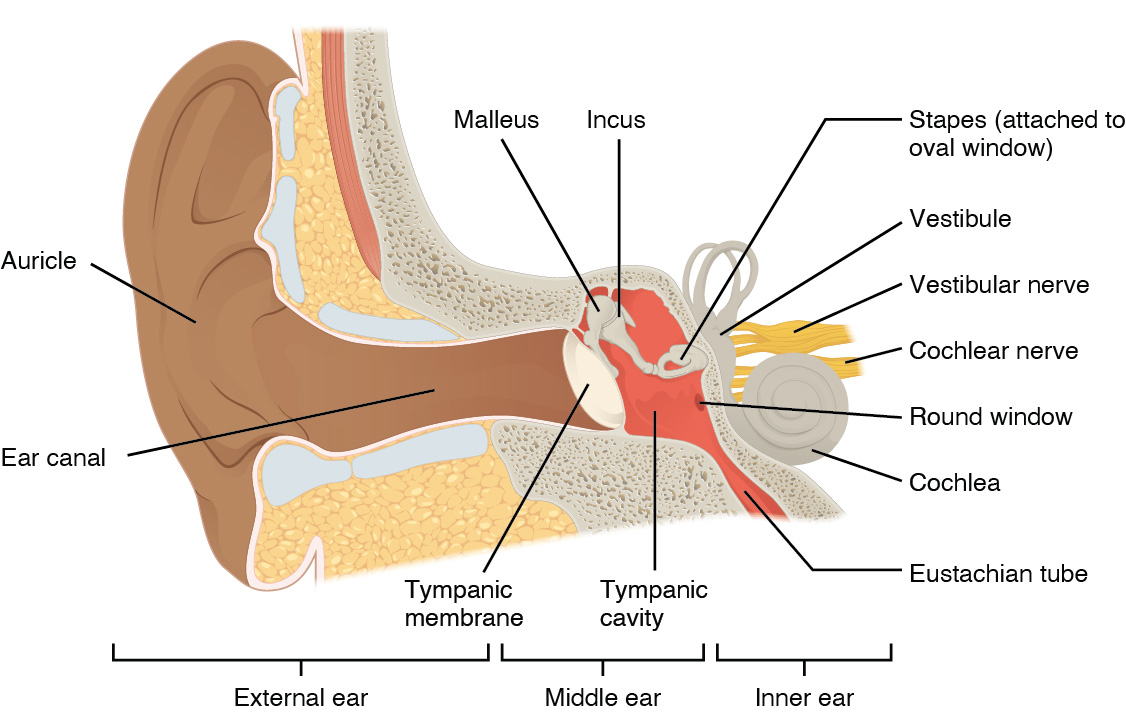
Ear (Hearing)
Transduction of sound waves into a neural signal that is made possible by the structures of the ear
Consist of the: Outer, Middle, and Inner divisions
Pinna → External auditory canal → tympanic
membrane → ossicles (malleus, incus, stapes) →
cochlea → auditory nerve → brain
The Outer Ear
Also known as the External ear. Consists of the: Auricle, Auditory, Tympanic membrane
Auricle (Pinna)- the visible portion of the ear. The C-shaped curves of the auricle direct sound waves toward the auditory canal. The canal enters the skull through the external auditory
Auditory- Lined with glands that secrete a wax-like substance called cerumen that protects the ear. The canal leads to the tympanic membrane.
Tympanic Membrane- or ear drum, which vibrates after it is struck by sound waves.
Middle Ear
Consists of a space spanned by 3 small bones called the Ossicles. The three ossicles are the Malleus, Incus, and Stapes.
Connected to the pharynx through the Eustachian tube, which helps equilibrate air pressure across the tympanic membrane.
Malleus (Hammer)- attached to the tympanic membrane and articulates with the incus.
Incus (Anvil)- articulates with the stapes
Stapes (Stirrup)- attached to the inner ear, where the sound waves will be transduced into a neural signal
Eustachian Tube- equalizes the air pressure in the middle ear with the outside atmosphere.
Inner Ear
often described as a bony labyrinth, as it is composed of a series of canals embedded within the temporal bone.
Contains the: Cochlea, Cochlear duct, Organ of corti, Semicircular canals
Cochlea- spiral shaped organ of hearing that contains the cochlear duct.
Cochlea duct- the central cavity of the cochlea that contains the sound-transducing neurons. filled with fluid that vibrates when sound waves are transmitted by the stapes.
Organ of corti- which transduce the wave motion of the two scala into neural signals. Contains delicate, hair-like cells inside the cochlear duct, picks up vibrations of fluid and transmits them as a sensory impulse along the auditory nerve to the brain.
Semicircular Canals- three structures in the inner ear that contain liquid set in motion by head and body movements. These impulses are sent back to the cerebellum to maintain balance or equilibrium. (nothing to do with hearing)
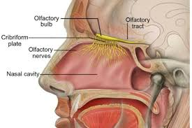
Nose (Smelling)
The sense of smell, or olfaction, is also responsive to chemical stimuli. The olfactory receptor neurons are located in a small region within the superior Nasal Cavity
Limbic System- generates our basic emotions such as affection, aggression, fear
Cilia- Small hair-like projections that trap larger dust particles entering the nose.
Nasal septum- partition that divides the nose into right and left sides.
Turbinates- Bones in the nasal cavity that are covered in mucous. Increases surface area of the nasal cavity & helps with filtration of the air breathed in.
Olfactory Bulb- structures located at the bottom of each cerebral hemisphere that process information about odors.
The nose and its structures provide for our sense of smell. Smell accounts for 90% of our sense of taste.
Olfactory Receptors- In the nasal cavity, there is a patch of tissue (about the size of a postage stamp) called the olfactory epithelium, which has a plentiful supply of nerve cells with specialized receptors. These receptors send signals to adjoining olfactory bulbs, an extension of the brain.
When particles travel through the air and enter your nose, those chemicals come into contact with nerve endings in our nose, those molecules stimulate olfactory receptors that will in turn send information to the *temporal lobe* of the brain (where smells are interpreted)
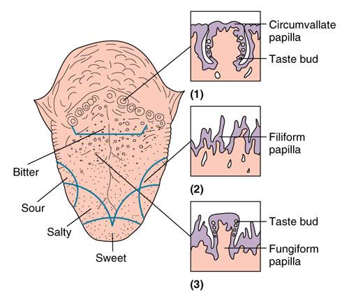
Tongue (Taste)
The tongue is a mass of muscle tissue. Contains of 4 Tastes- Sweet, Sour, Salty, Bitter
Taste is one of the weakest senses, with only 10,000 taste buds inside our mouths; even on the roofs of our mouths.
Everyone’s tongue print is different (like a fingerprint)
Along with the rest of the oral cavity, it is lined by a stratified squamous epithelium. Along with raised bumps (papilla)
Papila- Bumps on the surface of the tongue that contain the taste buds.
Taste buds- contain specialized gustatory receptor cells for the transduction of taste stimuli.
Receptors in taste buds send stimuli through 3 cranial nerves to the cerebral cortex for interpretation.
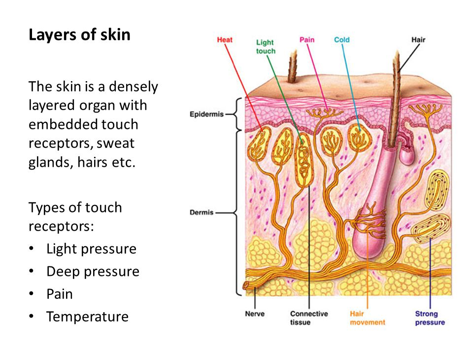
Skin (Touch/ Sensory Receptors)
Stimuli in the environment activate specialized receptor cells (specialized sensory nerves) in the peripheral nervous system (PNS).
The Epidermis, Dermis, and Hypodermis contain specialized sensory receptors (there are multiple kinds)
In addition, these sensory nerves detect: Touch, Surface Temp., Pain
Structural Receptor Type
receptors can be classified based on their location relative to the stimuli. (there are 3 but only talk about Proprioceptors)
Exteroceptor, Interoceptor, Proprioceptor
Proprioceptor- a receptor located near a moving part of the body, such as a muscle, that interprets the positions of the tissues as they move. (found in tendons, muscles, joint capsules)
without these we wouldn't be able to fundamentally move (feed/ clothing ourselves)
Detects changes in muscle length and tension
Functional Receptor Types
However the main Receptor cells mentioned are: Mechanoreceptors (Touch), Thermoreceptors (Temp.), Nociceptor (Pain)
Mechanoreceptors (Touch receptors)- Physical stimuli, such as pressure and vibration, as well as the sensation of sound and body position (balance), are interpreted by this receptor.
Reveals info about things that you actually come in direct contact with
Thermoreceptors (Temperature receptors)- either sensitive to temperatures above (heat) or below (cold) normal body temperature.
pass this information to the hypothalamus which then triggers changes needed to ensure body temperature remains constant.
Nociceptor (Pain receptors)- pain is primarily a chemical sense that interprets the presence of chemicals from tissue damage, or similar intense stimuli (can also be caused by mechanical, thermal, chemical stimuli)
Sharp pain = “quickly move away”
Dull pain = “avoid till healed”
Over 3 Million pain receptors, found in the skin, muscles, bones, blood vessels, some organs
Disorders
Only the most important parts. Not all symptoms/ treatments
Eyes
Cataracts- condition where the Lens gradually become cloudy
Common in people greater the 70 Yrs
Painless, gradually blurring and loss of vision. Pupil from black to milky white
Causes- aging, injury changes the tissue that makes up the lens, genetics
Signs/Symptoms- clouded, blurred, dim vision
Treatment- Surgical removal of the lens and replacing it with artificial lens
Color Blindness- inability to distinguish colors (misty from red, greens, blues)
Cone cells are affected by this
Causes- genetics, anything that messes with your body
Symptoms- trouble seeing colors and the brightness of colors in the usual way
Treatment- none. Special contact lenses and glasses may help
Conjunctivitis- Pink eye. inflammation of the conjunctiva. Highly contagious
Causes- viruses, bacteria, allergies
Symptoms- redness, pain, swelling, and discharge in one or both eyes
Treatment- antibiotic eye drops, good hygiene
Detached Retina- where the retina pulls away from the layer of blood vessels that provides it with oxygen and nutrients.
Longer untreated = higher permanent vision loss %
Causes- a hole or tear in the retina allows fluid to pass through & collect underneath the retina, pulling retina away from underlying tissues. Most common is Aging
Symptoms- sudden appearance of of many floaters, blurred vision, reduced peripheral vision
Treatment- surgery
Diabetic Retinopathy- complications of diabetes that affects the eyes
Can result in blindness blindness
Causes- damage of the blood vessels of the retina
Symptoms- spots/ floaters, blurred vision, dark/ empty areas in vision
Module 12- The Excretory System
Overview of The Excretory System
Excretory System-
The body's way of getting rid of waste that it cannot use. This system does not work alone but gets carried out with other systems like the;
The Respiratory System (gas)- The Integumentary System (sweat)- The Digestive System (solid waste)- and the Urinary System
Functions
regulation of blood pH, a function shared with the lungs and the buffers in the blood.
Regulation of blood pressure, shared with the heart and blood vessels.
eliminates metabolic waste (Things like CO2, extra water, and nitrogenous wastes)
Regulates blood volume and fluid balance
helps to maintain homeostasis
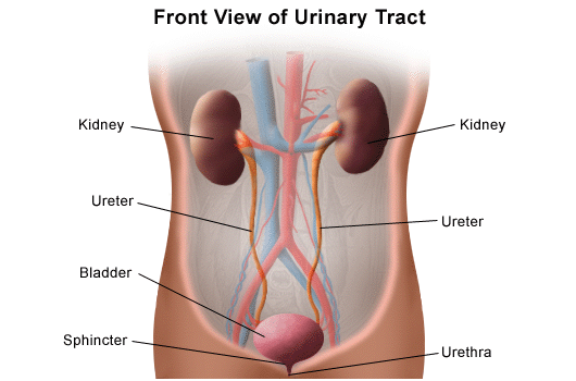
The Urinary System
The urinary system is made up of 4 specific organs: The 1. Kidney, 2. Ureters, 3. Urinary Bladder, 4. Urethra
The Kidneys (Renal)- Composition
The kidneys filter the blood as well as produce urine. They are 2 “bean-shaped organs” located in the lower back, just above the pelvis.
The Convex side (the outer curve part of a bean) faces laterally (away from the origin)
The Concave side (the inner curve of a bean) faces medially (close to the origin)
Renal Hilum- is the indent (or the double curve on the concave side of the bean) there are the exit and entry sites for structures of the kidney; renal vessels (the renal vein and artery), as well as the renal nerve and ureter to enter the kidney.
Renal capsule- Tough fibrous layer surrounding the kidney
Sections of The Kidneys (Inner Structures)
There are 3 regions in the kidney: Renal cortex, Renal medulla, and Renal pelvis.
Renal Cortex- the frontal section through the kidney that reveals an outer region. It is a smooth, outer portion of the kidney that contains blood vessels and collecting ducts.
Renal Medulla- the inner region of the kidney, contains Renal Pyramids, which are cone-shaped tissue that contains Tubules for the transportation of urine and Nephrons.
Renal Pelvis- emerges from the hilum, located above the ureter. It has cup-like calyces that collect urine before sending it to the urinary bladder.
Renal Column- located between renal pyramids. They are extensions of the renal cortex. Support and separate Renal pyramids
Renal Calyces- cup-shaped structures in the kidney that collect and channel urine through
Can be Minor (small) or Major (large) based on the picture
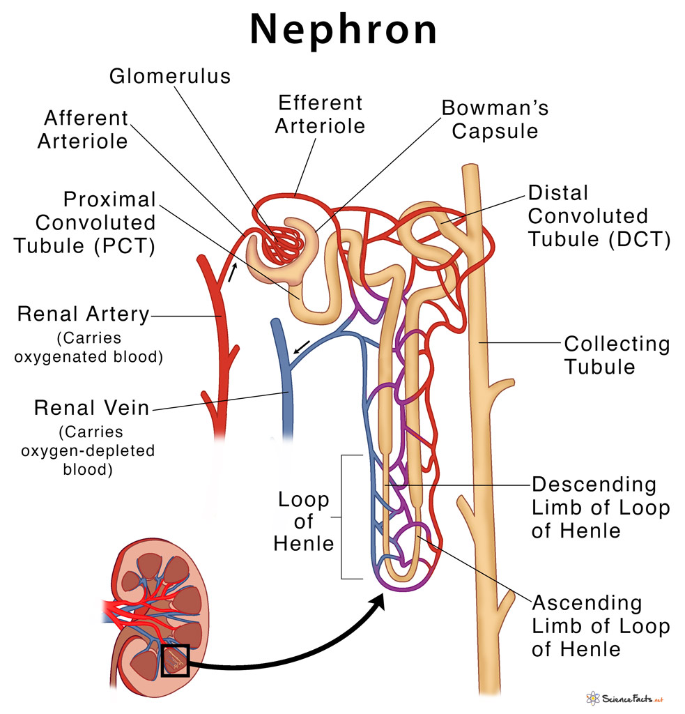
Nephrons
Both the Medulla and Cortex contain the nephrons.
They are the “functional units” of the kidney; they do the kidney’s job of:
Cleaning/ filtering the blood,
Selectively reabsorbs nutrients the body needs into the blood and secretes the ones we don't need or have too much of into the urine.
To balance the plasma to homeostatic set points
They do this in 3 principal functions: Filtration, reabsorption, and secretion.
Consists of 2 components, the Tubular Component (Renal Tubule), and the Vascular component (Renal Vasculature)
Tubular Component (Renal Tubule)
This component is responsible for the further filtration of the blood after being filtered by the glomeruli within the Bowman’s capsule. In other words, the second filtration
Consists of:
Bowman's Capsule- a cup-like shape that forms around the glomerulus, its purpose is to filter blood
Proximal Convoluted Tubule (PCT)- absorbs all the glucose and amino acids that we need
Distal Convoluted Tubule (DCT)- determines the amount of reabsorption based on what the body needs. If there are no hormones present, the DCT is impermeable,
Loop of Henle- has 2 parts, ascending and descending limbs, the major site of salt/ water balance in the body.
Ascending limb- only salts are absorbed here, part of the loop of Henle
Descending limb- only water is absorbed here, part of the loop of Henle
Collecting Tubule- a microscopic tube that carries urine from the nephrons to the renal pelvis and ureters
Vascular Component (Renal Vasculature)
This Component focuses on the filtration and the movement of absorption and secretion of blood filtering
Consists of:
Renal Artery- the first step in blood filtration, the site where blood enters to be filtered, connected with the kidney, blood enters through this, and the renal artery branches into afferent arterioles
Afferent Arterioles- branches from the renal artery in the kidney, they carry blood to the filtration site
Efferent Arterioles- this drives the filtration, making pressure to drive the blood through filtration from pressure, because the efferent is smaller than the afferent arteriole.
Glomerulus- the area where blood is filtered the most, a simple capillary bed formed by a branching afferent arteriole, filtered here, and everything is forced into the tubular Bowman’s capsule
Peritubular Capillary Bed- a network of tiny blood vessels in the kidney that filter waste from the blood and reabsorb nutrients, and excrete the useless ones
Renal Vein- the main blood vessel that carries blood from the kidney and ureter to the heart from the lower part of the body. Each kidney has 1
Ureters
From the renal hilum, the renal pelvis creates a narrow tube, which, being the ureter, its purpose is to transfer urine into the bladder. Each kidney has a ureter that connects to it via the renal pelvis. 10-12” long
Urinary Bladder
From the end of the ureters is the bladder. Where urine is delivered to, called a “dome and hollow”. In females, the bladder is anterior to the uterus, while posterior to the pubic bone and anterior to the rectum during late pregnancy. Since it's meant to hold urine until it's voluntarily let out, the “dome” expands when it's filled with urine.
Has 2 sphincter muscles that control the release of waste: Internal, External
Internal Sphincter- Involuntary, smooth muscle
External Sphincter- Voluntary control
Can hold 400mL (Normal Capacity)
Urethra
The end stage of the urinary system, where urine is transported out of the body for disposal. Vagina for women, penis for men. Connected to the bladder that releases waste.
Urinary meatus
External opening of the urethra to the outside of the body. Where waste comes out of.
The Excretory System (Urinary included) has 3 steps: Filtration, Reabsorption, Secretion. (and Excretion)
Filtration
Parts used- Glomerulus, Renal Artery (Afferent and Efferent Arterioles), Bowman’s capsule.
Blood enters the kidney via the Renal Artery and begins filtration at the Glomerulus.
The renal artery branches into afferent arterioles that carry the blood to the glomerulus
At the glomerulus, blood gets filtered, and everything gets forced into the Bowman's capsule via pressure from the efferent arterioles.
Back-up blood is released, which releases the pressure, and then the substances go to the tubule. In there, blood takes back all substances that the body doesn't want excreted as urine; they move from the tubule into the blood. Substances like water, ions, glucose, and amino acids
However, substances like electrolytes, drugs, excess vitamins, salts, and sugars are what the body doesn't need, so they are secreted in the urine. They move from the blood to the tubule. This is called Tubular Secretion.
Reabsorption
Parts used: PCT, Loop of Henle (Ascending, Descending limbs), DCT, collecting ducts
Most water is recovered in the PCT, loop of Henle, DCT, and 10% reaches the collecting ducts
Reabsorption and secretion are performed before the urine becomes urine
Reappropriation is taking the “good stuff”
First, the PCT will absorb all the glucose and amino acids the body needs
While the loop of Henle absorbs water (in the descending limb) and salt (in the ascending limb)
The DCT measures how much reabsorption the body needs to do.
2 things can happen, it can stop reabsorption or continue (to certain substances)
If there are no Hormones present, the DCT becomes impermeable, and nothing else can get through
If Hormone *ADH* is present, the DCT becomes permeable to water, which will move into the blood
If Hormone *Aldosterone* is present, the DCT becomes permeable to salts, which will move into the blood
Aldosterone- Regulates the balance of water and electrolytes in the body.
Encouraged the kidney to excrete potassium into the urine and retain sodium, thereby retaining water.
1 of 2 Hormones that control urine production
ADH (Antidiuretic hormone)- regulates water balance in the body by controlling how much water is excreted by the kidneys, leading to water retention and potentially higher blood pressure
1 of 2 Hormones that control urine production
Secretion
Parts involved: Tubule
Secretion occurs in the entire length of the tubule.
Excess materials we don't need or have enough of are moved into the tubule and finally made into urine.
The urine makes its way to the bladder via the ureters and into the bladder, where it's stored until it’s released from the body.
Excretion (Not part of the Urine formation)
Process of removing nitrogenous waste material, salts, and excess water from blood.
If not, toxic waste will build up and cells will suffocate and poison themselves.
Blood Pressure *NOT NEEDED.
The excretory system also monitors blood pressure; if the blood pressure is low, then blood cannot be transported to the Bowman’s capsule.
The kidneys monitor blood pressure with something called the juxtaglomerular apparatus.
If the blood pressure is low or the chlorine ion concentration is low, the Juxtaglomerular gets triggered secretion of an enzyme, “Renin” (maybe needed)
When renin mixes with blood, it triggers the release of angiotensin II, which causes Vasoconstriction (blood vessels get smaller), which will make the blood pressure increase.
Increased blood pressure stimulates the adrenal cortex to produce aldosterone.
This also stimulates the reabsorption of salt, which will make the body reabsorb water and increase blood pressure.
Normal and Abnormal Urine Characteristics
Dilute Urine- Urine that has a higher than average water content.
Diuretics- drugs that increase the passing of urine. (caffeine)
Average urinary output: 1,500 mL per day.
What constitutes an abnormal urinalysis: Color, UTI, Increased levels of protein, Odor
Abnormalities of Urine:
Increased level of protein (small amounts ok)
Sugar: normal levels too low to detect. Any amount of sugar that is detected calls for follow-up testing for diabetes.
Ketones: like diabetes, any ketones in urine could be a sign of diabetes.
Bilirubin: product of RBC breakdown. May indicate liver damage or disease.
Evidence of infection: if leukocytes present in urine, may indicate UTI.
Blood: could be a sign of kidney damage, infection, kidney/bladder stones, kidney or bladder cancer, or blood disorders.
Bacteria/yeasts: infection
Crystals: kidney stones
Infections and Diseases
Urinary Tract Infection (UTI)- infection and inflammation of the urethra and sometimes even the urinary bladder.
Much more common in women, due to their short urethra and located close to the anus, bacteria that are harmless in the GI tract can cause more problems in the urinary tract.
Cause pain and burning urination, frequent, urgent urination, cloudy or bloody urine, fever, and weakness
Cystitis- an inflammation of the urinary bladder. Can be caused by fecal bacteria entering the urethra
Usually brings fever, frequency, urgent, burning urination, bacteria, blood, pus, and/ or leukocytes in the urine.
May have lumbar or medial thigh pain and tenderness above the pubic bone
Incontinence- inappropriate, involuntary passage of urine. (Enuresis- Bedwetting. Occurs between 5-6 Yrs)
Stress incontinence- happens when coughing or straining. Very common after childbirth
Genuine stress incontinence- when a specific physical cause of the incontinence can be identified in women
Overflow incontinence- occurs in older men when a full bladder leaks
Urge incontinence- the leakage of urine that accompanies an intense desire to urinate with failure of restraint
Kidney Stones (Renal Calculi)- form in the renal pelvis when the urine contains excess calcium, uric acid, magnesium, etc. Usually small (3mm in diameter)
Often, no symptoms occur until the stone begins to move down the ureter, which can cause abdominal and groin pain, nausea, and vomiting.
Can be passed through the urethra on their own (with excessive pain). Larger ones may need to be surgically removed.
Glomerulonephritis- a rare inflammation of the glomeruli, which makes it stop functioning properly
Dark or rust colored urine, foamy urine, or bloody urine are common with glomerulonephritis.
Later symptoms include renal failure, systemic edema, fatigue, headaches, decreased urine output, and discoloration of the skin.
Pyelonephritis- an inflammation of the nephrons, typically due to an infection in the kidneys
Can be accompanied by cystitis, back pain, fever, chills, nausea, and vomiting
Can be cleared up with antibiotics
Renal Failure- when a kidney does not function properly
A LIFE threatening condition
Causes reduced urine output, systemic edema, and changes in mental state (due to toxins in the blood building up)
Treatments
Humans can live with only one kidney, most are born with one kidney. However, if one malfunctions, it needs to be looked at immediately.
2 types of treatment: Transplant surgery, or Dialysis
Transplant surgery- where a person receives a new kidney. 2 types of transplant surgery: living donor and non-living donor
Living Donor- whose tissue matches the patient, a living person willingly donates 1 kidney.
Non-living donor- someone who has died and agreed to donate their organs (kidneys, for example) to help others
Dialysis- performs the job of healthy kidneys by removing waste, controlling blood pressure, and maintaining homeostasis.
Can be done internally or externally. (In the Kidneys or a Machine [Hemodialysis])
This cannot cure kidney disease. Depending on the severity of the disease, dialysis may have to continue until the end of life (forever
For Kidney stones, during severe cases there are multiple ways to remove/ pass kidney stone(s):
Percutaneous nephrolithotomy- removes kidney stones through small incisions in the back. (Treatment)
Urestroscopic stone removal- uses a scope inserted through the urethra to access and remove stones in the ureter or kidney. (Treatment)
Lithotripsy- shock waves that break down stones to passable sizes (Treatment)
Module 13 - Reproductive System
Men and women structure and functions.
Gonad- the gender specific reproductive organs that produce sex cells, and their specific hormones.
Male, gonads are “Testes” that produce sperm and testosterone.
Female, gonads are “Ovaries” that produce eggs and estrogen.
Sex cells- sperm, eggs. They contain 23 chromosomes (half of body chromo.S./ somatic cells) meaning they are Haploid cells. And are produced during Meiosis (sex cell production)
Once a sperm cell reaches a egg they combine making them 46 chromosomes and then Meiosis begins and development starts
Reproductive System (Male)
The male reproductive system consists of the Testes and Penis.
Scrotum- highly pigmented, muscular sack that extends from the body behind the penis and covers the testes.
Control the temperature of the testes by contracting. Cold- shrink, Hot- relax
Testes- the male “Gonads”. The reproductive organs that produce Sperm and Androgens (group of hormones) like Testosterone.
Males have 2
About size of an egg
Seminiferous Tubules- form the bulk of each testes. Composed of developing sperm cells and where formed sperm are released into the duct system
The production, maintaindence, storage site of sperm
Each testis contains 1-4 minute, convoluted tubules
Supported by a type of tissue (interstitial tissue) which produces testosterone
Epididymis- coiled tube attached to the posterior and lateral part of the testes where newly formed sperm continue to mature.
Stores and transports sperm from the testes
Sperm- Male gamete,74 days to mature
Fertilize the egg and transmits information
Vas Deferens- a thick muscular tube that connects testes with urethra and carries sperm out of testes
Storage site and excretory duct of testis
Vasectomy- vas deferens is cut to produce sterility (permanent birth control)
Pathway of Sperm: testes→ epididymis→ vas deferens→ ejaculatory duct→ urethra.
What causes lower sperm count/ poor function: smoking, alcohol, stress, overweight, meds, disorders