Male Anatomy and Physiology
The primary purpose of the male reproductive system in mammals is to deliver the male’s genetic contribution to the offspring, in the form of spermatozoa, to the female tract in hopes to result in a conception
Male Reproductive tract three main divisions:
primary sex organs
accessory glands and ducts
external genitalia
“The male reproductive system is just as intensely regulate by hormones as the female system”
What every producer should know:
understand structure and function of male reproductive tract, to understand how it can impact their business
understand the process of making spermatozoa, or sperm, and what characteristics to look for when evaluating an animal for fertility
Fertility of a herd sire can have significant impact on the success of a breeding operation
Quality of sire, from a genetics standpoint and a reproductive soundness standpoint is as critical to a successful operation as assuring that dams quality
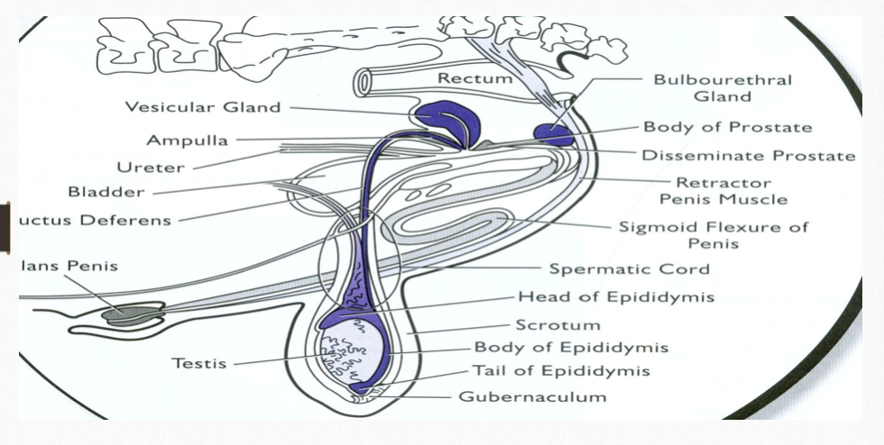
The Male Primary Sex Organ
primary sex organ of the female is the ovary; primary sex organ of the male is the testicle
Testes: paired structure that produces steroid hormones and sperm
located outside of the body cavity, as a male animal grows, the testes move from near the kidneys through the inguinal canals into the scrotum
Scrotum: lobed structures that support testes, temperature control, protection
supports, protects, and regulates temperature
Inguinal canals: the two passages in the anterior abdominal wall which in males convey the spermatic cords
The descent of the testes is caused by a ligament that extends through the inguinal canals and attached to the tail of the epididymus
Descent regulated by androgen hormones and testicular factors
Cryptorchidism: failure of one or both testes to descend into the scrotum (sometimes the testes fail to descend into the scrotum due to a defect in development)
Bilateral cryptorchidism: neither testes has descended, and the animal is infertile
Unilateral cryptorchidism: where only one testis has descended, and the animal is fertile from the descended testes
Genetic component to cryptorchidism, animal’s offspring are likely to be cryptorchidic
Functional Structure of the Testes
the size of the testes is species-dependent
the testes of livestock species and humans are structurally similar
the testes are covered by the tunica vaginalis
Tunica Vaginalis: a serous tissue that is an extension of the peritoneum obtained when the testes descended into the scrotum during development
Tunica Albuginea: the outermost layer of the testes proper is a thin white membrane of elastic connective tissue
Parenchyma: the yellowish functional layer of the testes, divided into segments by connective tissue
within these segments are the seminiferous tubules
site of spermatogenesis
makes up 80% of testis weight
Functional layer of the testes
brownish/red in boars
yellowish/orange in bulls
Seminiferous tubules: Site of spermatogenesis, Makes up 80% testis weight, long, estimated to be between 3 and 5 kilometers, convoluted tubes, lined with Sertoli cells.
germ cells (spermatogonia): eventually become sperm
sertoli cells (nurse cells): are responsible for forming the blood–testes barrier by the tight junctions between Sertoli cells as they line the basement membrane.
Isolate developing sperm from the immune system, otherwise sperm as foreign to body and be and destroyed.
provide nutrients for growing sperm
produces androgen binding protein and inhibin
Leydig cells (interstitial cells): produce testosterone
Center of the seminiferous tubules is a lumen.
fully formed sperm are released, flow to a network of tubes that collects sperm called the rete testis.
The rete testis connects to the efferent ducts, which connects to the head of the epididymis
Temperature control
testes need to be 4o to 10o F lower than normal body temperature (~91o to 97oF)
exposure to body temperature makes cryptorchids sterile
Hot & cold weather
Sweat and sebaceous glands
Pampiniform plexus
Arterial blood is cooled by venous blood
Venous blood is warmed by arterial blood
In hot weather, the spermatic cord lengthens providing more surface area for heat exchange
The male reproductive system has several methods to help maintain the temperature of the testis.
The scrotum: visible from the outside, two-lobed sack that encloses the testes, located in the inguinal region between the hind legs of most animals.
same embryonic tissue as the labia majora in the female
The outer most layer is thick skin covered with sweat, and sebaceous glands allow for evaporative cooling of the testes.
Under the outer layer is a layer of smooth muscle called the tunica dartos: expand and contract, cooling or warming in the testes, splits the scrotum into two pouches, and is attached to the tunica vaginalis at the bottom of the pouches to keep the testes in position in the scrotum
The spermatic cord: Paired structure, that connects the testes to the rest of the body with connective tissue and muscles.
Connects the testes to the urethra
Supports testes
Temperature regulation
Carries nerve and blood supply to testes
The cremaster muscles: a muscle that covers the testis and the spermatic cord.
The pampiniform plexus
Contained in the spermatic cord.
Arterial blood is cooled by venous blood
Venous blood is warmed by arterial blood
In hot weather, the spermatic cord lengthens providing more surface area for heat exchange
The spermatic cord contains the pampiniform plexus. A network of arteries and veins that helps to cool blood before entering the testes, this helps to maintain the testes at approximately 2°C to 5°C (4°F–10°F) below body temperature.
Sperm and Semen Transportation
once sperm cells are made in the testes they are not yet ready to fertilize an oocyte
undergo concecntration
maturation processes
be transported from the testes through the rest of the male reproductive tract
need fluids for survival to create semen
Epididymus
divided into three parts;
capcut to epididymus (head)
closer to testes
corpus epididymus (body)
cauda epididymus (tail)
farthest from testes
larger lumen than any other portion of the epididymus due to its role in storing mature sperm cells prior to ejaculation
The efferent ducts come together merge together near the top of the testes where 12 to 15 ducts merge together into a single duct, the epididymis.
A paired structure , first external duct that connects the testes to the outside of the male body.
Long, convoluted tube that measures around 34 meters in the bull!! longer in rams, boars, and stallions!!!
The three layers: a tunica serosa comprising the outer most layer, followed by a smooth muscle layer, and the innermost layer is an epithelial layer
Three sections:
1) caput epididymis: first section, “head”, a flattened portion at the top of the testis where the efferent ducts empty into a single duct.
2) corpus epididymis: the middle portion, “body”
3) Cauda epididymis: final portion, “tail” , has a larger lumen than any other portion of the epididymis due to its role in storing mature sperm cells.
Three major functions :
1) transportation of sperm
2) maturation of sperm
3) concentration and storage of sperm.
Sperm cells spend between 9 and 15 days in the epididymis; species-dependent.
May be shortened by 10% to 20% with frequent ejaculation.
Transport: several factors affect the movement of sperm through the epididymus
Pressure: production of sperm, as new sperm are formed in the seminiferous tubules theya re forced through the rete testis and efferent ducts and on into the epididymus, thus pushing older sperm through the epididymus
“pressure of perm production will force previously made sperm through the epididymus”