AMYGDALA PODCAST (need to study)
What the amygdala is
The amygdala is a large nuclear complex located in the anterior part of the medial temporal lobe, lying just anterior to the hippocampus.
It is not a single nucleus, but a collection of ~13 subnuclei
These nuclei are grouped into three major functional regions
It sits next to the temporal horn of the lateral ventricle, which makes it easy to identify anatomically
Because of its position, it is ideally placed to receive:
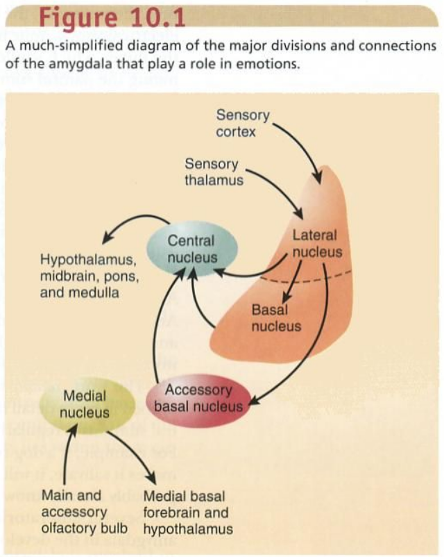
Sensory information from cortex and thalamus
Contextual information from hippocampus
Modulatory input from brainstem and basal forebrain
In short: the amygdala is a hub linking sensory input, memory, and physiological output systems.
Major functional regions (overview)
Although there are many subnuclei, they are usually organised into three functional groups:
Basolateral complex
Main input region
Receives information from cortex and thalamus
Cortical nucleus
Receives olfactory input
Evolutionarily older
Centromedial group
Main output region
Projects to hypothalamus and brainstem
This organisation explains how the amygdala can receive information, process it internally, and generate outputs.
What the amygdala does (core idea)
The amygdala’s central role is to evaluate the emotional significance of events.
It asks: Is this relevant? Is it important? Is it potentially dangerous or rewarding?
It is especially responsive to threatening, fearful, or salient stimuli
However, it is involved in both negative and positive emotional valence
It does not generate emotions on its own; instead, it biases perception, attention, learning, and bodily responses.
The amygdala as a “danger detector”
The amygdala is often described as a danger detector, but this is a simplification with a useful anatomical basis.
When activated, it can orchestrate:
Somatic responses
Freezing, startle, muscle tension
Autonomic responses
Heart rate, sweating, blood pressure
Endocrine responses
Via hypothalamic activation
Its outputs are fast because they do not require full cortical processing. This allows rapid responses when time matters.
Role in attention and perception
The amygdala does not just react — it feeds back to cortex.
Enhances attention toward emotionally salient stimuli
Biases sensory cortex to prioritise certain inputs
Influences prefrontal regions involved in evaluation and decision-making
This is why emotionally charged stimuli are hard to ignore.
Role in social behaviour
The amygdala plays a major role in social cognition, particularly in interpreting others.
Contributes to reading:
Facial expressions
Eye gaze
Body language
Important for detecting social threat, trustworthiness, and relevance
Evidence:
Lesions in primates lead to markedly abnormal social interactions
Human imaging and lesion studies show altered social perception when amygdala function is disrupted
Role in learning and memory
The amygdala is tightly linked to memory systems, especially the hippocampus.
Critical for fear conditioning
Enhances storage of emotionally significant memories
Does not store episodic memories itself, but modulates how strongly they are encoded
This explains why emotionally intense events are remembered more vividly.
1. Where the amygdala sits (spatial logic)
The amygdala is a collection of nuclei buried in the anteromedial temporal lobe.
Lies just anterior to the hippocampal head
Forms part of the roof and anterior wall of the temporal horn of the lateral ventricle
Because of this position, it is ideally placed to:
Receive sensory information from cortex and thalamus
Communicate easily with hippocampus, hypothalamus, and frontal cortex
Think of it as a junction box between sensory cortex, memory structures, and output systems.
2. Nuclear organisation (this explains everything else)
The amygdala is not one structure functionally. Connectivity follows nuclear divisions.
A. Basolateral complex
(lateral, basal, accessory basal nuclei)
Largest part of the amygdala
Main input region
Receives highly processed information from:
Cerebral cortex
Thalamus
Sends outputs:
To other amygdala nuclei
Back to cortex
This group handles detailed sensory representations.
B. Cortical nucleus
Small, phylogenetically older region
Receives direct olfactory input
Closely connected to:
Piriform cortex
Entorhinal cortex
This explains why smell has privileged access to emotional processing.
C. Centromedial group
(central + medial nuclei)
Main output nuclei
Poorly connected to cortex
Strong connections to:
Hypothalamus
Brainstem
Controls downstream responses via long projection systems
This group converts amygdala processing into bodily and behavioural outputs.
3. Afferent connections (inputs to the amygdala)
A. Brainstem inputs (diffuse modulatory systems)
Origin:
Locus coeruleus → noradrenaline
Raphe nuclei → serotonin
Ventral tegmental area → dopamine
Characteristics:
Broad, diffuse projections
Target multiple amygdala nuclei
Modulate excitability rather than carrying specific sensory content
These inputs adjust gain and arousal, not detailed information.
B. Basal forebrain
Cholinergic (acetylcholine) projections
Widely distributed across the amygdala
Particularly strong to basolateral nuclei
This input enhances attention and learning-related plasticity.
C. Olfactory input (special case)
Olfactory bulb and tract
Projects directly to the cortical nucleus
No thalamic relay
This is unique among sensory systems and explains why odours can evoke strong emotional responses very rapidly.
D. Thalamic inputs (two pathways)
1. Direct thalamic route
Sensory thalamic nuclei (e.g. pulvinar)
Projects directly to the basolateral amygdala
Fast and coarse
2. Cortical route
Thalamus → primary sensory cortex → association cortex → amygdala
Much more detailed and slower
Both routes converge on the basolateral complex.
E. Cortical inputs (largest source)
Major cortical contributors:
Anterior cingulate cortex
Orbitofrontal and medial prefrontal cortex
Anterior insula
Lateral temporal association cortex
Entorhinal and perirhinal cortices
Key point:
These inputs terminate mainly in the basolateral nuclei
They carry processed, contextual, and multimodal information
4. Intra-amygdalar flow (internal logic)
There is a consistent internal direction:
Cortex / thalamus → basolateral complex → central nucleus
The basolateral nuclei integrate information, then influence the central nucleus, which handles outputs.
5. Efferent connections (outputs from the amygdala)
A. Stria terminalis
Long, C-shaped fibre bundle
Origin: central and medial nuclei
Course:
Follows the lateral ventricle
Arches around the thalamus
Targets:
Septal area
Hypothalamus
Bed nucleus of the stria terminalis (BNST)
This pathway is slow, sustained, and modulatory.
B. Ventral amygdalofugal pathway
Shorter, more direct route
Origin: basolateral and central nuclei
Passes through basal forebrain
Targets:
Hypothalamus
Thalamus
Brainstem
Prefrontal cortex
This is a fast, direct output pathway.
C. Cortical efferents
Amygdala projects back to:
Medial and lateral prefrontal cortex
Anterior cingulate cortex
Insula
Occipito-temporal association cortex
These projections arise mainly from the basolateral complex and allow emotional information to influence cortical processing.
6. Clean wiring summary (memorise this)
Inputs in → basolateral nuclei
Olfaction → cortical nucleus
Integration → basolateral complex
Outputs out → central nucleus
Exit routes → stria terminalis + ventral amygdalofugal pathway
Worked example: spiders 🕷
Imagine you see a spider on the wall.
Visual information reaches visual cortex and, in parallel, a fast thalamic route
This information converges on the basolateral amygdala
The amygdala evaluates the stimulus as emotionally salient
The central nucleus activates output pathways:
Hypothalamus → autonomic changes (heart rate, sweating)
Brainstem → startle and freezing responses
Feedback projections bias:
Attention toward the spider
Prefrontal cortex toward avoidance behaviour
The hippocampus helps link this moment to past experiences with spiders
By the time you consciously think “that’s a spider”, your body is already reacting. The wiring explains the speed.