Neural Circuits and Neural Networks
1/42
There's no tags or description
Looks like no tags are added yet.
Name | Mastery | Learn | Test | Matching | Spaced |
|---|
No study sessions yet.
43 Terms
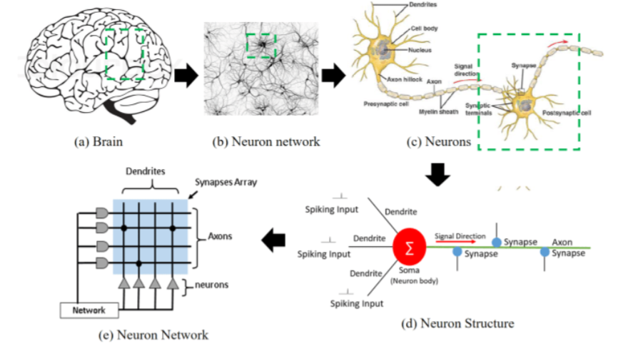
Overview of Neural Networks
Brain → Neuron network → neurons → neuron structure → neuron network
Neural Circuits
A population of neurons interconnected by synapses to carry out a specific function when activated. Neural circuits help us collect information, interpret it, and formulate an appropriate behavioral response.
Brain networks
Multiple neural circuits are interconnected with one another
Two key features of neural circuits
Connections between an identified group of neurons. The capacity to process a distinct type of information.
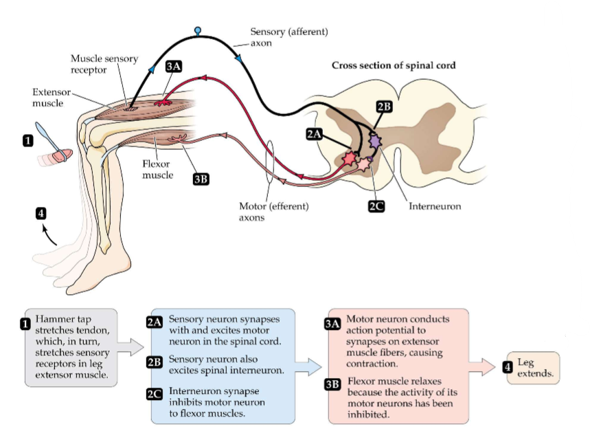
The myotatic, or knee-jerk, reflex is controlled by a simple neural circuit
Pathway A: Hammer tap causes muscle to stretch and activate stretch/sensory receptors in the muscle spindle. APs of the innervating sensory neurons travel to spinal cord and cause NT release at synapse between sensory neuron and motor neuron. Motor neuron becomes depolarized, AP travels down axon, causing a contraction and your leg jerks forward.
Information Processing in our brains requires us to ask the following:
1.What information is being represented? 2. How is the information encoded? the brain uses coding schemes. 3. How is the information decoded? decoded by a receiver - neuron or circuit. (how does brain actually interpret it) 4. What are the computations? The rules that determine how input representations are transformed to output representations. A representation must be transformed in a meaningful way. (not a single answer)
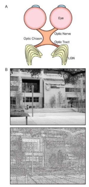
Neural Representation: Visual Cortex
For information to be processed by the brain the information must be represented in the brain. Example: Since the lateral geniculate nucleus (LGN) of the thalamus is anatomically connected to the eyes then the LGN represents visual information.
Neural Representation: Topographic Maps -The homunculus; encoding: place code
Information can also be represented in topographical maps of a particular stimulus. Information about the value of a stimulus may be encoded by anatomical location of the neuron within the representation - PLACE CODE
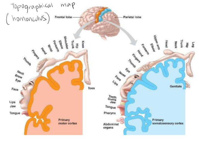
Place code
information represented by the location within a structure
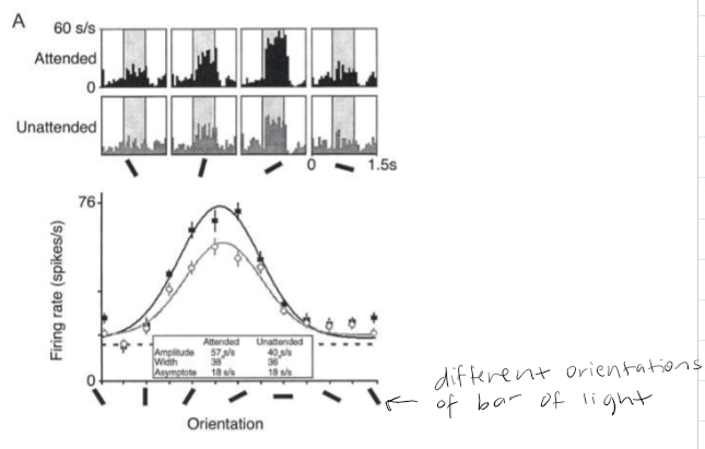
Encoding: Rate Code
The value of the attribute being represented by a neuron is encoded by the firing rate of the neuron measured over some discrete time interval. Example: Visual cortex - the orientation of the bar of light is encoded by the firing rate of the neuron. Neurons in visual cortex tend to be ‘tuned’ to a particular orientation.
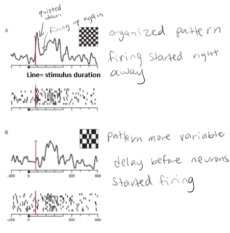
Encoding: Temporal Codes
Uses the timing of neural activity to encode information about a stimulus. Example in temporal cortex of primates - checkerboard like stimuli. (A) the cell fired an initial transient burst, was quieter for ~100 ms then fired again. (B) same cell, different pattern. For pattern B the cell did not produce the initial transient burst but instead fired only during the second interval after a delay of ~100 ms relative to pattern A.
Decoding
Downstream neurons that receive action potentials from upstream neurons must decode that signal to perform processing and create a new representation, which must be encoded into the firing of that neuron. In other words, how a neuron interprets the responses of its afferents to determine what original stimulus generated those responses.
Encoding and decoding are complementary operations
Encoding uses stimuli to predict activity while decoding uses activity to predict information about stimuli.
What are iconic neural circuits?
Iconic neural circuits are what create the larger neural networks. Green neurons are excitatory and red neurons are inhibitory.
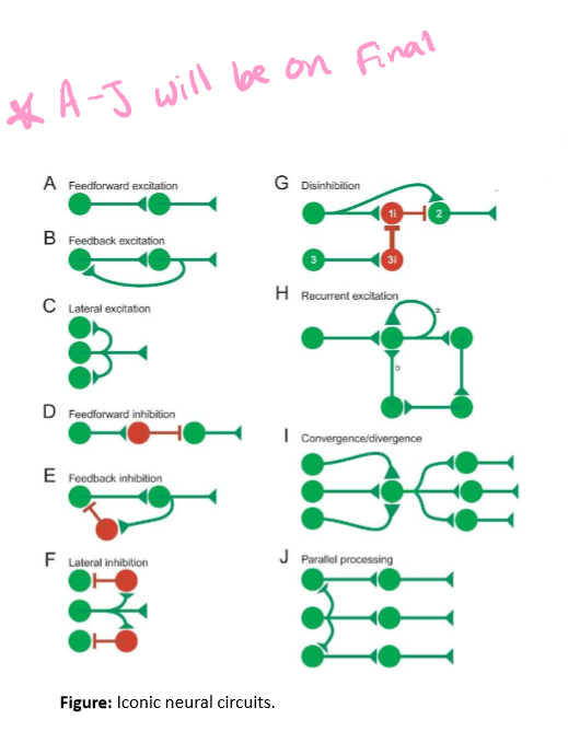
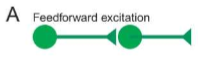
Feedforward excitation
The stretch reflex. Sensory neuron makes synapse with motor neuron (A). One neuron excites the next neuron Ex. glutamate
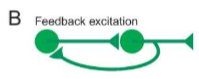
Feedback excitation
A connection that goes from a higher order neuron to a lower order neuron (B). Allows output of a circuit to influence input neurons. A excites B and B gives feedback to A (stimulating A) creating loop.
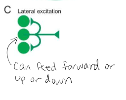
Lateral Excitation
Neurons within a level can communicate through both local and long-range lateral connections (C). Allows for computation to occur within a particular level of a circuit before output is transmitted to the next level via feedforward or feedback connections.
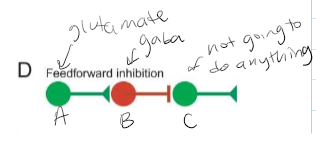
Feedforward inhibition
When the output of one level of a circuit requires the activity of the next level to be decreased (D). A synapse on B causing the next level to decrease then neuron C is not going to be activated
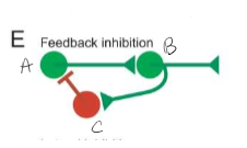
Feedback inhibition
The output of a higher-order level of a circuit is needed to shut down the input activity to that circuit (E). A excites B then B can either excite another neuron or send inhibitory signal to A shutting down activity. Can do both send signal forward and send inhibitory signal to A
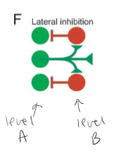
Lateral inhibition
The activity of one set of neurons can shut down the activity of other neurons at the same level. (F). Levels go from left to right
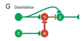
Disinhibition
A neuron may receive excitatory and inhibitory inputs but may be silent because the inhibitory input is stronger than the excitatory input. However, if the inhibitory interneuron (G1i) is inhibited by another neuron (G3i) the excitatory input then causes the neuron to fire-disinhibition of neuron 2. Get multiple inputs on one neuron
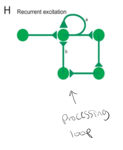
Recurrent Connections
Hybrid between feedforward, feedback, and lateral circuitry. The output neurons make connections with themselves (Ha). Chains of feedforward connections loop backward and eventually connect back onto the initial processing stages, forming a processing loop (Hb)
Convergence
The degree to which a neuron receives input from large numbers of other neurons. Multiple neurons synapsing on the same neuron.
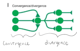
Divergence
The degree to which a neuron projects to a large number of target neurons. One neuron synapsing on multiple neurons

Parallel Processing
Can be contrasted to a serial computer in which the execution of each line of code must be complete before the next line of code can be executed.
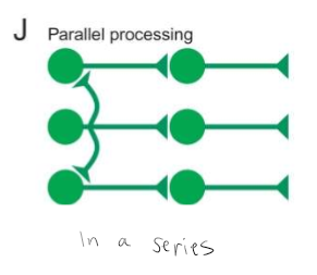
The brain works more like a parallel processor in which many lines of code are executed simultaneously.
Occurs in all sensory and motor systems of the brain. Allows a task to be divided and processed with specialized circuitry appropriate for the task. Example: visual system - info processed first by retina but then simultaneously by the LGN visual cortex and then higher cortical areas to leave you with a perception of what you see. Need all these circuits to go in order to process what you see in front of you.
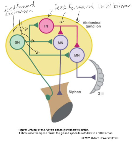
Example: Aplysia
The circuit contains feedforward excitation AND inhibition. Similar to the stretch reflex in humans. ~24 sensory receptor neurons innervate the siphon. These sensory receptor neurons synapse on motor neurons that innervate the gill and siphon. When stimulated both the gill and siphon withdraw. However there are also the inhibitory interneurons that modulate this circuits activity. SN = Sensory neuron, IN = interneuron, MN = motor neuron.
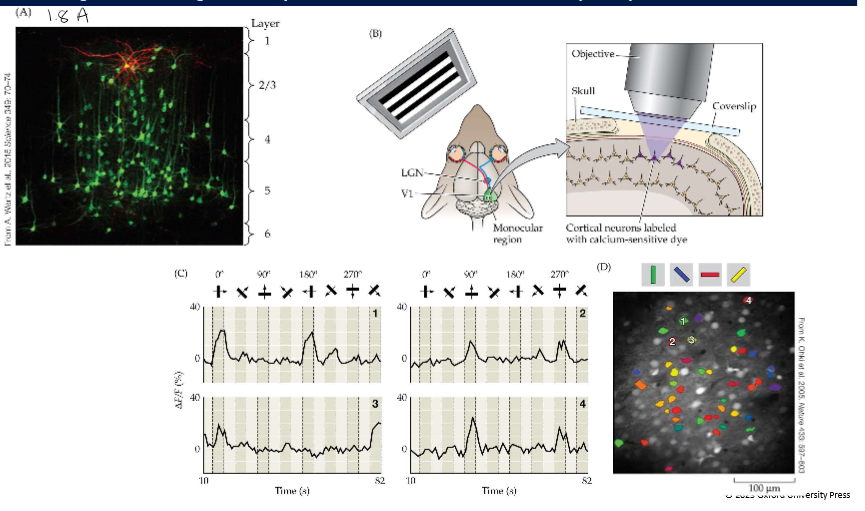
Visualizing and analyzing neural circuits
Genetically engineered virus to visualize pre- and post-synaptic targets. (Image 1.8A) Ca2+ imaging: using Ca2+ dye labels.(image 1.8 B-D)
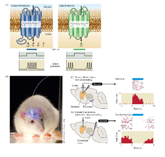
Optogenetics
A technique for controlling the communication between neurons. Genes for light-sensitive proteins (e.g. opsins) are introduced into certain neurons to monitor and control their activity in response to light signals → chemical signals
Neural Systems: Circuits that process related information constitute a neural system
Organization of individual neural circuits according to the type of information they carry into broader networks and larger volumes so that more complex behaviors can be generated. Unity of function. Neural systems are organized into representations of information that are processed in parallel pathways, culminating in topographic maps. Additional information processing based on time or order of input: Computational map.
Functional analysis of neural systems
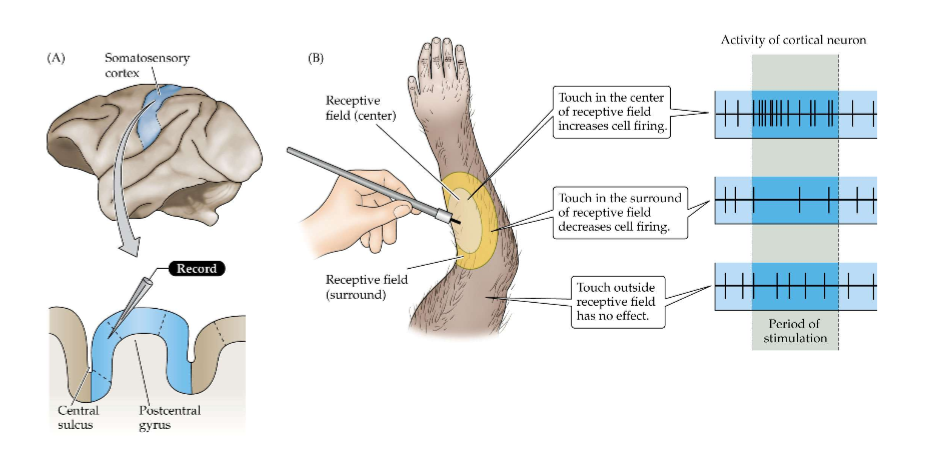
Determine what neurons do from moment to moment in a neural system. Receptive field. Using single-unit recording, it is possible to find the relationships between peripheral stimuli and neuronal responses.
Receptive field
The region in sensory space to which a neuron will respond
Defining relationships between peripheral stimuli and neuronal responses using single-unit recording
Neural Circuits and system can be analyzed in the human brain
Used for discerning various anatomical structures. Used for diagnoses of medical problems, such as tumor or stroke. Used to characterize the activity of the brain as it performs complex tasks such as problem solving, recall, speaking, singing, and so much more.
Minimally invasive analysis of human brain function
Electroencephalogram (EEG) and transcranial magnetic stimulation
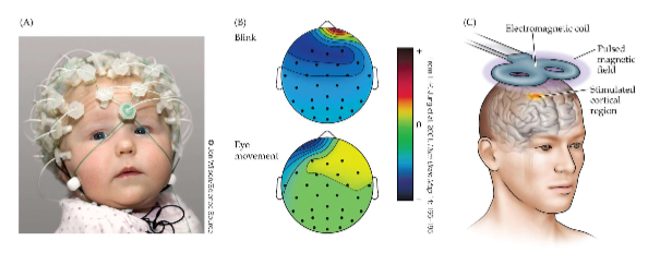
Electroencephalogram (EEG)
Can map brain according to event-related potentials (ERPs)
Transcranial Magnetic Stimulation
Used to correlate broad areas of the brain with ongoing behavior; used for neurologic and psychiatric conditions
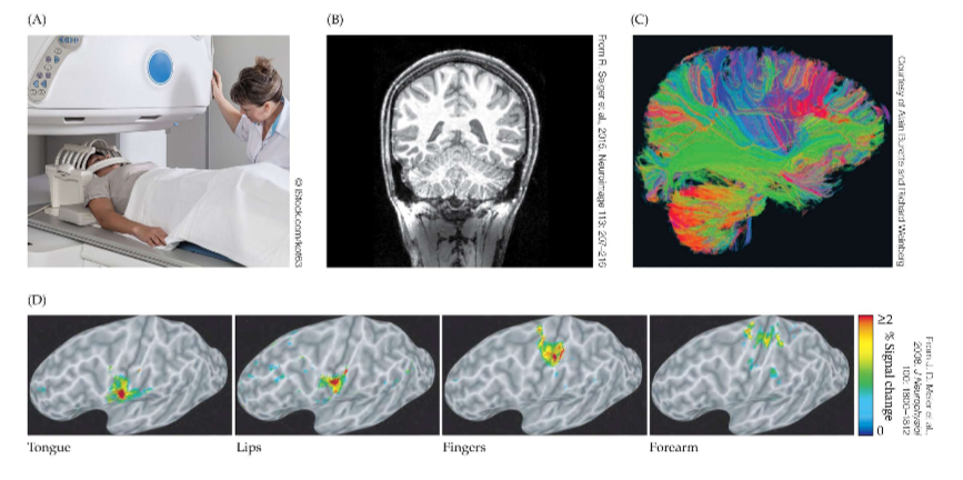
Neural Circuits and Systems can be Imaged and analyzed in brain
X-rays, computerized tomography (CT), Positron emission tomography (PET), MRI, Diffusion-tensor imaging (DTI), fMRI
Computerized Tomography (CT)
x-rays narrowed into a tube sweeps across the brain providing detailed anatomical structure
Positron Emission Tomography (PET)
Patient is injected with a radio-labeled molecule such as dopamine, which is then taken up by certain neurons (e.g. those of the basal ganglia)
MRI
When placed in a magnetic field, a patient (and his protons (H+)) will lineup with the magnetic field and spin at a frequency that depends on the field strength.
Diffusion-tensor Imaging (DTI)
Detected white matter tracts because water molecules can diffuse in limited directions along the length of the axon; useful for diagnoses.
fMRI
Based on metabolic activity (oxygen-dependent) of the brain