RADR 2401 Exam 2 special views/procedures
1/15
There's no tags or description
Looks like no tags are added yet.
Name | Mastery | Learn | Test | Matching | Spaced | Call with Kai |
|---|
No analytics yet
Send a link to your students to track their progress
16 Terms
Esophagram - LAO
Prone, 35-40 degree LAO, places esophagus between spine and hilar region of lungs. Looks for specific constricted areas and pathology
Esophageal reflux - breathing (Valsalva method)
Patient takes in deep breath, then bears down like they need to poop
Alternative - Mueller method - patient exhales then tries to inhale against closed glottis.
Esophageal reflux - water test
Pt in LPO position, drinks some water through a straw while fluoroing.
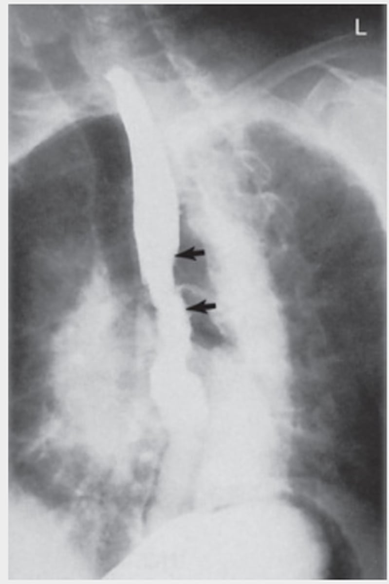
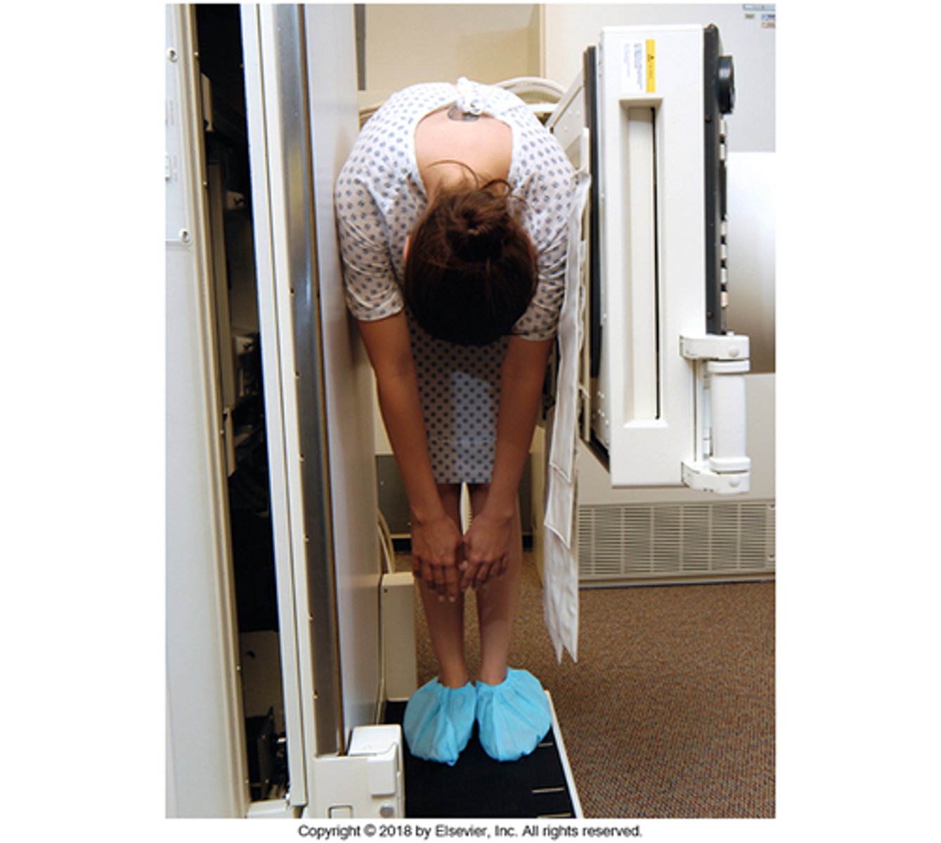
Esophageal reflux - toe touch
Pt lateral, leans down as if to touch toes. Can show reflux and hiatal hernia.
Esophageal reflux - compression paddle
Pt prone, compression paddle inflated under stomach. Can create reflux.
Small bowel - enteroclysis
Double contrast exam. Catheter inserted and advanced to duodenaljejunal flexure. Uses thin barium and air or methylcellulose.
Clinical indications: ileus, Crohn's, malabsorption syndrome, intestinal strictures, tumors, polyps, unexplained GI bleeding.

Small bowel - intubation
AKA small bowel enema. Single contrast, single lumen NG tube advanced to proximal jejunum while pt is RAO. Can use thin barium or iodinated water-soluble contrast.
BE - ventral decub
Double-contrast studies only! Pt prone, uses horizontal beam. CR at ASIS and MCP.
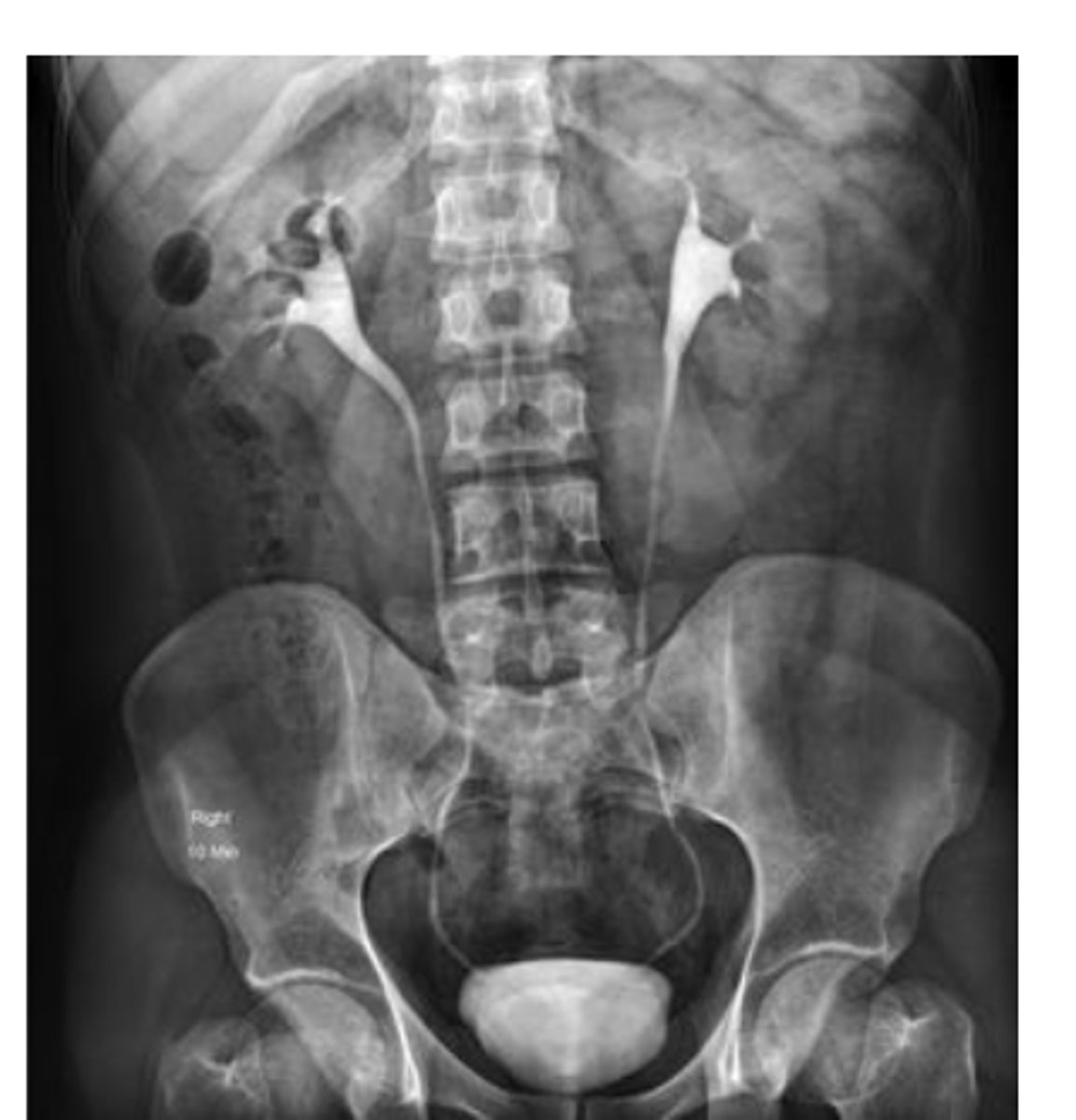
IVU (intravenous urogram)
Functional exam to visualize the collecting portion of the urinary system, and assess the function of the kidneys, bladder and ureters.
Clinical indications: back pain, kidney stones, kidney dysfunction, hematuria, abdominal/pelvic mass, UTI, hypertension
IVU contraindications
1.Hypersensitivity to iodinated contrast media
2.Anuria
3.Multiple myeloma
4.Diabetes, especially diabetes mellitus
5.Severe hepatic or renal disease
6.Congestive heart failure
7.Pheochromocytoma (fe-o-kro″-mo-si-to′-mah)
8.Sickle cell anemia
9.Renal failure, acute or chronic
IVU images and timing
Basic
-AP scout (CR at the Iliac Crest)
must include kidneys
-Injection of contrast (Note time at beginning of injection)
Must include timed markers
-Nephrotomography (1 min following injection)
-AP (5 min -10-15 min)
-RPO and LPO (20 minutes)
-AP postvoid (recumbent or erect)
Special
-AP ureteric compression
Nephrotomogram
Special imaging that is used to obtain images of specific layers of kidney tissue; improves demonstration of renal parenchyma. Uses special equipment attached to the IR and bucky. Three usually taken at one minute intervals. To set fulcrum: measure patient thickness, divide by three.
VCUG (Voiding Cytourethrography)
Functional study of the bladder and urethra. Performed after cystogram when catheter is removed.
Female - AP
Male - 30 degree RPO
Cystogram/Cystography
nonfunctional study of the bladder ONLY. Retrograde study; contrast is injected through catheter.
AP (10-15 degree caudal) and RPO/LPO (45-60 degrees).
Retrograde Urography
Nonfunctional study of the urinary system. Performed to evaluate location of renal stones/obstructions. Done in OR, contrast injected through catheter.
Retrograde Urethrogram
Nonfunctional radiographic study of the male urethra. 30 degree RPO, uses Brodney clamp, rarely done anymore.