Basic principles and techniques
1/94
Earn XP
Description and Tags
L2 and 3
Name | Mastery | Learn | Test | Matching | Spaced | Call with Kai |
|---|
No analytics yet
Send a link to your students to track their progress
95 Terms
What do nearly all cells of the mammalian body all derive from?
from 3 germ layers : ectoderm, mesoderm, endoderm
What derives from the ectoderm?
nervous system and epidermis - neural tube + crest, epidermis (skin, hair, nails, sebaceous glands), sensory organs eg lens of eye and inner eye
What derives from the mesoderm?
skeleton, skeletal muscles, connective tissue, blood, kidneys, gonads, reproductive ducts
What derives from the endoderm?
epithelial lining of GI and respiratory tracts, glands and organs like the liver, pancreas, gallbladder, thyroid gland
What does a zygote become through development?
blastocyst then gastrula eventually becoming the different cells from the 3 distinct germ layers + the germ cells
At which stage do vertebrates exhibit remarkable similarities through development?
pharyngula stage
Which structures are present in vertebrates at the pharyngula stage?
pharyngeal pouches
somites
notochord
hollow neural tube
post-anal tail
Why is the vertebrate stage termed phylotypic and what explains this?
stage typical of vertebrates through which they all go during development - a bottleneck that can not be bypassed, lack of it results in death
Which co-ordinated biological processes underlie embryonic development?
pattern formation, morphogenesis, cell differentiation and growth
Definition of pattern formation
process by which cells are organized in space and time to produce a well-ordered structure within the embryo
Where does the term morphogenesis come from?
morphe (shape) and genesis (creation)
Define morphogenesis
cell / tissue movements and changes in cell behaviour that give the developing embryo or organ its shape in 3D, limbs and digits start to form
Define cell differentiation
Process by which cells become different from each other and acquire specialized functional properties. Governed by changes in gene expression, which dictate the repertoire of proteins synthesized
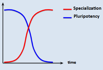
How do specialised vs pluripotent cell numbers change during development?
specialised increase, pluripotent decrease with time
What discrete steps are involved in the differentiation of embryonic/stem cells?
stem cell / progenitor cell → specification → determination → differentiation → post-mitotic maturation
Is it true that once a cell reaches specialisation this is its final state?
not necessarily, eg if cell is moved and under different extrinsic cues can sometimes change fate/differentiation
Define growth
increase in mass or size
Why is growth termed a continuous process?
growth can be embryonic, fetal, post-natal, adult so continues throughout life
What does growth rate depend on?
rate depends on age and organ
What are the 3 processes involved in growth?
mainly cell proliferation also cell enlargement and ECM production
What type of growth occurs in adults?
eg gut lining continuously regenerated
What processes does morphogenesis involve?
cell migration, cell adhesion, cell shape, cell death
Describe differentiation and its relation to pluripotency as well as its steps
progressive restriction of pluripotency and the progressive gain of specialized properties. It is a step-wise process: cell specification, determination, differentiation, maturation.
What are the main animal models used in developmental biology?
drosophila, fish (zebrafish), amphibians (frogs), birds, mice
What are 3 experimental methods to determine where and when a gene is expressed in the embryo?
in situ hybridisation
reporter lines (transgenic)
RNA-seq
Describe in situ hybridisation
shows which areas of a cell express a gene - probe is lab made as an antisense rna so very specific and takes advantage of rna hybridising, binds to rna in drosophila embryo, anti-dig binds the probe and alkaline phosphatase binds the transparent target substrate making it blue
blue lines are the areas of the cell where the gene is being expressed
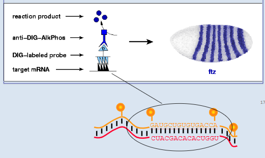
Limit to in situ hybridisation
sample has to be fixed so cell must be dead
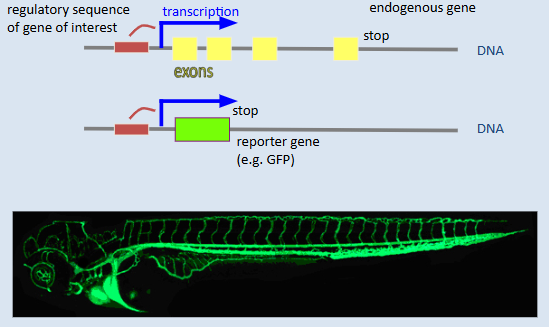
Describe reporter lines as a way to study gene expression
transgene made where reporter gene eg gfp is added on - protein will absorb electron exciting it at a lower (475nM) wavelength than it is then emitted at (510nM), the gene is under the action of the same regulatory sequence as our gene of interest so regulator activated = gene and gfp activated
Describe RNA-seq as a technique to study gene expression
RNA is isolated, converted into complementary DNA (cDNA) via reverse transcription, and then sequenced. The resulting reads are mapped back to a reference genome or assembled de novo to identify and quantify transcripts.
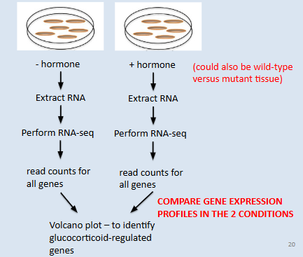
What is RNA-seq used for?
measuring expression levels of all genes in different samples (eg different tissues or with different mutations or with different chemical treatments) and can identify all genes exhibiting differential expression between samples
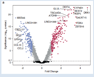
What does a volcano plot inform on gene expression within a cell? Which technique can a volcano plot be made from?
identifies genes exhibiting statistically significant changes in transcript abundance caused by exposure to hormone.
Blue dots identify down-regulated genes, Purple dots identify up-regulated genes
made from RNA-seq
What polymer is studied by in situ hybridisation, reporter lines and RNA-seq and what do they aim to study?
all aim to quantify and localise gene expression through studying the mRNA polymer = temporal and spatial distribution of transcripts for one or more genes
Which technique reveals expression pattern of one gene vs all genes in a biological sample?
in situ hybridisation and reporter lines // RNA-seq
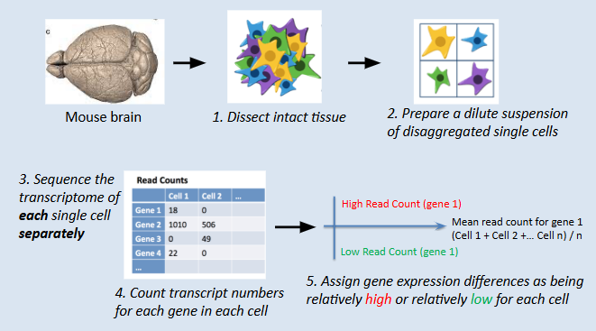
Describe how single cell RNA-seq can describe molecular phenotypes and define the cellular composition of complex tissues
intact tissue dissected eg from mouse brain, disaggregated/dissociated single cells suspended in their own droplet/dilution then transcriptome of each single cell is sequenced separately
transcript numbers counted for each gene in each cell - isolated sequencing reaction and gene expression differences assigned as relatively high or relatively low for each cell
bar coded gene expression profile made to visualise and compare gene expression between individual cells
cells clustered based on similar read count profiles = cell types
entire cell divided into a visual representation of gene expression
When performing a single cell RNA-seq, what scale of data is expected in terms of transcribed genes per cell and cell per tissue?
25 000 genes transcribed and usually 10s or 100s of 1000s of cells looked at
Dumb down single cell RNA-seq
basically cells are separated in a tissue and their entire transcriptome is determined in isolation of one another - shows cells with high vs low expression of different genes allowing for creation of cell clusters in terms of function and expression
How are cluster cells grouped together when performing single cell RNA-seq?
using statistical method (principal component analysis) so neurons naively put together based on similarities which allows cell identification
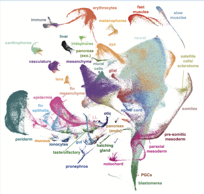
What is a visual result of a single cell RNA seq for a whole zebrafish eg?
cells with similar gene expression clustered and identified
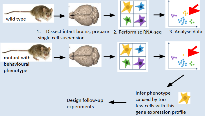
How can single cell RNA-seq be used to compare mutant and wild-type tissues and what is the aim of this?
differences in gene expression of cells can be highlighted, mutant phenotype will be linked to this different gene expression profile allowing follow-up experiments to investigate function of the isolated gene
essentially aim: understand molecular and cellular basis of complex phenotypes
What are 2 techniques to analyse protein epxression?
immunohistochemistry and visualisation of fluorescent fusion protein
Which proteins allow the spatio-temporal analysis of distribution of proteins in embryos experimentally?
tagged antibodies
How are specific antibodies made eg in rabbits to use in immunohistochemistry for example?
by injection animals with protein that we want to detect - this will be detected by immune system as foreign and specific antibody will be produced in response and bind with very high specificity
What is the small region of specificity on an antibody called?
epitope
What is a tagged antibody?
antibody with a dye or enzyme attached (conjugated) to it to determine its location
What are 2 interests in a protein’s location when using fluorescently tagged antibodies in an organism?
either sub-cellular localisation or general location in organism ie which tissues
What are 2 commonly used enzyme conjugates used to analyse the spatio-temporal distribution of proteins in embryos?
alkaline phosphatase and horseradish peroxidase
When using alkaline phosphatase, how is the bound substrate modified?
turns blue
When using horseradish peroxidase, how is the bound substrate modified?
turns brown
What is the aim of making a sandwich antibody?
amplifies the signal bc many secondary antibodies bind each primary antibody
When making a two antibody sandwich, would the antibodies be made in the same organism or two different ones? Why?
2 different ones eg 1ary in rabbit and 2ary in mouse
2ary ones compatible with host, allows specific recognition of primary antibodies from a different species (if same species would not bind specifically enough)
What is the function of a 1ary vs a 2ary antibody in experimental techniques?
1ary recognises protein of interest // recognises primary antibody and carries the tag
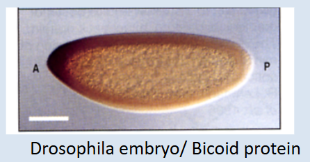
Which technique has been used here and what can be inferred from it?
immunostaining - shows localised bicoid protein through use of horseradish peroxidase enzyme conjugate binding at the anterior side suggesting high bicoid levels vs low ones at the posterior axis - conclusion: more bicoid protein at anterior than posterior axis
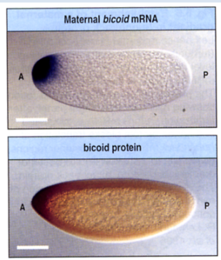
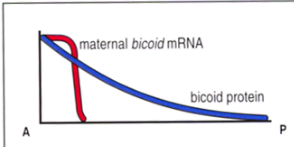
What difference is highlighted here by bicoid in situ hybridisation vs antibody staining and what this means?
top photo is an in situ which shows mRNA levels in the cell - only present at the anterior axis
bottom is an immunostaining showing a gradual gradient established across the cell with high bicoid protein at anterior axis and low at posterior axis
Describe the process by which a fusion protein construct can be used to analyse the spatio-temporal distribution of proteins in embryos
GFP added onto gene of interest which tags the subsequently transcribed mRNA and translated protein - allows following of the protein of interest through the cell
Give an example of using a fusion protein construct to understand a mechanism in c elegans
gfp tagged protein followed throughout cell division before finally becoming nuclear localised - shows journey of protein to nucleus in c elegans embryo development
What are Muller’s morphs?
coined terms describing mutations based on their behaviour and dominance (amorph, hypomorph hypermorph, antimorph)
Define an amorphic mutation
complete loss of gene function (null mutation/knock-out). Most genes are haplosufficient in diploid organisms so these are usually recessive.
Define hypomorphic mutation
reduction of wild type function. Usually recessive.
Define antimorphic
competitive inhibitors. Also called dominant negative.
What are the 3 types of loss-of-function mutations that can occur?
amorphic, hypomorphic and antimorphic
What is a type of gain-of-function mutations that can occur?
hypermorphic
Which of Muller’s morphs are dominant ?
antimorphic and hypermorphic
Which of Muller’s morphs are recessive?
usually amorphic and hypomorphic in diploid organisms
Define forward genetics
Phenotype to gene - seeks to identify a gene whose mutation caused a particular phenotype
Define reverse genetics
Gene to Phenotype - seeks to characterize the phenotype of particular mutated gene, by targeted mutagenesis
Define hypermorphic
increased activity of the gene product. Dominant.
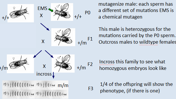
Describe the process of forward genetics
1) randomly mutate the genome (chemicals like ENU or EMS)
2) look for interesting phenotypes in the offspring
3) identify the gene that causes the defect
What is a limit to forward genetics?
random - most phenotypes produced are not relevant to the research being done so luck of the draw if it does work, many mutagenised animals would have to be studied for specific phenotypes of interest
Which animal models are mainly used in forward genetics and why?
c elegans, drosophila and zebrafish - forward genetics is essentially luck of the draw, mutagenesis has to be done in very high numbers to get an interesting phenotype so these organisms are bets
Name some types of genetic screens
loss of certain cells or tissues, disease-like phenotype, biochemical abnormalities, loss of hearing or vision, behaviour, drug addiction, etc, etc.
Describe the process of a forward genetic screen in drosophila
takes 3 generations of flies to make the mutation homozygous so to identify genes that affect a specific process like visual function, thousands of F2 families need to be screened
In what animal models do reverse genetics tend to be done?
mice and chick
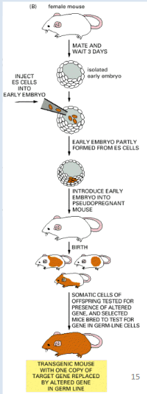
Describe the process of reverse genetics
Knock-out the gene in a mouse embryonic cell line, selected cell line is then reintroduced into mice embryos
first generation are mosaic (a mixture of cells from the stem cell line and the mother). Their gonads are also mosaic
mosaic animals are bred to generate non- mosaic carriers of the transgene (2nd generation)
carriers are then interbred to create homozygous mutant animals (3rd generation)
Compare forward to reverse genetics
mutant found in a forward genetics screen - start with only a phenotype, we do not know what the gene encodes // reverse genetics we already know the gene and want to find the function
Forward genetics: function (phenotype) ----> gene
Reverse genetics: gene ----> function (phenotype)
What is a way to find a mutation in the genome and identify the gene in forward genetics ?
positional cloning - maps a gene to a specific chromosomal region (no indication of function)
What are 2 ways to manipulate tissue interactions in embryos?
embryology tissue manipulation (graft, ablation) and signalling pathway manipulation (drugs, transfection/electroporation, genetics, bead implants)
What is the aim of tissue manipulation in embryos?
demonstrate inductive function of one tissue on another
How is tissue manipulation in embryos carried out?
tissue ablation/graft/transplantation (surgical) or bead/cell implantation (signalling molecules and drugs)
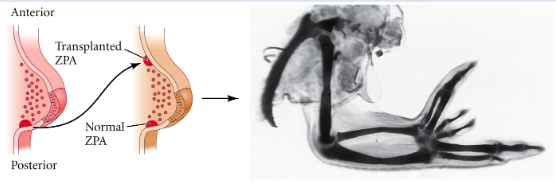
How has the tissue been manipulated in this chick by John Saunders and Robert Riddle?
shows signal in embryo responsible for digit formation later on.
through transplant ie surgical - wing formed in chick has a mirrored axis so posterior digits found both posterior and anterior and anterior ones found in the middle
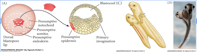
Describe the spemann and mangold experiment as seen in this image and what type of tissue manipulation was carried out
shows tissue (dorsal blastopore lip) responsible for organising/structural signal allowing dvpt of entire organism and leads to twin
dorsal blastopore lip - fragment from the presumptive notochord and somites moved to presumptive epidermis in blastocoel - resulted in a siamese twin body axis showing the dorsal blastopore lip as the organiser or specific tissue mvt and organism dvpt
What are 2 techniques for fate mapping/lineage tracing in embryos?
embryology labelling with dye and genetics labeling with a reporter plasmid or recombinant retroviral vector expressing GFP
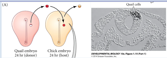
In what model animals is embryology labeling with a dye done to trace fate/lineage in embryos ?
chick/quail chimera
What is fate mapping/lineage tracing used for?
to determine which later developmental structures arise from a particular group of cells (that may express a gene of interest, for example) in an early embryo.
What are methods to carry out fate mapping/lineage tracing?
- Cell/tissue transplantation (Spemann and Mangold)
- Cell/tissue labeling with dye
- Cell/tissue labeling genetically (electroporation, GFP transgenic lines)
How is a gene of interest investigated in terms of its role in development?
techniques of developmental genetics are used to test whether a gene of interest is essential for development
What do tissue manipulations essentially investigate?
which developmental signals regulate expression of gene of interest
which tissue produces signals regulating gene of interest
Technique to use if trying to understand the molecular and cellular basis of complex
phenotypes.
comparative analysis of single cell RNA-seq data from WT and mutant tissues
Technique to use if trying to describe the molecular phenotypes and define the cellular
composition of complex tissues
single cell RNA-seq
Technique to use if trying to find out where and when the product of a gene is expressed
Immunofluorescence and GFP-protein fusion transgenes
Technique to use if trying to test whether a gene of interest is essential for development.
techniques of developmental genetics
Technique to use if trying to determine which developmental signals regulate the expression of a gene of interest and which tissue produces them
tissue manipulations eg ablations, transplantations, bead implantations
Technique to use if trying to determine which later developmental structures arise from a particular group of cells (that may express a gene of interest, for example) in an early embryo.
fate mapping and lineage tracing techniques
What does this allow the understanding of?
donor quail cell from specific region of embryo is now known to be responsible for the development of those types of cells - stand on among the chick cells
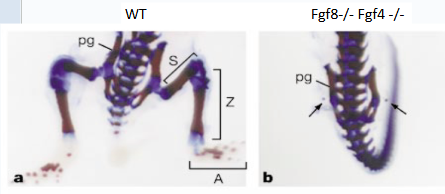
What does this reverse genetics knockout show?
role of fgf8 and 4 beads in limb development
What is shown here ?
creation of transgenic mouse through selection of mice carrying the introduced transgene to create homozygous mutant animals with specific gene of interest