Strokes n' Shiiiii
1/16
There's no tags or description
Looks like no tags are added yet.
Name | Mastery | Learn | Test | Matching | Spaced | Call with Kai |
|---|
No analytics yet
Send a link to your students to track their progress
17 Terms
General- Ischemic
Clot of blockage → most common type 80% of CVA
Cerebral thrombosis: blood clot w/in the cerebral arteries
Cerebral embolism: traveling bits of matter produce occlusion (from other locations)
General- Hemorrhagic
Rupture or leakage→ less common, worse prognosis
Hematomas can lead to hemorrhage
Epidural hematoma
between the skull and dura
Caused by a tear of the meningeal arteries
Brain is affected by the compressed fluid
Subdural hematoma
midline shift; crescent shape, between dura and arachnoid space
Tearing of bridging veins b/t brain and dural sinus (venous)
Acute onset accompanied by headaches and AMS
C/L hemiparesis
May have epilepsy
Subarachnoid
Unable to identify lateral ventricles; between arachnoid and pia
bleeding into brain (fills)
Thunderclap headache
worse prognosis (often leads to death)
All gray; cannot see any ventricles
TIA- Transient Ischemic Attack
Temporary interruption of blood supply to the brain
May last a few minutes to several hours < 24 hours
Precursor to susceptibility for cerebral or myocardial infarction
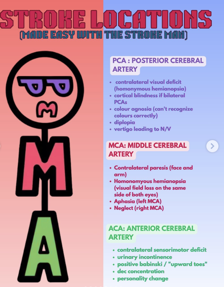
Anterior cerebral artery (ACA)
Area of brain
Anterior frontal lobe
Medial surface of frontal lobe
Medial surface of parietal lobe
S/S
C/L loss of LE motor and sensory function
Loss of bowel and bladder fxn
Aphasia, apraxia, agraphia, akinetic mutism
Middle cerebral artery (MCA)
Area of brain
Cerebrum
Basal ganglia
S/S
UE more affected
C/L weakness and loss of face sensory function
Wernicke aphasia, apraxia, anosognosia
Homonymous hemianopsia
Internal Carotid artery (ICA)
Area of brain
parietal lobe
S/S
Supplies both the MCA and ACA, so s/s can include all those that would occur with infarcts of either artery
Lesions involving both MCA and ACA can lead to massive edema, brain herniation, and death
s/s of MCA involvement with reduced consciousness
Warning signs: Hx of TIAs and temporary fading of vision in I/L eye
Posterior cerebral artery
Area of the brain
Occipital lobe
Midbrain
Thalamus
S/S
C/L hemiplegia
C/L loss of pain and temperature sensation
Prosopagnosia- inability to recognize faces
Vertebral basilar artery
Area of the brain
Cerebellum
Medulla
Pons
S/S
Locked-in syndrome: patient cannot move or speak but is A&O
No horizontal eye mvmt, but vertical eye mvmt/ blinking intact
Pseudobulbar palsy d/t CN V-XII paralysis
Tetraplegia
Rapid progression from hemiparesis to tetraplegia or quadriplegia
Internal Capsule
S/S
supranuclear palsy
weakness of C/L lower half of face and C/L extremities
Decreased corticobulbar tract function
Differentiate b/t SCI and CVA
An SCI never affects the face, so if the face is affected you know the CVA is above the midbrain
Brain stem
S/S
Both I/L (face) and C/L (limb) impairments
R stroke → R sided face, L sided arm weakness
Any injury above the medulla with present with only C/L loss
Cerebellum
S/S
I/L impairments
Ataxia, dysdiadochokinesia, dysmetria, dyssynergia, dysphagia
Side specific s/s- Right CVA
s/s on left
L-sided weakness or paralysis
L-sided neglect → spatial and perceptual problems
poor judgement/ impulsiveness
overestimates abilities
difficulty perceiving emotions
rigidity in thought
short attention span/ poor memory
cognitive problems
Think toddler
Side specific S/s- Left sided CVA
S/s on right
Right-sided weakness or paralysis
Aphasia: wernicke's or broca’s
Cautious, slow, insecure
Aware of impairment; frustrated
depressed; sad
difficulty processing information in a linear and sequential manner
difficulty understanding new info
Think old person
Stroke Man
ACA- LE deficits
MCA- UE and face
PCA- Visual