Adult Health II: Exam #1 (Neuro & Critical Care)
1/125
There's no tags or description
Looks like no tags are added yet.
Name | Mastery | Learn | Test | Matching | Spaced | Call with Kai |
|---|
No analytics yet
Send a link to your students to track their progress
126 Terms
Skull & Spinal Radiography
- x-ray reveals fractures, bone erosion, dislocation, etc.
CT
- cross-sectional images to revel tumors, internal bleeding/clotting, bone fractures, abnormalities (cancer), etc.
Non-Contrast CT
first step for stroke
Contrast CT
- better & more detailed
Nursing Education
* pt NPO for 4 hrs prior
- radiation harmful to fetus
* monitor BUN & creatinine
MRI
- cross-sectional image to reveal torn ligaments, bones, organs, joints, & tissue
- magnetic imaging
Nursing Education
* ask to remove metals or ask about implants (pacemaker)
LP/Spinal Tap
- CSF removed to detect cancers/malignancies, infections (meningitis, syphillis), etc.
Nursing Care
- pt must be in knee-chest position w/ neck flexed & head on pillow
- use 20-22 g inserted in L3/4 or L4/5
Cerebral Angiography
- visual cerebral blood vessels to reveal defects, narrowing, or obstruction of arteries
Nursing Education
* pt NPO for 6 hrs prior
- may have metallic taste/feel warm
* assess BUN & creatinine
Electroencephalography (EEG)
- assess electrical activity of brain (seizure activity, sleep disorders, behavior changes)
Nursing Education
- pt hair must be clean
- pt needs to be sleep deprived
* avoid stimulants (caffeine, etc) before test
Caloric Testing
- assess vestibular system by instilling warm/cold water into ear canal
Nursing Education
- abnormal result = NO rapid eye movement
ICP Monitoring
- device inserted into cranial cavity to record pressure
- shows waveform w/ pts who have head injury
- irrigate system to maintain potency & recalibrate
ex: intraventricular, subarachnoid screw/bolt, epidural sensor
Contrast Dye
Nursing Education
- withhold Metformin 48hrs prior to& 48 hrs after
- ask if allergic to iodine or shellfish
- ask about renal function (could lead to contrast-induced AKI)
Meningitis Pathophysiology
- inflammation of the meninges that affects the subarachnoid fluid space
Causes --> bacteria (most contagious; Neisseria meningitidis), viruses, fungi, parasites, amoeba, trauma, or meds
transmission --> high pop density (college dorms, prison)
* DROPLET PRECAUTIONS *
Meningitis Clinical Presentation Infants
- anorexia, vomiting, diarrhea
- irritability
- macular rash
- respiratory symptoms
Meningitis Clinical Presentation Adults
- fever
* severe headache
- stiff neck
- seizures
* photosensitivity
- N/V
Meningitis Diagnosis
* LP = most effective
* CSF proteins --> C & S = elevated
- blood or nasopharynx cultures
Glasgow Coma Scale
Nursing Assessment = notify provider of any significant changes
Prioritize Hypothesis
- lower = 3 pts (deep coma or death)
- highest = 15 pts (fully awake)
- score < 8 pts = severe head injury & coma
- score 9-12 pts = moderate head injury
- score > 13 pts = minor head injury
Migraines
Causes = genetics (#1), trauma, environment, foods/additives
Clinical Presentation = reoccurring headaches persisting for at least 72 hrs
- photophobia
- N/V
- unilateral pain
- stress
- anxiety
- phonophobia
Migraine Phases
1. Prodromal
2. Aura
3. Headache
4. Postdromal
Aura
- can tell it is coming
- visual field loss
- visual/sensory/motor disturbances
- tunnel vision
- blindness
- heavy limbs
- speech disturbances
- tingling
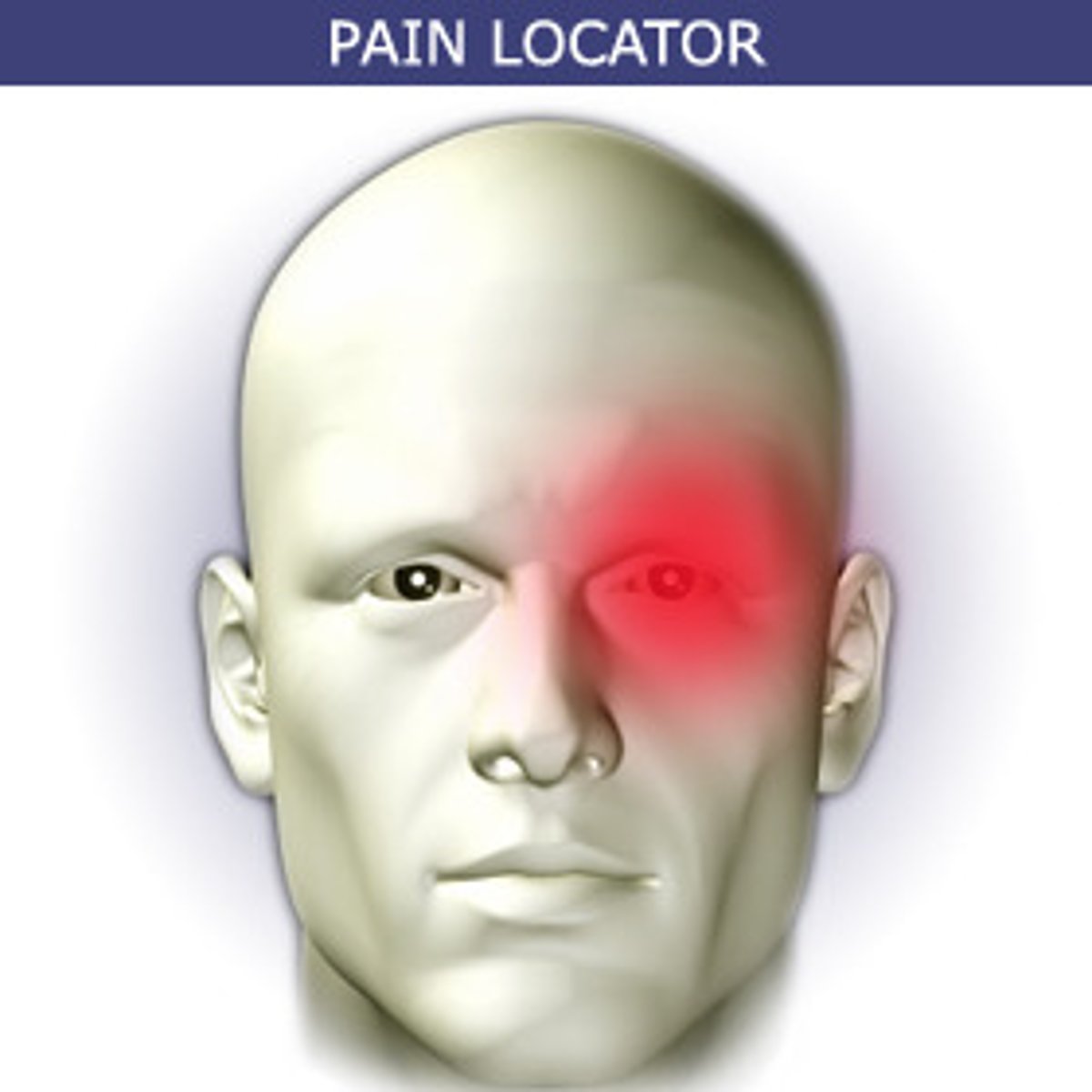
Cluster Headache
Causes = stress (#1), allergens, environment, tobacco/alcohol, meds
Clinical Presentation
- severe pain (excruciating & non-fluctuating) on one side of head & behind eye
- lacrimation, nasal congestion, eyelid/facial swelling
- 15 mins to several hours, several times a day
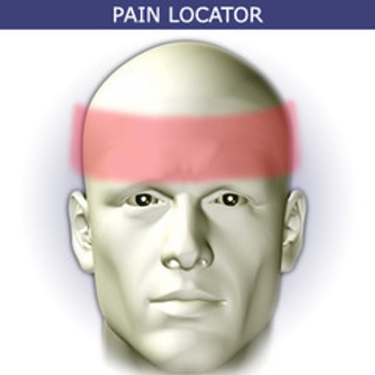
Tension Headache
Causes = stress (#1), posture, depression
Clinical Presentation
- mild or moderate pain (dull/band-like pressure) on both sides of head
- muscle tightness in shoulder, back, neck
- 4-6 hrs
Primary Headaches
migraine, tension, cluster
Secondary Headaches
Cause = underlying causes
ex: trauma, infection (meningitis), intracranial disorders, psychiatric
Seizure Pathophysiology
- abnormal, sudden, excessive discharge of electrical activity in the brain
Types
- generalized = tonic-clonic, absence, myoclonic, atonic, akinetic
- partial = simple or complex
Seizure Nursing Assessment
- seizure hx (type, occurrence, prodromal signs, aura, loss of motor activity)
- occurring during postictal states (headache, LOC, sleepiness, impaired speech or thinking)
Seizure Nursing Interventions
* Assess ABCs & LOC
* remain w/ the client (NEVER leave alone)
- time & duration (document)
- assess behavior at onset of seizure (if aura, what kind)
- call an ACT/rapid
- if standing or sitting --> lay on floor, left side, protect head
* protect airway from injury --> do NOT stick anything in mouth & have O2/suction ready
-do NOT restrain (loosen restrictive clothing)
- note the type, character, & progression of the movements during the sz
- monitor for incontinence
* IV access & admin medication IV push
* seizure precautions (fall/aspiration)
- monitor behavior following sz (LOC, motor ability, speech)
Seizure DC Teaching
- avoid alcohol, excessive stress, fatigue, & strobe lights
- community resources (Epilepsy Foundation of America)
- encourage the client to wear medical alert bracelet
* instruct client about importance of lifelong medication & need for follow-up to determine medication blood levels
Seizure Medications
* 1st line = benzodiazepines
- carbamazepine = 3-14
- clonazepam = 20-80
- divalproex = 50-100
- ethosuximide = 40-100
- lorazepam = 50-240
- phenobarbital = 15-40
- phenytoin = 10-20
others = gabapentin, lacosamide, lamotrigine, levetiracetam, oxcarbazepin, pregabalin, tiagabine, topiramate, zonisamide, vigabatrin
Flumazenil
* reverse effects of Benzos
- do NOT give to pt w/ IICP or status epileptics (cause seizure)
Phenytoin
- give slowly to prevent hypotension or cardiac dysrhythmias
- can decrease effectiveness of BC
Seizure Medication Education
** do NOT dc --> status epilepticus may occur
- take w/ food
- avoid alcohol, OTC meds, antacids
- wear medical alert bracelet
- caution = performing activities w/ alertness
* toxicity --> follow-up w/ periodic blood studies
- monitor serum glucose levels
- urine = may have harmless pink-red or red-brown color
* REPORT --> sore throat, bruising, nosebleeds (indicate blood dyscrasia)
- inform HCP of bleeding gums, N/V, blurred vision, slurred speech, dizziness
Transient Ischemic Attack (TIA) Pathophysiology
- a mini stroke
- caused by inadequate blood flow due to stenosis (narrowing of blood vessels) or an occlusion (most commonly clot)
* resolves completely
TIA Manifestations
- slurred speech
- dizziness
- headache
- blurred/double vision
- weakness
- aphasia
- dysphagia
- ataxia
- increased BP
FAST --> facial drooping, arm weakness, speech disturbance, time to call 911
TIA Risk Factors
- HTN, CVD, DM, HLD
- physical inactivity, obesity
- smoking, excessive alcohol intake
- low SES
TIA DC Education
* pt at risk for stroke or TIA for next 48 hrs
- decrease risk for aspiration (due to dysphagia)
- educate how to recognize manifestations
- encourage regular exercise to lose weight
- smoking cessation, decrease alcohol intake
- reinforce diet & medication for DM
Ischemic Stroke
- sudden loss of brain blood flow --> O2 deprivation
Causes
- embolism
- stenosis
- plaque buildup in arteries (atherosclerosis)
Hemorrhagic Stroke
- blood vessels leak or rupture --> increased pressure in brain
Causes
- small cerebral arteries that have been damaged overtime by HTN
- hemorrhages or aneurysms --> lead to brain death
Stroke Modifiable Risk Factors
- HTN, HLD
- smoking
- physical inactivity, obesity, unbalance diet
- drugs
- sleep apnea
- oral contraceptives
Stroke Non-Modifiable Risk Factors
- age, sex
- genetics, family hx
- sickle cell
- race, ethnicity
Stroke Manifestations
BE FAST
- balance = dizzy, loss of balance/coordination
- eyes = difficulty seeing
- face = severe headache, numbness, facial drooping
- arms = numbness on one side of arms & legs
- speech = aphasia, slurred, confused, difficulty understanding
- time = call 911
Neuro = mental status change & seizure
Stroke Diagnosis
* CT = primary test (non-contrast to differentiate b/t ischemia & hemorrhagic) -- w/in 25 mins
- ASAP = CT, CT angiography, MRI
- ultrasound to determine occlusion
- ECG & chest x-ray to detect cardiac abnormalities
Stroke Safety
* NPO until swallow test --> consult SLP
- fall precautions
- bleeding precautions
Stroke Education
- manifestations of stroke
- medication regimen
- assistive devices/rehab
- community resources
- modifiable risk factors
tPa
indication = Ischemic Stroke
time window = < 4.5 hrs (cannot give if no witnessed time change)
side effects = significant risk of bleeding (intracranial, internal, site), drop in BP, N/V, & dizziness
tPA Exclusive Criteria
- head trauma/prior stroke in prev. 3 months
* subarachnoid HEMORRHAGE
- arterial puncture in prev 7 days
- hx of previous intracranial hemorrhage
- intracranial neoplasm, AVM, or aneurysm
- recent intracranial or intraspinal surgery
* BP > 185 or > 110
- active internal bleeding
- CT demonstrates multilobar infarction (hypo density > 1/3 cerebral hemisphere)
Acute Bleeding Diathesis
- platelets < 100,000/mm^3
- heparin w/in 48hrs w/ abnormally elevated aPTT
- current anticoagulant w/ INR > 1.7 or PT > 15 sec
- current direct thrombin inhibitors or direct factor Xa inhibitors w/ elevated sensitive lab tests (e.g. aPTT, INR, platelet count, ECT, TT, or appropriate factor Xa activity assays_
* BG < 50 mg/dL (2.7 mmol/L)
Stroke Aspiration Precautions
- NPO until completion of swallowing eval to decrease risk of choking or aspirating --> then pick appropriate diet
- assess for changes in neurological status (LOC) to prevent aspiration
- assist in upright position when eating, promote small bites, no straws, check cheeks for food pocketing
Stroke Anticoagulants (Warfarin)
* do NOT give for hemorrhagic stroke
- common in pt w/ AFIB
- monitor PT/INR (4x 1st week) --> normal = 2-3
- onset = 24-72 hrs
- peak = 5-7 days
* antidote = vitamin K
Cerebral Edema Pathophysiology
- swelling of the brain that causes increase ICP
- restricts blood & spinal fluid from entering or leaving the brain
Vasogenic Cerebral Edema
- interruption of blood brain barrier (something wrong w/ vessel)
- common in = strokes, brain tumors, high altitude
Cellular/Cytotoxic Cerebral Edema
- influx of Na+ & then water into cells --> brain cells lose ability to maintain normal fluid balance, leading to edema
- common in: TBIs & strokes
Interstitial Cerebral Edema
- CSF moves from intraventricular space into interstitial space of brain --> fluid increases pressure in interstitial
- common in = hydrocephalus & meningitis
Osmotic Cerebral Edema
- brain cells take water from circulating plasma
- common in = DKA & hyponatremia
Cerebral Edema Nursing Assessment
Manifestations
- sensory & mental status alterations
- visual changes
- weakness
- seizures
- headache (early manifestation)
- confusion, lethargic
- dizziness
- N/V
Diagnosis --> CT & MRI
Assess --> changes in LOC & mental status
Head Injury Pathophysiology
- any damage to the head due to a traumatic event
- does not always result in a brain injury but can
Traumatic Brain Injury (TBI)
concussion or subdural hematoma --> temporary or permanent alteration in neurological function
Concussion
- mild
- may or may not cause temporary loss of consciousness, headache, confusion, difficulty concentrating, N/V, dizziness, blurred vision, & amnesia
Subdural Hematoma
- may be asymptomatic
- change in consciousness, alteration in pupillary response, hemiparesis, HTN, bradycardia, bradypnea, coma
Common Causes of Head Injury in Older Adults
* hitting head during FALL --> decreased visual acuity, weakness, chronic HTN, poly pharmacy (increased dizziness), lack of safety devices (stair rails, shower bars), throw rugs, electrical cords, floor clutter
- increased incidence of SDH --> natural atrophy of brain tissue allows more room for bleeding inside cranial vault
- abuse from caregivers
Head Injury Nursing Assessment Conscious
subjective
- mechanism of injury
- time of injury
- pain level, location, & characterstics
- any manifestations/symtpoms that began at or immediately following the injury
- hx of prior head trauma
- hx of head surgeries
objective
- thorough head exam for signs of injury
- full neuro assessment (reflexes, sensation, strength, visual acuity)
- determine GCS score to assess severity
Head Injury Nursing Assessment Unconscious
- thorough head exam for signs of injury (lacerations, edema, ecchymosis)
- neurological exam
- determine clients Glasgow Coma Scale (GCS) score to help assess injury severity (< 8 = severe injury)
- Assessment: unarousable, primitive or no response other than painful stimuli, altered respirations, decreased cranial nerve & reflex activity
- Interventions = assess airway, VS, watch for CO2
expected vs unexpected
Nursing Care of Unconscious Head Injury
* assess latency of airway --> maintain & have emergency equipment readily available
- monitor BP, pulse, & heart sounds
- assess respiratory & circulatory status
* do NOT leave client unattended if unstable
- maintain patent airway & ventilation (high CO2 = increases ICP)
* assess lung sounds for accumulation of secretions --> suction as needed
* assess neurological status --> LOC, pupillary reactions, motor/sensory function, using coma scale (GCS)
- place client in semi-fowler's position
- change position of client q2 hrs, avoiding injury when turning
- use side-rails unless contraindicated or protocol
- assess for edema
- monitor dehydration
- monitor I/Os & daily weight
* maintain NPO until conscious (check gag & swallow reflex)
- maintain nutrition as prescribed (IV or enteral), monitor fluid/electrolye balance
- monitor status of skin integrity & prevent skin breakdown
- provide freq mouth care
- remove dentures & contact lenses
- assess eyes for presence of corneal reflex & irritation & instill artificial tears
* monitor drainage from ears or nose for the presence of CSF
- assume the unconscious pt can hear
- avoid restraints
- initiate seizure precautions if necessary
- provide ROM exercises to prevent contractures
- initiate PT as appropriate
- use footboard or high-topped sneakers to prevent foot drop & use splints to prevent wrist deformities
Cerebral Aneurysm Pathophysiology
- dilation of the walls of a weakening cerebral artery --> ruptures & increases ICP
Unruptured CA Manifestations
typically asymptomatic until rupture or leak
Ruptured CA Manifestations
-sudden, severe headache due to small amount of leaking blood (considered subarachnoid hemorrhage)
- similar to stroke manifestations (hemiparesis) = N/V, stiff neck, blurred or double vision, sensitivity to light, seizure, loss of consciousness, confusion, & tinnitus
Leaking CA Manifestations
- extremely severe headaches that may last several days & up to 2 weeks
Cerebral Aneurysm Nursing Intervention
- patent airway, O2, VS (HTN & dysrhythmias), avoid rectal temperature
- surgical interventions: microsurgical clipping, artery bypass & occlusion
- end-vascular interventions: coiling, flow diversion w/ stents
Parkinson's Disease Pathophysiology
- degenerative, progressive condition caused by gradual loss of cells in the substantial nigra --> stops producing dopamine & NE
Parkinson's Disease Manifestations
- shaking
- stiffness
- freezing in place
- loss of balance
- shuffling gait
- stooped posture
Parkinson's Stage 1
- manifestations are mild
- client can provide self-care duties w/out assistance
- tremors & other involuntary movements occur unilaterally
- changes in walking & facial expressions
Parkinson's Stage 2
- tremors affect both sides of the body & interfere with/ self-care activities
- change in posture
- balance is uncoordinated
- clients develop difficulty walking
- activities of daily living become more difficult to complete
Parkinson's Stage 3
- mid-stage w/ mild to moderate disability
- client experiences loss of balance & slow movements
- motor manifestations continue to worsen
Parkinson's Stage 4
- manifestations severely disabling
- self-care activities require assistance due to severe tremors & stiffness
- client can walk but may need to ambulate w/ a can or walker to maintain safety & prevent falls
Parkinson's Stage 5
- most advanced & debilitating
- client are wheelchair or bed bound
- require maximum assistance w/ self-care activities
Parkinson's Nursing Interventions
Fall Precautions = assistive devices, non-slip socks, bed/chair alarm (reduce wandering)
Nutrition = aspiration precautions, appropriate diet, small bites, eat upright, thickened liquids, soft foods, high-fiber, fluids
Safety = locks on doors, use signs, remove rugs & wires, adequate lighting
Calm Environment = calming music, reduce loud noises
- adherence to medication & treatment
- plan of care focuses on self-care activities, safety, social activities
- promote resources/support groups
- promote physical, occupational, speech therapy
Carbidopa-Levodopa
- Parkinson's medication
- acts as natural chemical, dopamine, in the brain
Adverse Effects
- irregularities in BP
- dyskinesia
- confusion
- dry mouth
- constipation
- hallucinations
- headaches
Increased Intracranial Pressure (IICP) Manifestations
- Change in LOC
- headache
- seizure
- eyes = papilledema, pupillary changes, impaired eye movement
- postural = decerebrate, decorticate, flaccid
- decreased motor function
- vomiting
- change speech
- increased systolic BP
- decreased pulse
- altered respirations
IICP Assessment
* LOC (most sensitive & earliest indication)
- headache
- abnormal respirations
* increase BP w/ widening pulse pressure
- elevated temp
- vomiting
- pupil changes
- bradycardia
- Late Signs = increased systolic BP, widened pulse pressure, low HR, motor function changes from weakness or hemiplegia, positive babinski reflex, decorticate or decerebrate, seizures, positive glucose testing
* be alert for Cushing's triad
*assess for CSF leakage
IICP Risks
- decreased cerebral perfusion
- hypoxia
- seizure activity
* CSF leakage
- risk for shunt failure or infection following VP shunt placement
IICP Generate Solutions
- monitor respiratory status & maintain ventilation
- maintain body temp & prevent shivering
- decrease environmental stimuli
- monitor fluid & electrolyte balance, acid-base balance, & I/Os --> limit fluid intake to 1200 mL/day
- instruct client to avoid coughing, sneezing, & valsalva's maneuver
* elevate HOB 30 degrees (avoid trendelenburg)
- prevent fixation of neck & hips
- administer prescribed medications = anti-seizure medications, antipyretics, muscle relaxants, antihypertensives, corticosteroids, IV fluids, hyper osmotic agents (mannitol)
IICP Nursing Interventions
- monitor CO2 levels as prescribes
- avoid administration of morphine sulfate to prevent hypoxia
- monitor renal function w/ mannitol therapy
- expect & monitor for diuresis
- VP Shunt = position supine, position from back to non-operative side, monitor for shunt failure, monitor for signs of infection
Spinal Shock
- complete but temporary loss of motor, sensory, reflex, autonomic function
- occurs immediately after cord's response to injury
Manifestations
- loss of sensory/motor function & reflexes below the injury
- hypotension
- hypothermia
- urinary retention
- fecal incontinence
Neurogenic Shock
- common in injuries above T6 (experienced soon after injury)
- vasodilation leading to pooling of blood in vessels, tissue hypo perfusion, & impaired cellular metabolism
Spinal Cord Injury Assessment
* assess level of spinal cord injury (lowest spinal cord segment w/ intact motor & sensory function)
- assess motor & sensory changes below level of injury
- assess total sensory loss & motor paralysis below the level of injury
- assess for loss of bladder/bowel control, urinary retention, & bladder distention
- assess VS
* assess respiratory status (changes), especially w/ C4 injuries
- monitor ABGs & ventilator status if ventilated
- monitor for respiratory infection
- monitor for dysrhythmias, hemorrhage or bleeding, shock, DVT, & orthostatic hypotension
* assess neurological status
- assess motor ability (squeeze hands, spread fingers, move toes, turn feet)
- assess for absence of sensation
* monitor for Autonomic Dysreflexia, Spinal Shock
* assess pain
- assess fluid & electrolyte balance
- assess skin integrity
- assess psychological status, including. feelings of anger, depression, loss, & sexual concerns
Spinal Cord Injury Risks
- respiratory failure
* Autonomic Dysreflexia
- spinal shock
- further spinal cord damage
- urinary retention
- impaired skin integrity
- complications related to immobility
Spinal Cord Injury Interventions
* always suspect spinal cord injury when trauma occurs until ruled out
* maintain airway --> prevent head flexion, rotation, or extension
- immobilize the client & maintain traction & alignment
- logroll client
- encourage deep breaths & use of IS
- monitor infection
- monitor for complication related to lack of movement
- maintain bowel program --> high fiber diet
- prevent urinary retention --> initiate bladder control program
- prevent UTIs & calculi
- Q2 turns
- promote rehabilitation & self-care measures
- set realistic goals based on client's potential functioning level
- utilize community resources
Autonomic Dysreflexia Etiological/Causative Factors
- occurs after spinal shock has resolved
- occurs when/ lesions or injury above T6 & cervical lesions
- most commonly caused by = visceral distention from a distended bladder or impacted rectum
- Bladder Triggers --> kinked/clogged foley, bladder spasms, bladder stones, UTIs
- Bowel Triggers --> pooping quickly, stool impacted in rectum, enema or manual removal of stool, gas & gas pain, constipation/diarrhea, Diverticulitis, Crohn's disease, anal fissure, hemorrhoids
- Skin Triggers --> rashes, bedsores, cuts/bruises, tight/lumpy/baggy clothing, itching that cannot be sense or scratched, ingrown toenails
- Others = neurological disease, stroke, brain injury, CNS tumor, MS, Guillian-Barre syndrome, med AE, severe head trauma, subarachnoid hemorrhage, stimulant drugs
Autonomic Dysreflexia Manifestations
- VS: high BP that rises rapidly, slow or fast HR
- severe, pounding headache
- N/V
- blurred or other vision changes
- stuff nose
- cold, dry, pale, clammy skin
- anxiety, apprehension, or uneasiness
- heavy sweating --> red, hot, flushed skin
- goosebumps or hair standing up
- tingling sensations
- narrowed blood vessels
- compare current BP to patient's usual BP
- review hx of spinal cord injury
Autonomic Dysreflexia Nursing Interventions
* neurological emergency
* FIRST --> raise HOB or sit up right & dangling
- remove tight clothing, jewelry, or devices
- monitor BP --> at least 2 hrs after it begins to decrease
* identify cause & fix
- check bladder drainage --> insert foley if no output
- check bowel or stool blockage --> remove blockage
- check skin for wrinkles, constrictions & tight clothing
- continue looking for & removing triggers if symptoms persist
- emergency tx = fast-acting medications (nitrates, hydralazine, labetolol, or nifedipine)
Respiratory Acidosis
Cause = hypoventilation (e.g. COPD)
pH < 7.35
PaCO2 > 45 mmHg
HCO3 = 22-26 mEq/L (normal)
Respiratory Acidosis Manifestations
- headache
- confusion
- drowsiness
- SOB
- hypotension
Respiratory Acidosis Nurse Management
- monitor ABG levels
- encourage deep breathing
Respiratory Alkalosis
Cause = hyperventilation (e.g. anxiety)
pH > 7.45
PaCO2 < 35 mmHg
HCO3 = 22-26 (normal)
Respiratory Alkalosis Manifestations
- dizziness
- tingling in extremeties
- chest pain
- confusion
- dry mouth
Respiratory Alkalosis Nurse Management
- address underlying cause
- coach controlled breathing
Metabolic Acidosis
Cause = increased acid production or bicarbonate loss (e.g. DKA, diarrhea)
pH < 7.35
PaCO2 = 35-45 mmHg (normal)
HCO3 < 22 mEq/L
Metabolic Acidosis Manifestations
* rapid breathing (Kussmaul respirations)
- nausea
- lethargy
- confusion
- abdominal pain
Metabolic Acidosis Nurse Management
- correct underlying cause (i.e. insulin for DKA)
- administer Bicarb
Metabolic Alkalosis
Cause = excess bicarbonate or acid loss (e.g. vomiting, diuretics)
pH > 7.45
PaCO2 = 35-45 mmHg (normal)
HCO3 > 26 mEq/L
Metabolic Alkalosis Manifestations
- muscle twitching
- dizziness
- paresthesia
- confusion
- hypoventilation
Metabolic Alkalosis Nurse Management
- replace electrolytes
- address volume status