Week 1 - Vertebral Column Notes
1/120
There's no tags or description
Looks like no tags are added yet.
Name | Mastery | Learn | Test | Matching | Spaced | Call with Kai |
|---|
No analytics yet
Send a link to your students to track their progress
121 Terms
Where is the vertebral column located?
Posterior side of the body
How has the vertebral column evolved for us?
Standing upright and for walking on 2 legs
Functions of the vertebral column:
Support for body weight and thorax
Transmission of forces
Flexibility of movement
Positioning and support for the head and upper limbs
Protection for the nervous system
Kyphosis
Concave anteriorly
Thoracic region
Sacral region

Lordosis
Concave posteriorly
Cervical region
Lumbar region
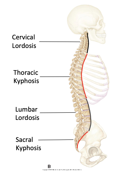
Curvatures present at birth
Primary Cruves (Thoracic and sacral kyphosis)
Curves not present at birth
Secondary curves (cervical and lumbar lordosis)
Cervical lordosis
Appears once the child gains head control and can start looking upwards
Essential to be able to stand or sit upright and move the head around in various directions
Lumbar lordosis
Appears once the child starts to stand upright and walk around
Essential to keep the centre of gravity behind the hip joints during erect standing or walking
How many vertebrae are there?
33, held together by intervertebral discs, ligaments and muscles
Seperate vertebrae
24 - 7 cervical, 12 thoracic, 5 lumbar
Fused vertebrae
9 - 5 sacral, 4 coccygeal
What are vertebrae in the cervical, thoracic, and lumbar regions joined by?
Intervertebral disc (starting C2-C3)
What joints do not have an intervetebral disc?
Atlanto-occipital and atlanto-axial
What type of joint is zygapophyseal joint?
Synovial plane joint
Allows gliding movements of adjacent vertebrae
Movements between individual vertebrae
Both the intervertebral and zygapophyseal joints move together as a functional unit.
Movement between individual vertebrae is small.
Over successive vertebrae the movement is cumulative and accounts for the overall movement of the spine
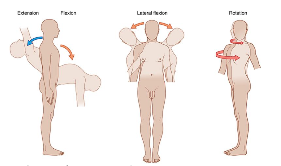
Movement of vertebral column
Extension, flexion, lateral flexion, rotation
Craniovertebral joints
Atlanto-occipital, atlanto-axial
Atlanto-occipital joint movement
Flexion/extension of head and neck. Head nodding when indicating “yes”
Atlanto-axial joint movement
Head rotation of the neck
About 50% of head rotation
Head shaking movements when indicating “no”
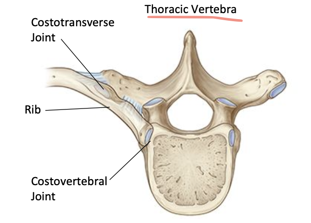
Costovertebral joints
Costovertebral joint: head of rib (vertebral joint)
Costotransverse joint: rib (transverse process)
Splenius capitis and Cervicis movement
Head and neck extension, head and neck rotation
Erector spinae muscle movement
Straighten spine from a flexed position. Control forward flexion of the spine.
Transversospinales muscle movement
Local extension, local rotation
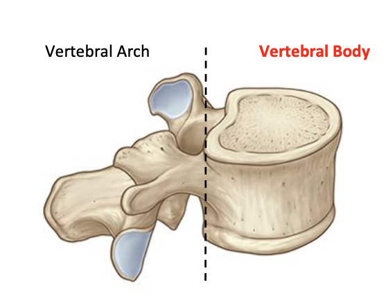
Vertebral body
Block shaped structure.
Forms the anterior part of the
vertebra.
Main weight bearing part.
Get progressively larger and thicker from the cervical region down to the lumbar region.
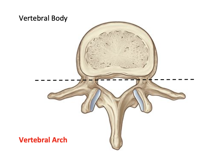
Vertebral Arch
Ring shaped structure.
Forms the posterior part of the
vertebra.
Protects the spinal cord and cauda equina.
Attachment for ligaments and muscles
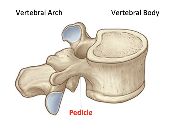
Pedicle
Attached vertebral arch to body
Lamina
Extend from each pedicle to meet and form the roof of vertebral arch

Transverse process
Extends postlaterally from where pedicle and lamina meet
Muscle/ligament attachment
Articulation of ribs in thoracic
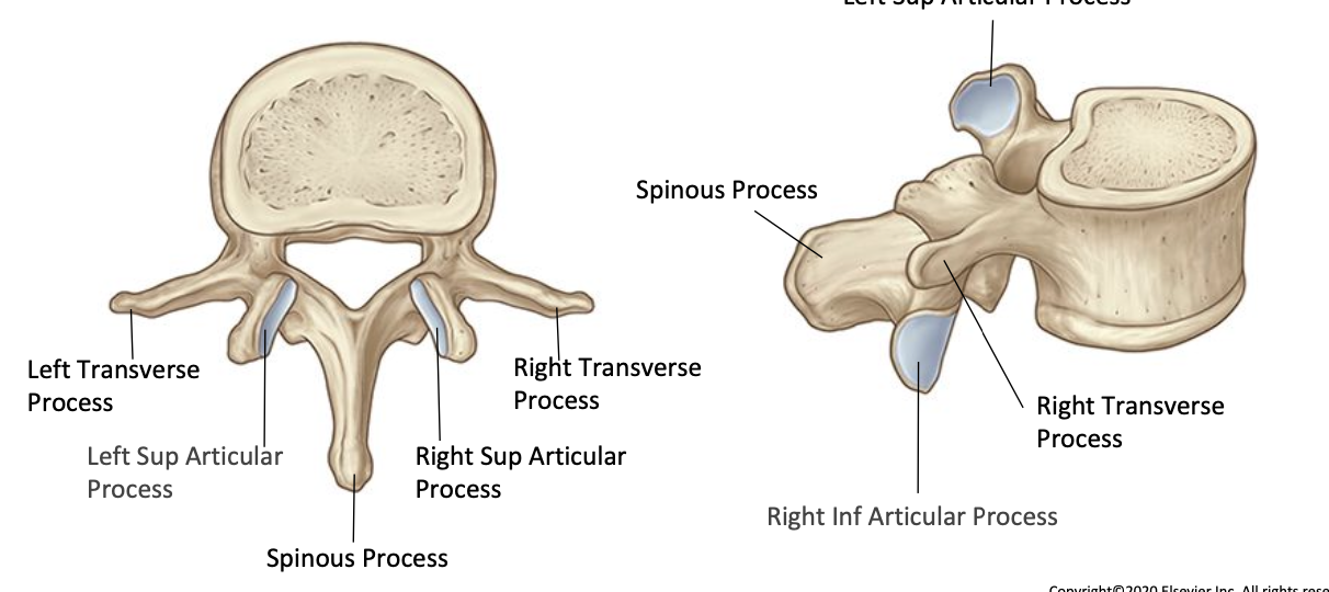
Spinous process
Posteriorly and inferiorly
From junction of 2 laminae
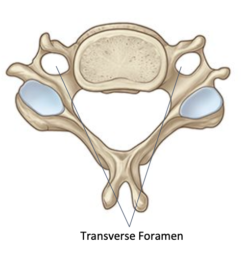
Cervical vertebrae - transverse foramen
C1-C6 have an opening in the transverse process called the tramsverse foramen
Only found in cervical vertebrae
Contains vertebral artery
What is the vertebral arch composed of?
Pedicles + lamina. Vertebral foramen. Superior and inferior articular processes. Transverse processes and spinous processes.
What type of structure does C1-C2 have?
Atypical
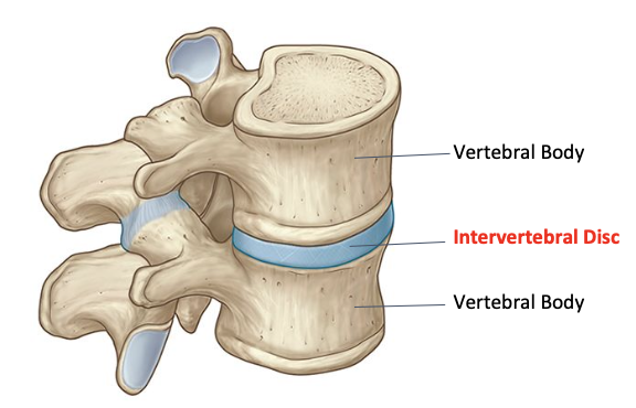
Intervertebral joints
Secondary fibrocartilaginous joints (symphysis)
Designed for weight bearing, shock absorption, and strength
Intervertebral joint location
All the vertebrae in the cervical, thoracic and lumbar regions are joined by an intervertebral disc starting at C2-C3.
The atlanto-occipital and atlanto-axial joints do not have an intervertebral disc
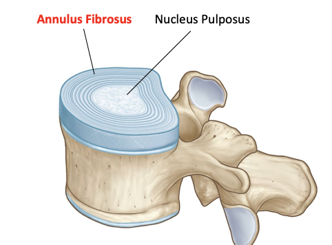
Annulus fibrosus
Multiple layers of fibrocartilage and elastic fibres.
Arranged into concentric rings (lamellae).
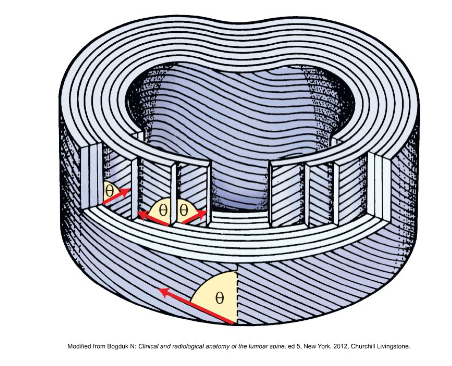
Annulus fibrosus - orientation of layers
Fibers are arranged in multiple concentric layers.
Every other layer runs in identical directions.
The orientation of each layer (depicted as θ) is about 65 degrees from the vertical.
Resists twisting and buckling.

Nucleus Pulposus
Nucleus pulposus is a gel like consistency which is about 80% water.
Contained within the annulus fibrosus but there is no clear division between the nucleus pulposus
Nucleus Pulposus Function
Compression force (straight arrows) raises the hydrostatic pressure in the nucleus pulposus. In turn, the increased pressure elevates the tension in the annulus fibrosus (curved arrows).
The increased tension in the annulus inhibits radial expansion of the nucleus. The rising nuclear pressure is also exerted upward and downward against the vertebral endplates.
The pressure within the disc is evenly redistributed to several tissues as it is transmitted across the endplates to the adjacent vertebra
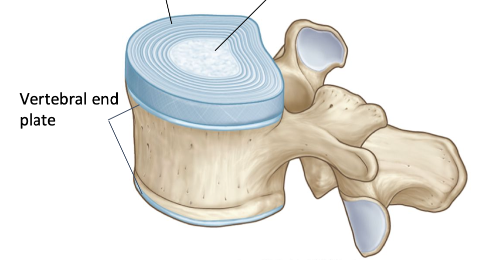
Vertebral end plate
● 1mm layer of cartilage which covers the upper and lower surface of the vertebral body.
● Anchors the intervertebral disc.
● Facilitates fluid exchange between the disc and vertebral body
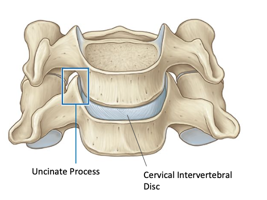
Cervical discs - uncinate process
● The cervical intervertebral discs do not extend across the entire width of the vertebral bodies.
● They extend to the uncinate process.
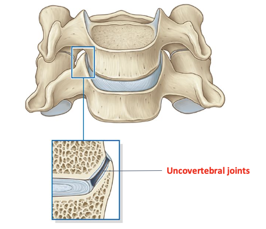
cervical discs - uncovertebral joints
● Synovial joints with defined articular surfaces, a joint capsule and synovial membrane.
● Limit excessive rotation of the cervical intervertebral joints
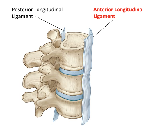
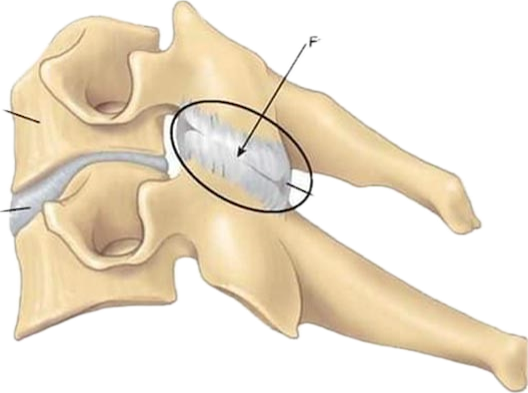
Intervertebral joint - anterior longitudinal ligament
● A broad thick ligament.
● Spans the anterior aspect of the
vertebral bodies.
● Runs the entire length of the vertebral column from the sacral region to C1.
Resists separtion of the vertebral bodies during hyperextension of the spine

Intervertebral joints - posterior longitudinal ligament
● Not as broad as anterior longitudinal ligament.
● Spans the posterior aspect of the vertebral bodies.
● Runs the entire length of the vertebral column from the sacral region to C1
Resists separation of the vertebral bodies during hyperflexion of the spine
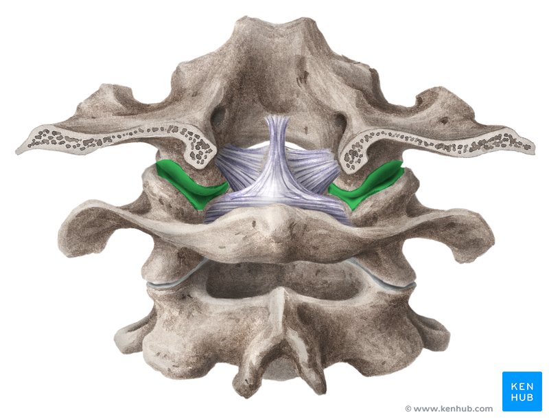
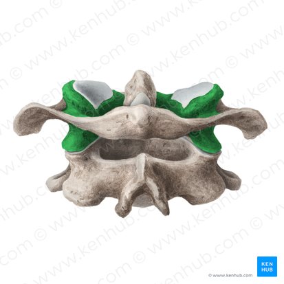
Zygapophyseal joints - joint surface
● Form a right and left zygapophyseal joint at each vertebral level.
● The superior articular process of the vertebra below articulates with the inferior articular process of the vertebra above
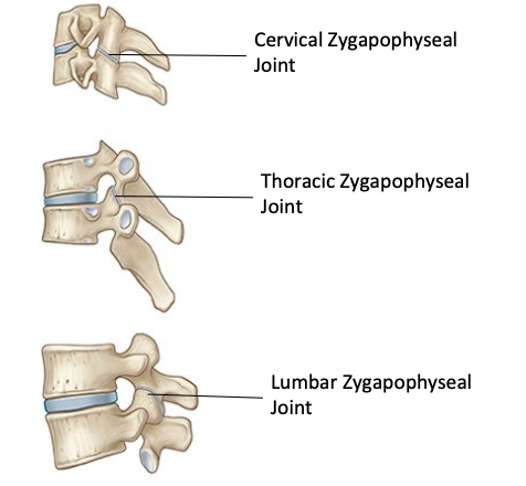
Zyagopophyseal joint capsule
Loose
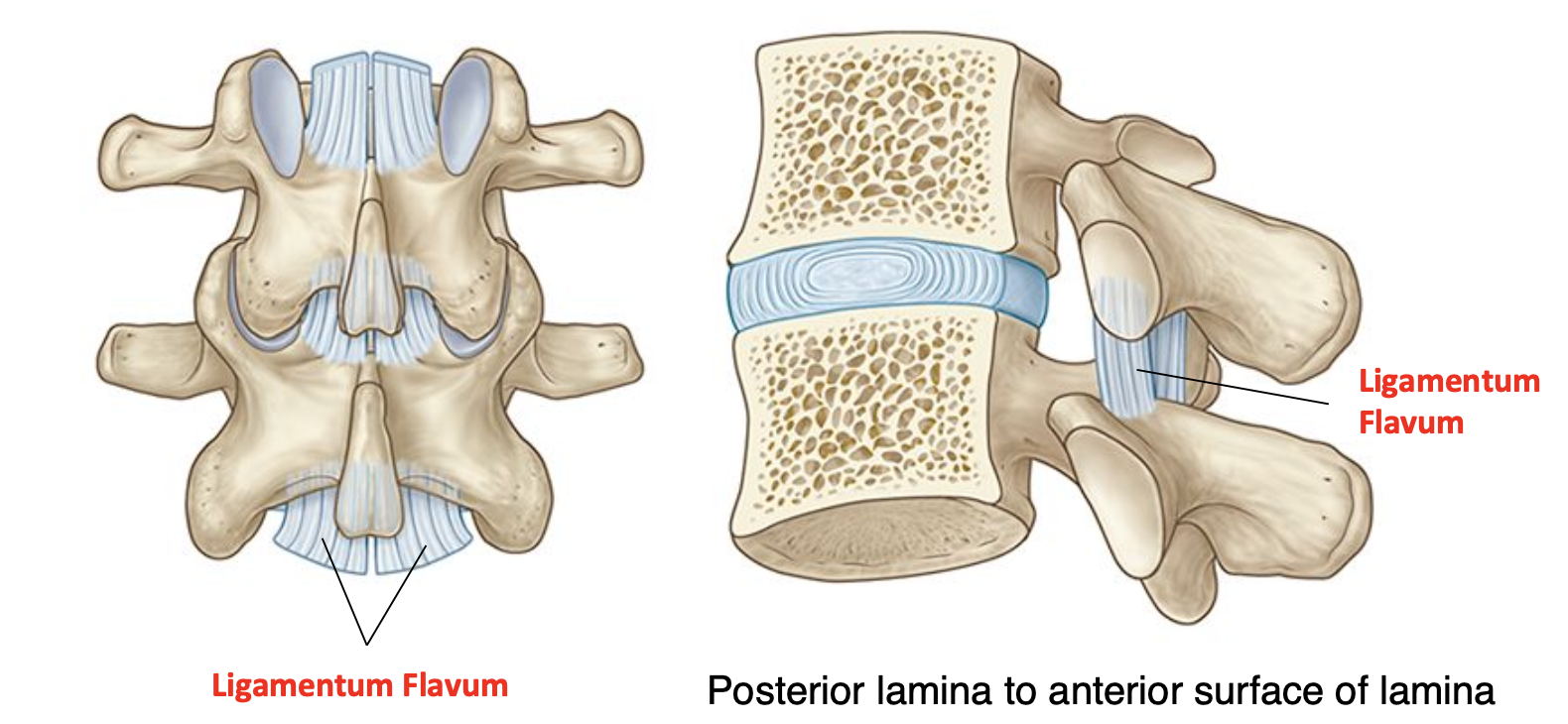
Ligamentum flavum
● Fibres are highly elastic.
● During forward bending the ligamentum flavum becomes stretched.
● Its stored elastic energy is released when returning back to an upright position

Ligamentum nuchae
● The supraspinous ligament becomes the ligamentum nuchae in the cervical region.
● Helps to return a flexed head back to upright posture.
● Attachment for cervical muscles
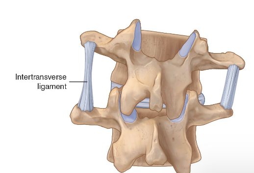
Intertramsverse ligament
● Thin bands that connect adjacent transverse processes
● Absent from the cervical spine and often more obvious in the lumbar region.
Determinants of movement
● Amount of movement determined by thickness of intervertebral discs in relation to the height of vertebral body.
○ Cervical spine ≅ 40%
○ Thoracic Spine ≅ 25%
○ Lumbar spine ≅ 33%
● Direction of movements guided by orientation of the Zygapophyseal joint surfaces
Cervical spine movement
Flexion/extension, right/left rotation, right/left lateral bending
Thoracic spine movement
Flexion/extension, right/left rotation, right/left lateral bending
Lumbar spine movement
Flexion/extension, right/left lateral bending, right/left rotation
What are the vertebral arches reinforced by?
Ligamentum flavum, interspinous ligament, supraspinous ligament, ligamentum nuchae, intertransverse ligament
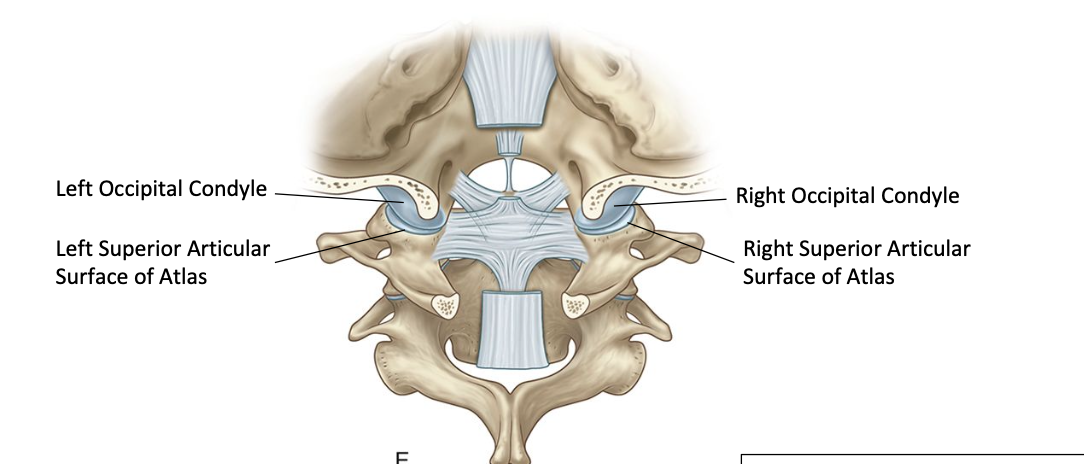
Classification of atlanto-occipital joint
Synovial condyloid joint

Classification of lateral atlanto-axial joint
Synovial plane joint
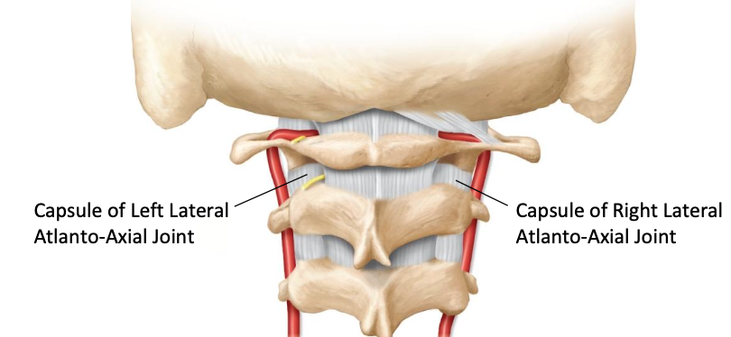
Lateral atlanto-axial joint capsule
● Joint capsules are thick and loose.
● Reinforced by a thickening called the accessory atlanto-axial ligament (not seen)
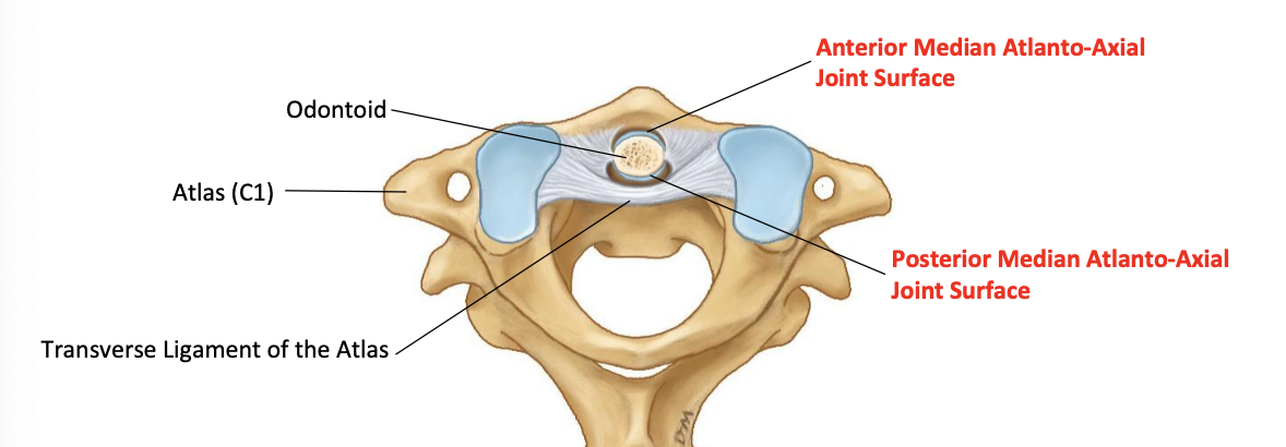
Median atlanto-axial joint classification
Synovial pivot joint
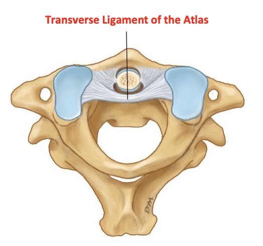
Function of tramsverse ligament of atlas
● Holds the odontoid firmly in place against the atlas
● Completes the median atlanto-axial joint.
● Prevents the atlas from sliding forward off the axis when bending the head forward
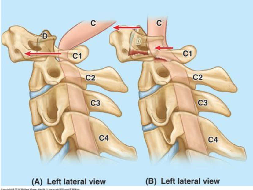
Transverse ligament tears - atlanto-axial instability
● Damage to the ligament results in excessive forward displacement of the atlas during head flexion (seen here by illustration A).
● This narrows the intervertebral foramen at C1 and potentially impinges on the spinal cord
Function of alar ligaments
● Prevent excessive rotation of the head and the atlas to the opposite side.
● Head rotation to the right will be resisted by the left alar ligament.
● Referred to as the “check” or “shoulder check” ligaments.
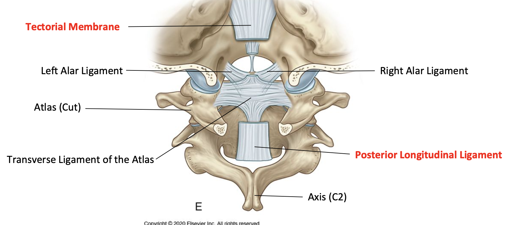
What is the tectorial membrane a continuation of
Posterior longtudinal ligament
Spinal cord
● Occupies the upper two thirds of the vertebral canal.
● Medulla oblongata to level L1-L2 interveternal disc.
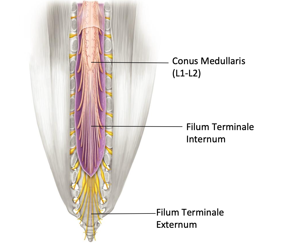
What is the filum terminal internum made of?
Connective tissue
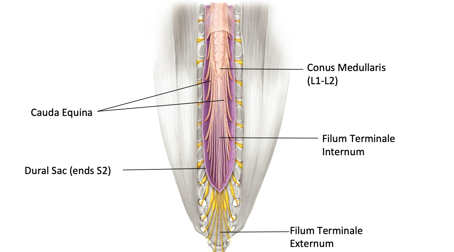
What is the cauda equina?
Nerve roots that hang freely
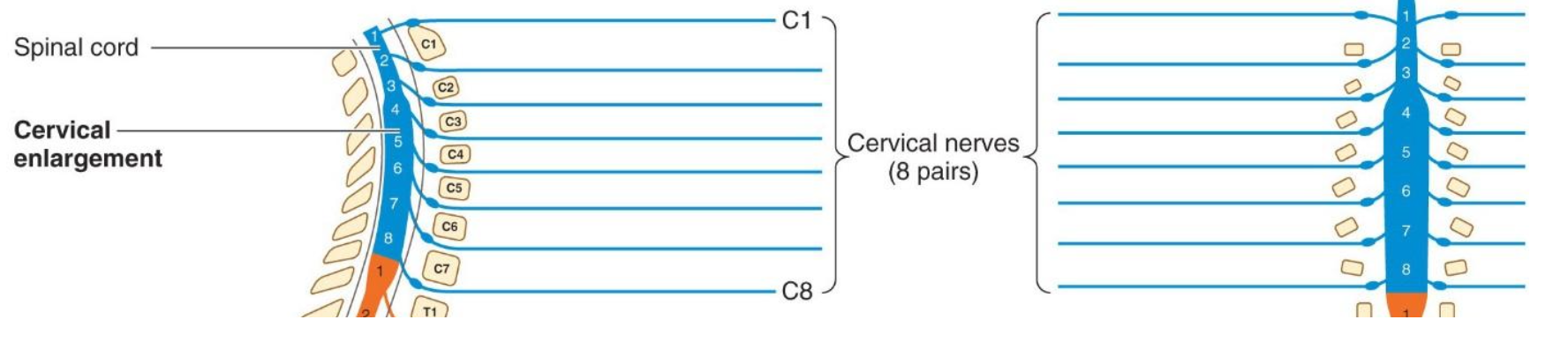
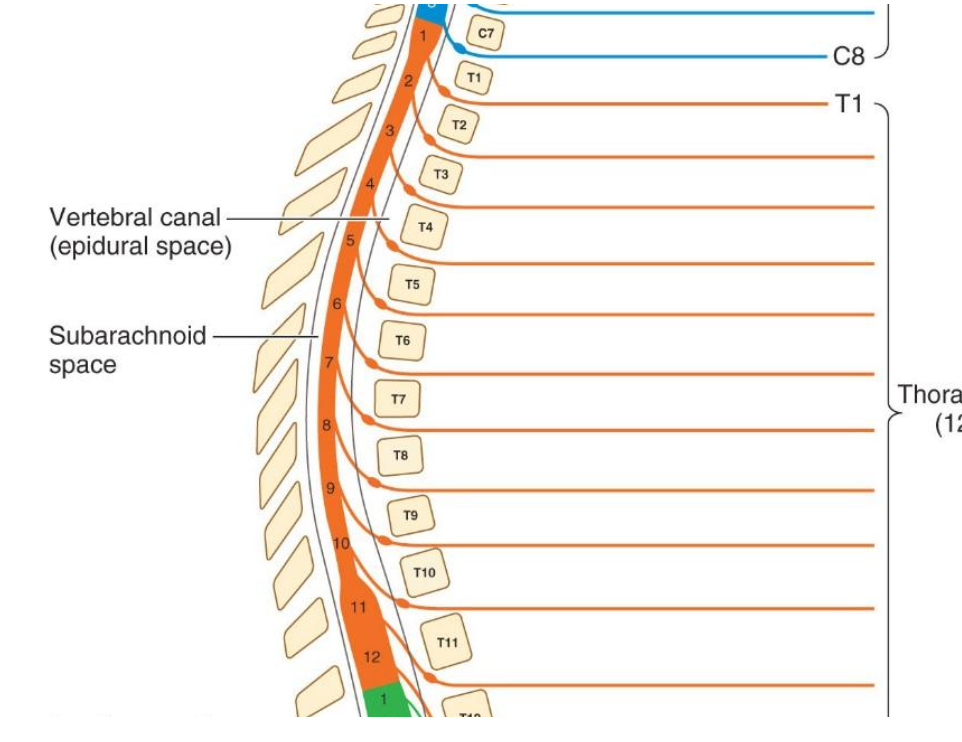
How many pair of spinal nerves are there?
8 cervical, 12 thoracic, 5 lumbar, 5 sacral, 1 coccygeal
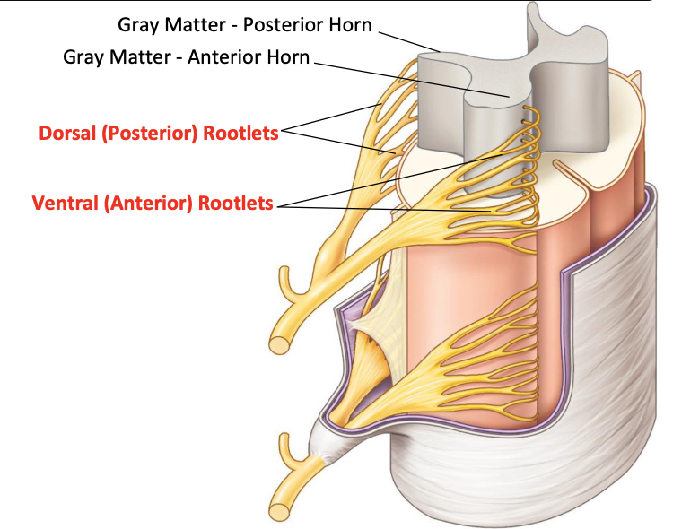
Spinal nerve rootlets
● Ventral rootlets - mainly motor fibres
● Dorsal rootlets - mainly sensory fibres
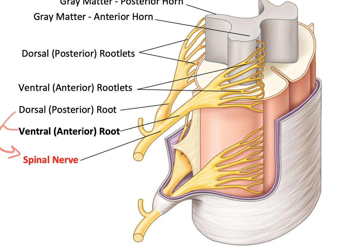
Spinal nerves
● Spinal Nerve - Motor and sensory fibres.
● Boundary between central and peripheral nervous system
Cervical spinal nerves
Cervical spinal nerves exit above their corresponding numbered vertebra
Thoracic spinal nerves
Thoracic spinal nerve exit below their corresponding numbered vertebra
Cauda equina
The lumbar, sacral and coccygeal nerve roots descend downwards in the cauda equina.
Lumbar and sacral spinal nerves
Lumbar and sacral spinal nerves exit below their corresponding numbered vertebra
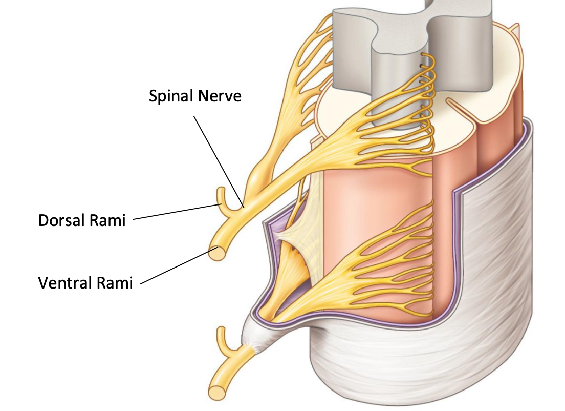
What does the dorsal (posteriior) rami supply
● Intrinsic back muscles
● Z-joints of spine
● Ligaments of spine
● Skin on posterior head,
neck, spine and pelvic girdle
What does the ventral (anterior) rami supply
● Cervical Plexus
● Brachial Plexus
● Lumbar Plexus
● Sacral Plexus
● Intercostal Nerves of
Trunk
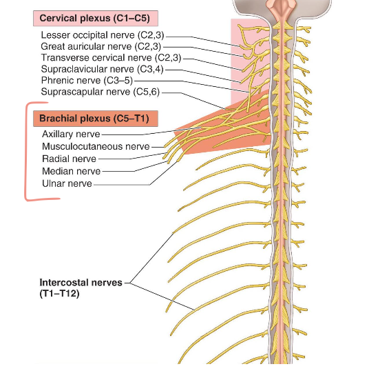
Intercoastal nerves - thoracic region
Each intercostal nerve formed by a single spinal nerve.
Supplies:
● Muscles and skin in
thoracic wall
● Segmental distribution
Recurrent meningeal nerve
Doubles back into the intervertebral foramen
Supplies
● Anterior dura and dural
sleeves
● The posterior
longitudinal ligament
● The posterior and
posterolateral portions of the intervertebral disc
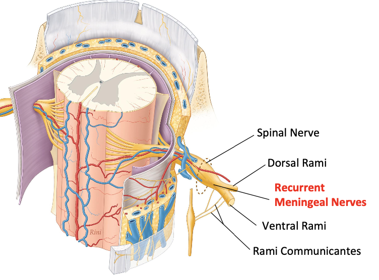

Rami communicates
Connects ventral rami with sympathetic chain of autonomic nervous system
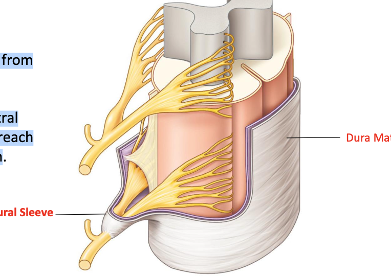
Dural sleeve
● Sleeve like projections from the dura mater.
● Covers dorsal and ventral nerve roots until they reach intervertebral foramen
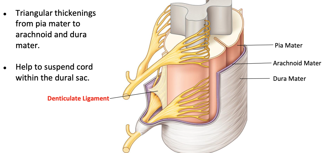
Arachnoid mater
● Thin shiny layer just underneath the dura mater.
● Has delicate spider web like appearance
Pia mater
Skin like layer that covers the spinal cord and brain
Denticulate layer
● Triangular thickenings from pia mater to arachnoid and dura mater.
● Help to suspend cord within the dural sac
Extrinsic back mucle
● Connect the spine with the upper extremity.
● Insert onto the scapula or the humerus.
● Move the scapula and shoulder
Intrinsic back muscle
● Lie underneath the extrinsic muscles on the posterior aspect of the vertebral column.
● Insert onto the vertebral column.
● Move the vertebral column

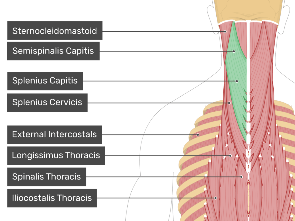
Superficial layer of back muscle
Splenius capitis and cervicis
Fibres travel obliquely upwards and laterally from the spinous processes to the transverse processes of the cervical vertebrae or the mastoid process
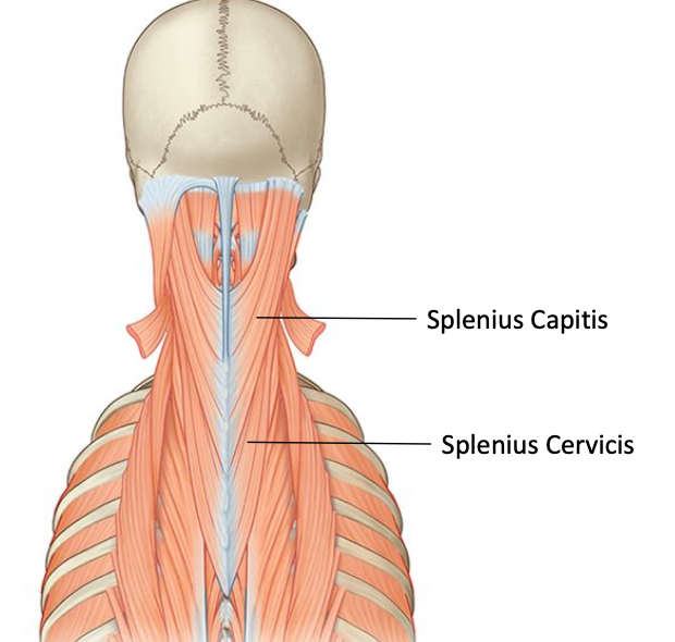
Splenius capitis
Origin: Ligamentum nuchae and spinous processes of T4 to C7
Insertion: Mastoid process

Splenius Cervicis
Origin: Spinous processes of T6 to T3
Insertion: Transverse processes of C3-C1
Nervse supply of splenius capitis and cervicis
Dorsal rami of adjacent spinal nerve at same spinal.
Action of splenius capitis and cervicis (bilatera)
Head and neck rotation, head and neck extension
Action of splenius capitis and cervicis (unilatera)
Rotate the head and neck to teh same side as muscle
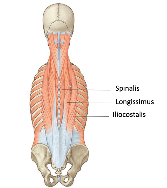
Erector spinae - common tendon
● All the erector spinae muscles arise from a thick broad tendon called the common tendon.
● Split into three muscle tendons around the upper lumbar region.

Erector spinae multisegmental
● All the erector spinae muscles span several segments.
● Not one single column.
● Instead, a series of overlapping fibres.

Erector spinae muscles - division
● Each erector spinae muscle has three divisions.
● Divisions named where the most superior fibres insert.
○ Lumborum - lumbar spine.
○ Thoracis - thoracic spine
○ Cervicis - cervical spine
○ Capitis - back of skull

Iliocoastalis
● Iliocostalis is the most lateral muscle.
● Fibres travel upwards from the common tendon to insert into the angles of the ribs and the transverse processes of C4 to C7.
● Iliocostalis has a lumborum, thoracis and cervicis division.

Longissimus
● Longissimus is the middle muscle and largest.
● Fibres travel upwards in a line between angle of ribs and transverse process to insert onto transverse processes of T1-T12, C2-C7, and the mastoid process.
● Longissimus has a thoracis, cervicis and capitis division.

Spinalis
● Spinalis is most medial muscle and smallest.
● Fibres connects sides of spinous processes and mostly found in thoracic region.
● Spinalis has a thoracis, cervicis and capitis division.
Nerve supply - erector spinae gourp
Dorsal rami of adjacent spinal nerve at same spinal.
bilateral contraction erector spinae action
Straighten spine from a flexed position, control forward flexion of the spine
Unilateral contraction erector spinae
Laterally bend the spine to the same side.
Erector spinae function
During one legged stance erector spinae on the non-weight bearing side contracts prevent the pelvis from dropping.
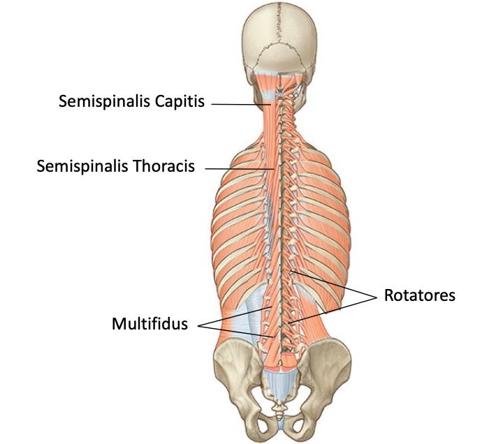
Deep layer tramsversospinales
● All three muscles are located underneath the longissimus muscle.
● Run in the gutter formed between the transverse processes and the spinous processes of the vertebrae