Neurobiology
Neurobiology of the mind is one of the fastest moving areas in psychology
Human nervous system can be divided into two
central nervous system, peripheral nervous system
Peripheral nervous system:
Includes both somatic and autonomic systems
Somatic nervous system:
Transmits signals to central nervous system from muscles, joints and skin via nerves. CNS sends signals through somatic nervous system to muscles, joints and skin to initiate, modulate or inhibit movement.
Autonomous Nervous System
Regulates internal environment of the body. Stimulates glands (i.e sweat glands), and organs (i.e heart, liver, lunges, pupils …). Nerves of ANS project signals from these targets to CNS
Is divided into sympathetic and parasympathetic systems. They are opposing systems in terms of outcome
Sympathetic signalling:
prepares body for action. Chronic stress leads to increased activity of this system
Dilates pupils, relaxes bronchi, accelerates and strengthens heartbeat, inhibits activity in intestines, contracts vessels
Parasympathetic signalling:
returns body to resting state
Contracts pupils, constricts bronchi, slows heartbeat, stimulates activity in intestines, dilates vessels
endocrine system
A commutation network that influences thought, behaviour and actions. It works together with the nervous system(preparing body to deal with perceived threats). Communicates through hormones.
Signals slower than nervous system. Uses hormones to influence breaking and body. These hormones are released into the bloodstream and can have wide-spread effects on the body and brain
Is primarily controlled by the hypothalamus via signals to the pituitary gland,, located at the base of the hypothalamus
Hypothalamus
Secretes hormones corticotropin-releasing hormone (CRH) that promotes pituitary gland to release adrenocorticotropic hormone (ACHT) which increases cortisol production of the adrenal gland
Cortisol, on the other hand, inhibits production of the CRH and ACHT ina negative feedback loop.
Central nervous system
Santiago Ramon y Cajal (1852- 1934)
Neuroanatomist who won Nobel prize in 1906, for establishing the neuron or nerve cell as the basic unit of the nervous structure
Consists of the spinal cord and brain
Building blocks of central nervous system are neurons and glial cells
spinal cord
Nerve cable that emerges from the base of the brain and extends down about two thirds of the length of the vertebral column
glial cells
Essential part of the nervous system allowing neurons to function. There are microglia and these are macroglia.
Microglia
protect CNS neurons. They are smaller than the other glial cells and are mobile within the brain. They can metabolize dead tissue and are involved in keeping the CNS healthy
macroglia types
Astrocytes: Link neurons to blood vessels, forming part of the blood-brain barrier. They engulf synapses (where neurons connect), regulating neurotransmitter release during synaptic transmission
Oligodendrocytes : Surround axons in the CNS, forming the myelin sheath that insulates axon, which allows the electrical signal that travels in the axon to travel faster.
Anatomy of neurons// neuron cycle
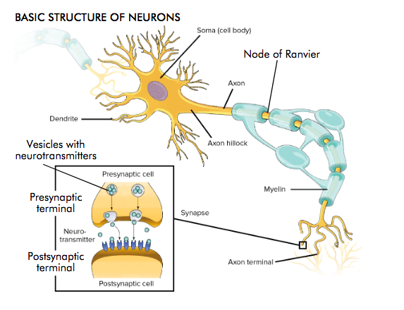
A signal is received at the dendritic spines at the post-synaptic terminals where the neuron synapses with the axon of another neuron.
This signal can produce an electric current that travels from the dendrite to the soma of the neuron
If the signal accumulating at the axon hillock in the soma is strong enough, the receiving neuron will “fire” i.e it will produce an electrical impulse at the axon hillock
This electrical impulse travels down the axon toward the terminal buttons 9the pre-synaptic terminals)
When the electrical impulse reaches the pre-synaptic terminal, it can produce a chemical signal: the release of neurotransmitters
When neurotransmitters reach the post-synaptic terminal of the receiving neuron and the cycle restarts.
The resting potential (part of the action potential)
Electrical potential refers to how much energy is stored up in a system
At rest, when a neuron is not active, the electrical charge inside and outside the neuron is different
This difference is called a potential. It is the potential electric charge that could be released
A neuron at rest has the resting potential of around -70millivolts (i.e there are more negatively charges inside the neuron than outside of it)
Change of the resting potential
If the electrical stimulation is strong enough, it exceeds the threshold of excitation and the axon of the stimulated neuron will fire an action potential
During the action potential, the neuron is briefly depolarizer so the membrane potential reaches about 40mV
Diffusion
Particles tend to move from a region of high concentration to a region of low concentration, eventually reaching equilibrium of equal dispersion
Diffusion results from Brownian movement which correlates with temperature
Diffusion is a force that pushes particles against their concentration gradient
Electrostatic pressure
Equally charged particles repel each other and differently charged particles attract each other. As applied to neurons: negatively charged molecules (anions such as Cl-) tend to move away from each other and so do cations (positively charged molecules like Na)
Anions and cations are moving towards each other
Resting potential: diffusion and electrostatic pressure
At rest, the concentration of negative ions inside the neuron is larger than outside, which has more positively charged particles than the inside. Unequal distribution of K+ and Na+ causes resting potential
In the extracellular space(outside the neuron) there’s a high concentration of Na and Cl particles in equal proportion and some K+ molecules.
In the intracellular space(inside neuron) there are many negatively charged large proteins 9organic anions), K+, and low amounts of Na+ and Cl-
As a consequence, during rest, the inside of the neuron has more negatively charged particles than the outside, which has more positively charged particles. This is why the resting potential of the neuron is negative
Dynamics of diffusion and electrostatic reassure to determine the resting potential
Cl- is in greatest concentration outside, diffusion forces it inside. However, because there are many negatively charged organic anions inside, electrostatic pressure pushes Cl- out
K+ is higher concentrated inside, diffusion pushes it outside. However the outside is positively charged therefore at the same time electrostatic pressure pushes K+ in
Na+ is in greater concentration outside, so diffusion forces it inside. At the same time, because it is positively charged and the inside has more negatively charged particles, electrostatic pressure pushes Na+ also inside. This is why there is the sodium potassium pump
Organic anions cannot leave the neuron
Sodium potassium pump
3 sodium ions bind to intracellular sites on the sodium potassium pump
Phosphate group s transferred to the pump via hydrolysis of atp
The pump undergoes a conformational change, translocating sodium across the membrane
Conformational change exposes two potassium binding sites on the extracellular surface of the pump
The phosphate group is released which causes the pump to return to its original conformation
This relocates the potassium across the membrane completing the ion exchange
Ion channels in the neuron membrane
There are proteins in the neuronal membrane that form little channels(pores) connecting the inside of the neurons with the outside. Some of these proteins allow certain types of ions to pass. These channels are called ion channels. There are for example sodium channels and potassium channels
Some ion channels open only under certain conditions, for example when the potential across the neuron membrane has a certain value. These channels are called voltage-dependent ion channels
voltage dependent channels
Sodium channels are voltage dependent channels: they open and Na+ rushes into neuron(driven by force of diffusion and electrostatic pressure). This depolarizers the neuron membrane potential
ion flow during action potential
Sodium channels are voltage dependent channels: they open and Na+ rushes into neuron(driven by force of diffusion and electrostatic pressure). This depolarizers the neuron membrane potential
When depolarisation reaches a point close to 0mV, potassium channels open. K+ leaves neuron due to force of diffusion (less K+ outside), and driven by electrostatic pressure from inside due to increase in positive charge from Na+ influx
When depolarisation reaches about 40mV the sodium channels enter a refractory state and close: no more Na+ can enter the neuron
The forces of diffusion and electrostatic pressure continue to force K+ out of the neuron. This reduces the positive charge inside the neuron, repolarising it i.e driving down the membrane potential
When potential reaches resting potential, k+ channels close
There is a slight hyperpolarisation the end, neuron reaches -70mV. Sodium-potassium pumps restore resting potential
agonist modulate neurotransmission
agonist drugs can increase how much neurotransmitter is made, so there is more inside each vesicle they can block the reuptake of neutransmitters they can mimic a particular neurotransmitter, binding to that neurotransmitter's postsynaptic receptors and either activating them or increasing the neurotransmitter's effect
eg. banzodiazepines (anti-axiety), agonist for GABA-A receptor
antagonist modulate neurotransmission
antagonist drugs can decrease the release of neurotransmitters so there are fewer inside each vesicle
they can help destroy neurotransmitters in the synapse they can mimic a particular neurotransmitter, binding to that neurotransmitter's postsynaptic receptors enough to block neurotransmitter binding
eg. ketamine(narcotic), antagonist for NMDA receptor
signal transmission summary of core events
When the action potential reaches the pre-synaptic terminal, it is converted from an electrical signal into a chemical signal. ‣ First, the action potential causes Ca2+ entry into the presynaptic terminal. This promotes that vesicles loaded with neurotransmitters (proteins) fuse with the presynaptic membrane. ‣ This causes neurotransmitters to be released into the synaptic cleft. There, they diffuse and eventually bind to receptors (proteins), that swim in the membrane of the post-synaptic cell. ‣ There are several different types of receptors in the post-synaptic membrane of neurons in the CNS. Each receptor can bind a particular neurotransmitter. For example, the AMPA and NMDA receptors are glutamate receptors, binding the neurotransmitter glutamate. ‣ Binding the neurotransmitter can cause a specific action of the receptor. For example, some receptors can form channels that allow electrically charged molecules (ions) to enter the post-synaptic terminal. Now the chemical signal has been converted back into an electrical signal. ‣ This influx of electrically charged molecules can cause a depolarisation of the post-synaptic neuron, which can then lead to the neuron firing an action potential. Some neurotransmitters can have the opposite outcome, they are inhibitory, not excitatory. ‣ Some receptors do not lead to changes in charge when neurotransmitters bind to them, but they influence biochemical processes in the neuron, which can change the structure or functioning of the neuron. ‣ Released neurotransmitters are removed from the synapse by enzymatic degradation or reuptake into the presynaptic terminal. ‣ Some psychopharmacological drugs influence these processes. SSRIs (selective serotonin reuptake inhibitors) for example artificially increase the amount of the neurotransmitter serotonin in the synapse, which is used as antidepressant treatment.
basic structure of neurons
soma- cell body dendrite axon hillock axon node of ranvier myelin axon terminal
inside synapse, vesicles with neurotransmitters, presnaptic terminal , neuro transmitter, postsnyaptic terminal
release and binding of neurotransmitters
Neurotransmitter binding, depending on the transmitter and the receptor can have a variety of outcomes. ‣ Some neurotransmitters can have an inhibitory effect (e.g., GABA): they make it less likely that the post-synaptic neuron will fire. Some have an excitatory effect (e.g., glutamate): they make it more likely that the post-synaptic neuron will fire
control of neurotransmitter release
synapse consists of: presynaptic termina, synaptic cleft, post-synaptic terminal. it also includes glial cells (astrocytes) that enclose three parts:
autoreceptor 2.reuptake 3.enzymatic degradation
autoreceptor
sense the amount of released neurotransmitter to regulate exocytosis
reuptake
a reuptake mechanism recycles neurotransmitter from the synaptic cleft, moving it back into the presynaptic terminal. some antidepressants interfere with this process for the neurotransmitter serotonin (SSRIs)
enzymatic degradation
enzymes in the synaptic cleft actively degrade released neurotransmitters
hippocampus
a complex neural structure consisting of grey matter and located on the floor of each lateral ventricle.
center for episodic memories. No new memories of events may be formed if there is damage to the hippocampus
logically set up. Neurons are perfectly lined up in the same way and the information flow goes through a loop
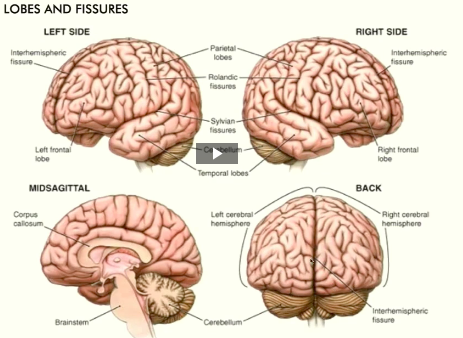
anatomy of the human brain
**MEMO THE PARTS OF THE BRAIN
two hemispheres, cerenellum, fissure, gyri, sulci.
folded structure fissure: (space between two hemispheres/ spaces within the nodes
size of the brain
reflects the size of the animal. receives input from all sides of the body so if u wanna represent the body u need a certain amount of neurons to do that. the bigger the animal the bigger the brain.
why is the brain folded
the brain is folded because it gets u more space. neurons are all at the outer layers so the more u fold it the higher the neuron count and sometimes this goes along with higher computational power
posterior view of the brain
frontal lobe olfactory bulb (synapse point of cranial nerve 1) optic chiasma optic nerve (2) optic tract mammillary body midbrain pons temporal lobe medulla oblongata cerebellum spinal cord
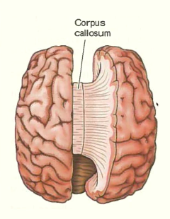
corpus callosum
Corpus callosum consists of millions of myelinated axons that connect the two hemispheres. ‣ Importance of this connection apparent in split brain patients
wide shape bc its axons. having epilepsy a treatment would be to cut the corpus callosum which prevents one hemisphere from talking to the other. but it also prevents that if u have epilepsy from one brain area to travel to the entire brain so if u have seizures they're much more reduced in severity because they can no longer become global seizures.
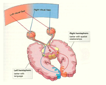
contralateral organization
in the eye, the part of the retina that gets the right visual field gets projected to the right hemisphere
corpus callosum transmits information from one hemisphere to the other so that both hemispheres get the input of what is being seen although only one side gets the input
right hemisphere better with spatial relationships left hemisphere better with language
because the hemispheres are somewhat specialized, in split brain patients two independent forms of knowledge exist
Left hemisphere critical for language: if split patient sees object with right eye, this is projected into the left hemisphere and therefore the object can be names
what patient sees with left eye(project into right hemisphere) cannot be named bc the right side does not have access to language system. however, patient can choose this item with the left hand (controlled by right side)
show patient a a picture of a spoon and a fork. ask what they see they say fork. ask to pick up the object with the left hand, will pick up a spoon
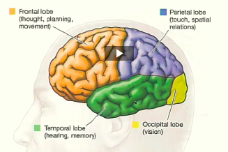
lobes
frontal lobe pariental lobe temporal lobe occipital lobe
frontal lobe functions
executive functions deals with cognition and memory ability to concentrate, judgement, consequence, analysis, problem solve, plan, personality(including emotional traits) control eye movement planning can inhibit certain actions easier
parietal lobe function
integrating information from several senses processes spatial orientation, some parts of speech, visual perception, and pain and touch sensations
visual processing pathway that gets split up and goes to occipital lobe- refered to as the what pathway and where pathway
occipital lobe function
visual processing center of the brain. contains most of what is referred to as the visual cortex is also the part of the brain where dreams originate. if this is lesioned u are blind even if ur eyes are perfectly functioning
vision isn't limited to here, on the temporal lobe (where objects are represented)
temporal lobe function
chief auditory receptive area and contains the hippocampus, which is the chief region where long-term memory is formed. also deals with high level visual processing (faces and scenes)
penfields mapping studies
cut open the brain, doesn't hurt, patient is conscious, have to go through the brain so before u cut stuff away you stimulate it with an electrode and see what happens
u find a path to the target that destroys as little essential functions as possible
wilder penfield did this kind of surgey and found out if u stimulate certain areas people respond with different experiences (i smell roses...). important bc linked brain activities to the state of experiences, so brain and mind have some sort of relationship because brain stimulation can lead to certain experiences
primary motor and sensory cortex
neurons devoted to the thumb compared to the rest of the hand is a larger area. human face has a lot of motor control in the mouth. u can see that the lips area of the face also has a high sensitivity
basically by analyzing how many neurons the cortex has u can see how sensitive/ how much motion an area on the body has bc more neurons devoted to that area to process it. u can see whats more important for humans
brain stem: basic survival functions
medulla oblongata - involuntary functions like sneezing, vomiting, respiration and other autonomic functions, breathing, some areas control of the heart, control of the sleep wake cycle
Brain stem is the superior end of spinal cord. ‣ Brain stem a main communication pathway between brain and body. ‣ Houses nerves that control basic functions, such as breathing, heart rate, swallowing, vomiting, urinating, orgasm
Reticular formation: projects into cerebral cortex, affects general alertness. Involved in sleep regulation
cerebellum
Cerebellum (“little brain”) critical for proper motor function. For example, damage causes head tilt, balance problems. ‣ Cerebellum essential for motor learning and motor memory. It operates independently. ‣ Cerebellum also involved in planning, event memory, language, emotions.
subcortial structures, thalamus
Thalamus is gateway to cortex. With the exception of odour information, it receives all other sensory modalities. Smell, the oldest and most fundamental sense, has a direct route to cortex. ‣ During sleep, thalamus partially shuts down incoming sensory stimulation.
hypothalamus
Hypothalamus (below thalamus). Indispensable for survival. ‣ Receives afferents from almost every body and brain region. ‣ Affects functions of many internal organs, regulates body temperature, blood pressure, blood glucose levels. ‣ Involved in motivated behaviours, e.g, thirst, hunger, aggression, lust.
basal ganglia
Basal ganglia are critical for planning and producing movement. ‣ Afferents from entire cerebral cortex. Efferents to motor centres of brain stem. ‣ Damage can cause tremors and rigidity in Parkinsons’s disease, or loss of movement control in Huntington’s disease. (no inhibition for the muscles to stop moving in huntingtons disease, almost all muscle functions are affected even swallowing -kills people) ‣ Nucleus accumbens is part of basal ganglia, and is important for reward processing and motivating behaviour. This involves dopamine activity in the nucleus accumbens.
limbic system
Hippocampus essential for episodic and certain spatial memories. ‣ Hippocampus and amygdala densely connected. Amygdala processes emotions, such as fear. ‣ Amygdala modulates processes in the hippocampus (and other brain regions), might signal importance of events, thus increasing their likelihood of being retained.
interaction between amydala and hippocampus can make certain memories stronger and more important, amygdala sends a signal and allows that space to be better remembered bc something significant happened there.
Psychophysics
the science of defining quantitative relationships between physical and psychological events. Research aimed at relating physical stimuli to the contents of consciousness
absolute threshold
level of stimulus intensity required to create a conscious experience
just noticeable difference
smallest magnitude of stimulus required to detect discrete stimuli (advanced in weber's law)
signal detection theory
accounts for individual biases
sensation
the ability to detect a stimulus. features of the environment that are used to create understanding of the world
perception
the act of giving meaning to a detected stimulus combining of sensations arriving from the sensory ssytem with prior knowledge
how do we assign meaning to incoming sensory information?
transduction
conversion of one energy to another. process where stimuli i.e light are converted to neural electrochemical energy
bottom-up processing
processing the elementary messages from the environment
top-down processing
applying memory, knowledge etc to understand and create a perception
extramission theory of vision
the proposal that visual perception is accomplished by rays of light emitted by the eyes, has been replaced by the intromission theory
plato - 4th century bc first proposed extromission theory of vision; that the eye sends out vision beams which seize objects
galen, 2nd venture AD further supported extromission theory, widely influence in European thinking for the next millennia
winer et al. 2002 found that 50% of American adults believe in extromission theory
intromission theory
has replaced extromission theory of vision. visual perception comes from some representation of the object entering the eyes
light
electromagnetic energy exists as both particles (photons) and waves
we only detect a small band ~400 - 700 nm
wavelength
perceived hue
frequency
cycle rate
amplitude
perceived intensity
things in the eye
cornea iris/pupil lens retina fovea
cornea
transparent tissue which allows light rays to enter the eye and focus on objects
iris/pupil
coloured part of the eye, consists of muscular diaphragm which regulated light entering the eye by expanding and contracting the pupil (center of the iris)
lens
crystalline lens inside the eye that enables the changing of focus (accomodation)
retina
contains photoreceptors (light-sensitive neurons)
neural signal pathway:
photoreceptors
bipolar cells
ganglion cells
fovea
small pit that cointains the highest concentration of colour sensitive light receptors- has the highest visual acuity in the retina
photoreceptors
transduce light into neural activity
two main parts: rods and cones
rods
Dim light (“night vision”) • Sensitive to all wavelengths of light • Black and white vision • Low resolution (one bipolar cell: many rods) • Around 100 million rods in each human eye
cones
Bright light (“day light vision”) • Sensitive to blue/ red/ green wavelengths of light • Colour vision • High resolution (one bipolar cell: one cone) • Around 5 million cones in each human eye
there are three types of cones
types of cones
s- cones m-cones L-cones
s-cones
short-wave length cones (blue)
m-cones
medium-wave length cones (yellow and greens)
L-cones
long--wave length cones (reds)
tetrachromats
humming birds also see uv
photoreceptors location in the retina
Rods are found in the periphery. • Respond to amount of light • Signal information about motion
Cones are found in the fovea (centre). • Respond to quality of light. • Signal information about detail.
blindspot
Where the optic nerve leaves the eye -> No photoreceptors in this area. • Visual system usually fills in blindspot with information from surrounding area
bipolar cells
Intermediate cells that determine the information flow from photoreceptors to ganglion cells.
two types: diffuse bipolar cells and midget bipolar cells
diffuse bipolar cells
In the periphery. • Respond to around 50 rods -> increase sensitivity but reduced acuity (1 diffuse bipolar: 50 rods • Convergence of information: Many rods:one diffuse bipolar cel
midget bipolar cells
Found in the fovea (centre). • Receive input from a single cone and pass on info to a single ganglion (1 midget bipolar: 1 cone)
ganglion cells (retinal ganglia)
final layer of the retina
m-cell and p-cell
Retinal Ganglion Cell (RGC) axons form the optic tract.
m-cell (large ganglion cell)
Mostly respond to rod cells via diffuse bipolar cells
P-cell (small ganglion cell)
Mostly respond to cone cells via midget bipolar cell
receptive field
the region on the retina in which visual stimuli influence neural firing rate
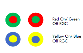
Receptive fields of individual retinal ganglion cells
ON-center, OFF-surround cell A ganglion cell that increases firing in response to increase in light intensity in it’s receptive field center. • When light hits the center - firing rate at maximum • When light covers full receptive field - firing rate returns to baseline • When light on surround -> baseline firing is repressed
off-center, on-surround cell retinal ganglia
A ganglion cell that increases firing in response to decrease in light intensity in it’s receptive field
function: Contrast in illumination help detect object edges, whether is it day time/ night time, whether indoors/ outdoors etc. Also thought to be involved in colour processing
Opponent process theory (colour detection)
P-cells fire rapidly to one wavelength and reduce to another- forms pairs of colours. • Red-Green (P-cell) • Blue-Yellow (P-cell) • Black-White (M-cell) (e.g. the reduction of red firing = green)
Trichromatic theory
Colour vision occurs by comparing the activation of the three different cones.
the visual pathway
Information from the retina leaves the eye via the optic nerve • Information from the optic nerve travels to the optic chasm (CROSS OVER). • Lateral geniculate nucleus (LGN) of thalamus. • Visual Cortex/Striate Cortex/V1
processing after visual cortex
what stream ventral stream - temporal lobe object recognition
where stream dorsal -> parietal lobe location of objects in space
gestalt principles
Refers to theories of visual perception developed by German psychologists in the 1920s • Gestalt psychology describes how people tend to organise visual elements into whole entities. Gestalt psychologists believe we are born with specific predisposed ways of organizing information so that it has utility. • Six Gestalt Principles