EM 4 Saccades
1/69
There's no tags or description
Looks like no tags are added yet.
Name | Mastery | Learn | Test | Matching | Spaced | Call with Kai |
|---|
No analytics yet
Send a link to your students to track their progress
70 Terms
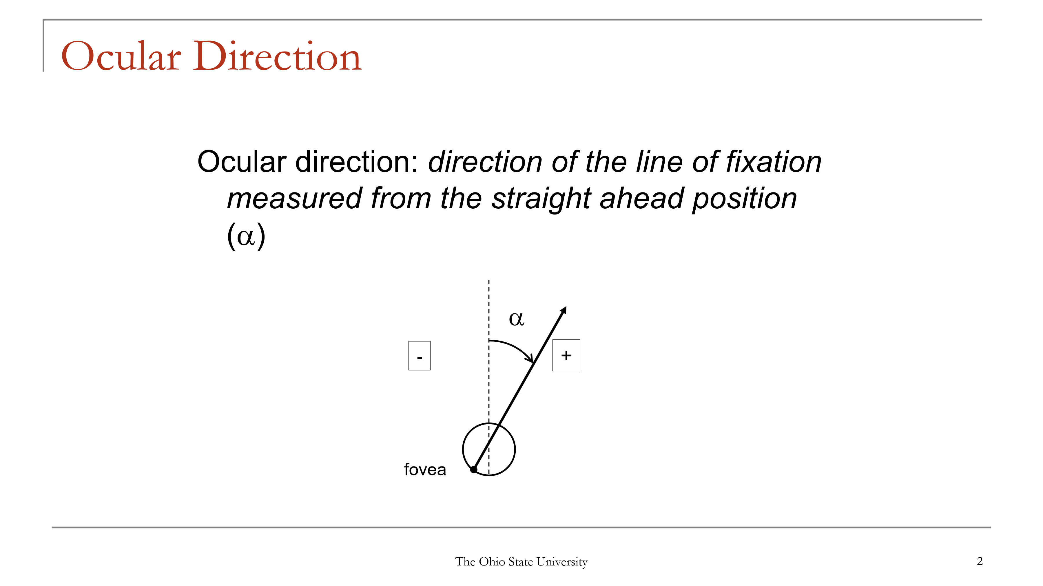
What is ocular direction (α), and how is it measured?
Ocular direction = the direction of the line of fixation measured from the straight-ahead position.
Symbol: α
It describes where the eye is pointing relative to straight ahead.
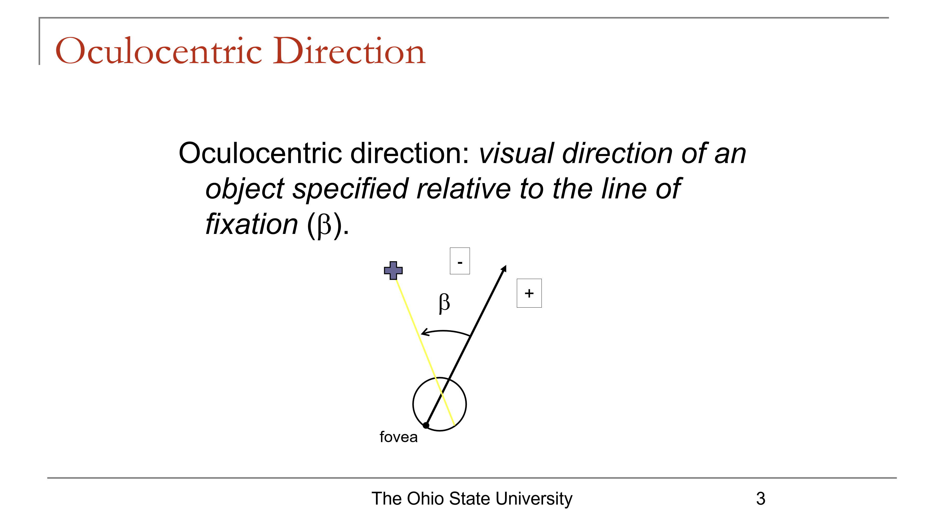
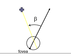
How do oculocentric direction (β) and egocentric direction (χ) differ?
Oculocentric direction (β): direction of an object relative to the line of fixation
“Object relative to where the eye is looking”
Egocentric direction (χ): direction of an object relative to straight ahead
“Object relative to the observer’s straight-ahead position”
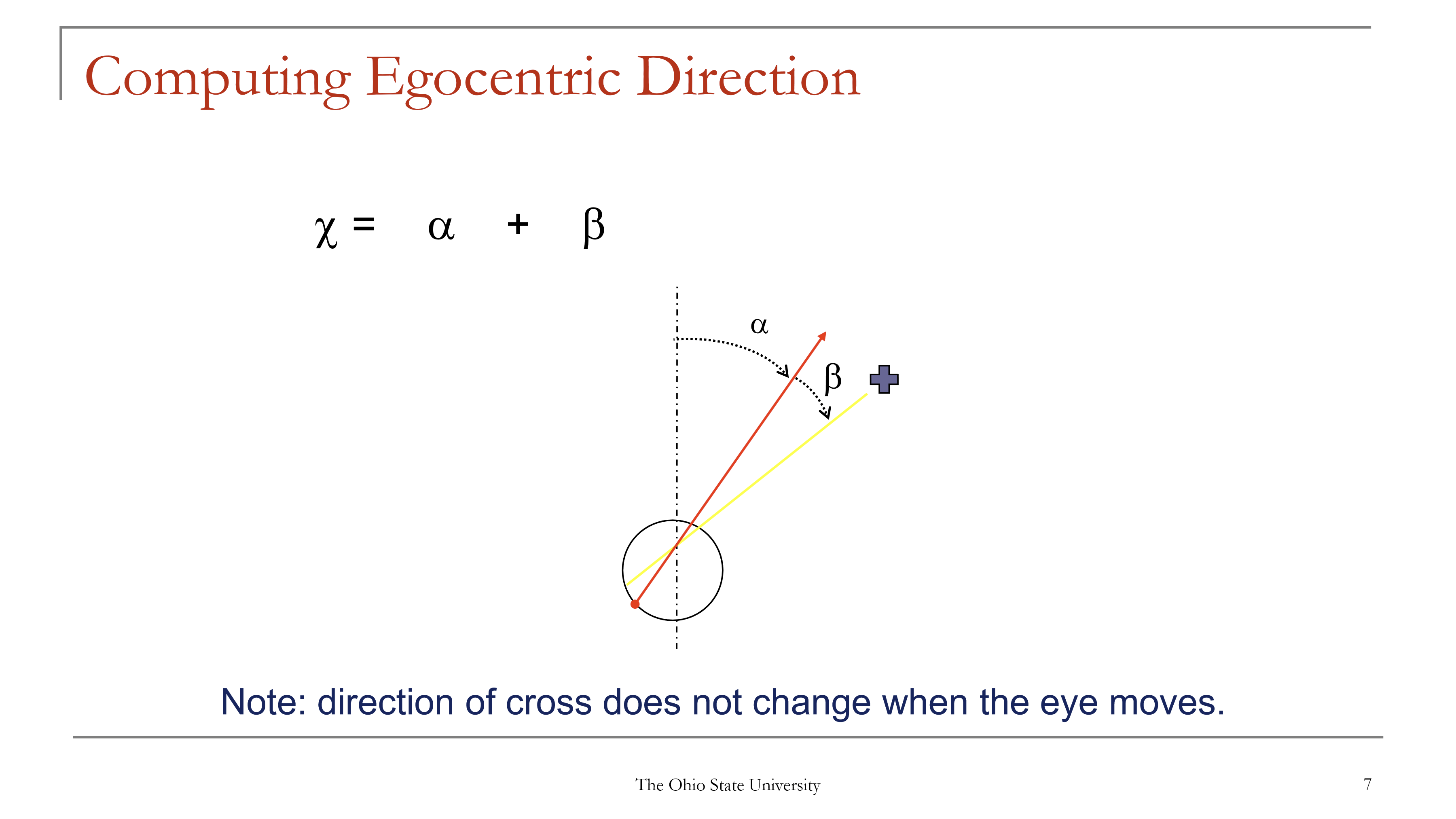
What is the relationship between ocular direction, oculocentric direction, and egocentric direction?
The formula is:
χ = α + β
Where:
α = ocular (eye) direction = where the eye is pointing
β = oculocentric (relative) direction = where the object lies relative to the fovea / fixation
χ = egocentric (absolute) direction = where the object is perceived relative to straight ahead
What is the sign convention for visual direction?
Right and up = positive (+)
Left and down = negative (−)
What is the key difference between egocentric direction (χ) and ocular/oculocentric directions (α, β) in terms of processing?
χ (egocentric direction) is cortically computed
α (ocular direction) and β (oculocentric direction) are sensed
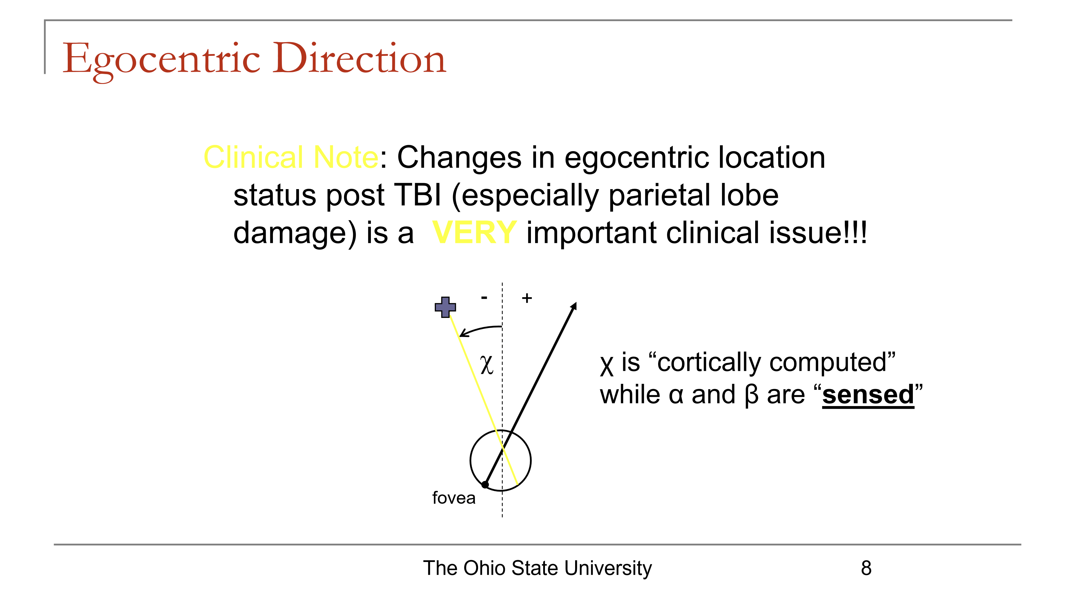
Why is egocentric direction clinically important after TBI, especially with parietal lobe damage?
Damage after TBI, especially involving the parietal lobe, can alter egocentric location processing.
This is clinically important because the patient may have impaired ability to judge where objects are relative to straight ahead / self.
High-yield association:
Parietal lobe → important for spatial localization
TBI/parietal injury → abnormal egocentric localization
A patient can sense eye position and object location relative to fixation, but still mislocalize where the object is relative to self. What process is likely impaired?
Impaired cortical computation of egocentric direction (χ).
How is oculocentric direction (β) sensed?
Oculocentric direction (β) is sensed from retinotopic information.
What is a retinal local sign?
A retinal local sign is the unique oculocentric direction associated with a functional retinal point.
How is ocular direction (α) sensed?
Ocular direction (α) is sensed from extraretinal information.
What are the 2 major theories for how ocular direction (α) is sensed?
Inflow theory
Outflow theory
Which type of information is used to sense ocular direction (α) versus oculocentric direction (β)?
Ocular direction (α) → extraretinal information
Oculocentric direction (β) → retinotopic information
Why is ocular direction (α) not determined by retinal location alone?
Retinal location tells where an object lies relative to fixation (β), but not where the eye itself is pointing.
So ocular direction (α) requires extraretinal signals.
What is the core computation in the model of egocentric direction?
The brain computes direction by combining:
Egocentric direction = α + β
Where:
α = ocular direction (eye position relative to straight ahead)
β = oculocentric direction (object relative to fixation)
Result: perceived object direction relative to the observer.
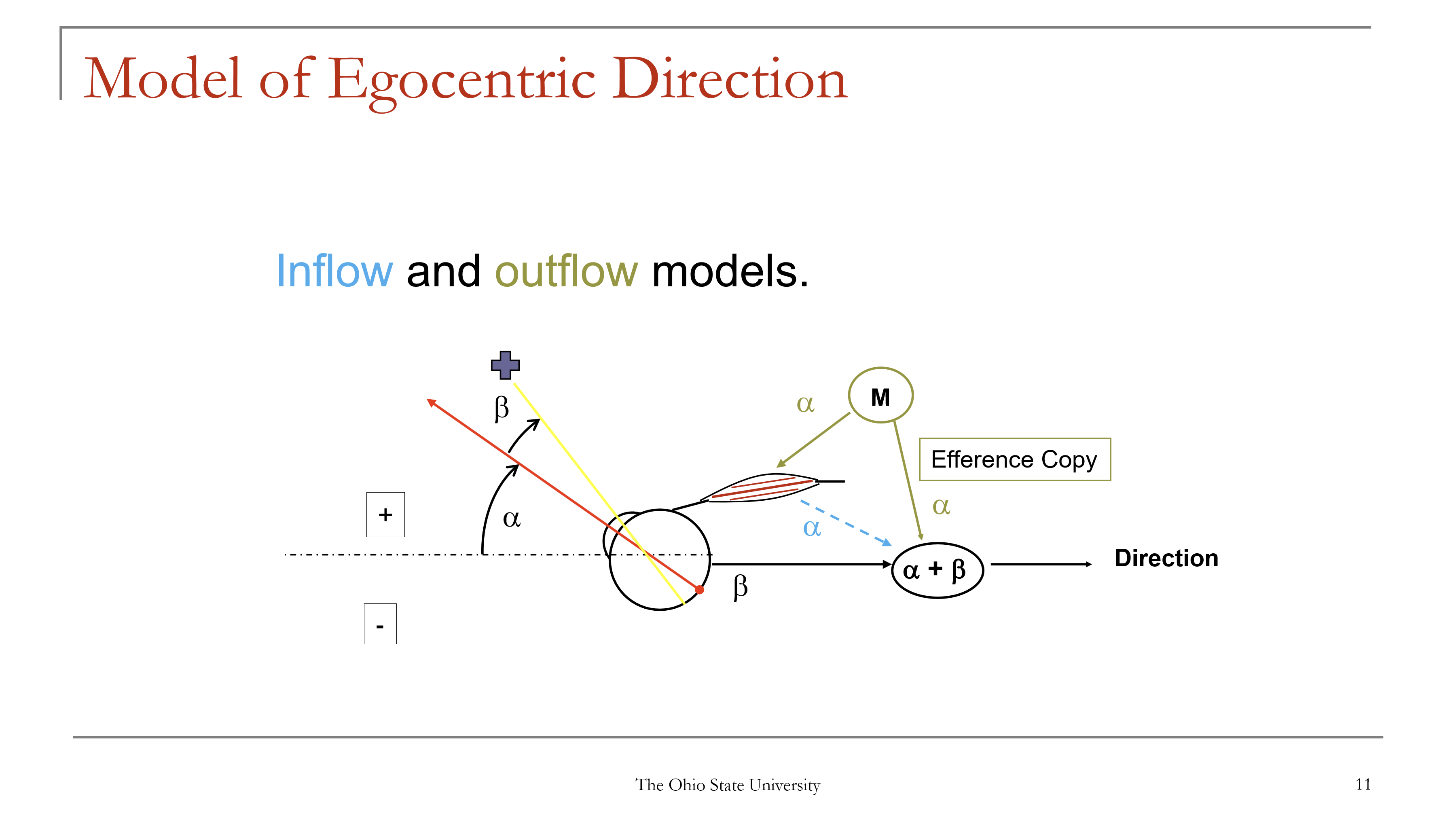
In the outflow model, what signal provides information about ocular direction (α)?
The outflow model says ocular direction (α) is derived from an efference copy of the motor command sent to the eye muscles.
High-yield association:
Outflow = motor command copy
Also called a signal related to intended eye movement
What is the role of efference copy in visual direction?
An efference copy provides the brain with information about the eye movement command, allowing the brain to estimate ocular direction (α) and combine it with β.
What is the purpose of the forced duction experiment in visual direction?
Forced duction tests how the brain senses ocular direction (α).
It compares:
Inflow theory = α from extraocular muscle proprioception
Outflow theory = α from efference copy of motor command
What happens to β when the eye is pushed upward during forced duction?
β becomes negative because the image no longer falls on the fovea and shifts in the negative retinal direction.
What does inflow theory predict during forced duction of the eye?
In inflow theory, the brain senses eye position from extraocular muscle proprioception.
So after the eye is pushed up:
perceived α becomes positive
β becomes negative
therefore α + β = 0
Prediction: the object should not appear to move.
What does outflow theory predict during forced duction of the eye?
In outflow theory, ocular direction comes from efference copy of the motor command.
If the eye is pushed externally:
no motor command was sent
so perceived α remains 0
β becomes negative
therefore α + β is negative
Prediction: the object should appear to move in the negative direction.
What is the actual perceptual result of forced duction, and what theory does it support?
During forced duction, the object is perceived to move in the negative direction. This supports outflow theory.
Why is forced duction evidence against a purely inflow-based account of ocular direction?
If inflow alone were correct, manual upward movement of the eye would make:
α positive
β negative
and these would cancel
So the object should appear stable.
Because the object instead appears to move, inflow alone does not explain the result.
How does forced duction illustrate the equation χ = α + β?
Forced duction changes retinal position (β) without a matching motor command for eye position (α) under outflow theory.
Thus:
β changes
α does not update
so χ changes
This makes a stationary object appear to move.
What is a saccade?
A saccade is a rapid gaze-shifting eye movement used to move the fovea onto an object of interest.
What does it mean that saccades are ballistic?
Ballistic means that once a saccade begins, it goes to completion and cannot be adjusted mid-flight.
What stimulus triggers a saccade?
A saccade is triggered when an object has a non-zero oculocentric direction (β ≠ 0) — meaning the object is displaced from the fovea.
Why are saccades important for vision?
Saccades place objects of interest onto the fovea, where visual acuity is highest.
What are the typical size characteristics of naturally occurring saccades?
About 85% of naturally occurring saccades are < 15°.
If a larger gaze shift is needed, eye movement is usually combined with head movement.
What are the key movement relationships that characterize saccades?
Saccades have characteristic relationships between:
Amplitude
Peak velocity
Duration
How fast can large-amplitude saccades become?
Large saccades can reach peak velocities of about 700°/sec (some sources cite up to 1000°/sec).
How does saccade peak velocity change as amplitude increases?
Peak velocity increases as amplitude increases, but the increase becomes much slower beyond ~20°.
Are saccades faster toward straight ahead or away from straight ahead?
Saccades are faster toward straight ahead than away from straight ahead.
How does saccade duration change with increasing amplitude?
Duration increases as amplitude increases.
In what situations are slower saccades reported in terms of peak velocity?
Slower saccades have been reported:
in infants
with alcohol use
with voluntary control
with fatigue
in neurologic disease
What neurologic conditions are associated with slower saccade peak velocity?
Disease conditions associated with slow saccades include:
Multiple sclerosis (MS)
stroke
tumor
aneurysm
What are the 2 parts of a saccade?
A saccade has 2 components:
Pulse
Step
What is the pulse component of a saccade?
The pulse is the brief, high neural command that drives the eye to a new position.
It overcomes the viscous resistance of the globe and orbital tissues.
What is the step component of a saccade?
The step is the sustained neural signal that keeps the eye in the new position after the saccade.
It overcomes the elastic forces that would otherwise pull the eye back toward its original position.
What mechanical problem does the pulse solve, and what mechanical problem does the step solve?
Pulse overcomes viscous resistance → allows rapid eye movement
Step overcomes elastic restoring forces → maintains the new eye position
What is a glissade, and when does it occur?
A glissade is a slow corrective movement that occurs when the pulse and step are not properly matched.
It occurs between the pulse and step
It helps bring the eye to where the step “wants” it to be
It is relatively slow (about 100 ms)
What happens if the pulse is too long during a saccade?
If the pulse is too long, the eye overshoots the target, then shows a glissadic return.
This is called a hypermetric saccade.
What happens if the pulse is too short during a saccade?
If the pulse is too short, the eye undershoots the target, then makes a glissadic correction.
This is called a hypometric saccade.
What is the typical behavior of large saccades (>20°)?
Large saccades almost always undershoot the target initially, then are followed by a corrective saccade.
After a large saccade undershoots, what type of correction usually occurs?
A corrective saccade usually occurs after the appropriate latency period.
How is a corrective saccade different from a glissade?
Corrective saccade: a separate, delayed saccade made after the initial movement misses the target
Glissade: a slow post-saccadic drift/correction caused by pulse-step mismatch
Can large saccades ever overshoot the target?
Yes. Although undershoot is more typical, large saccades can also overshoot and then require a corrective saccade.
What is the average latency of a saccade, and what is the usual range?
Average saccadic latency is about 200 ms, with a typical range of 120 to 350 ms.
What is saccadic latency?
Saccadic latency is the time from target presentation to the start of eye movement.
What is the duration of response for a saccade?
The duration of response is the time from when the eye starts to move until the eye is actually pointed at the target.
What processes contribute to saccadic latency?
Saccadic latency includes deciding:
whether a target is present, and
whether to look at it
What factor causes some of the greatest changes in saccadic latency?
One of the biggest influences on saccadic latency is giving subjects prior information about target movement.
How does knowing where a target will appear affect saccadic latency?
If the subject knows in advance where the target will appear, saccadic latency decreases compared with when the target could appear in one of two possible positions.
What are express saccades?
Express saccades are saccades with a very short latency, typically occurring when a warning period/cue helps the subject prepare for the target.
What is saccadic suppression?
Saccadic suppression = a temporary elevation in visual threshold before, during, and shortly after a saccade.
How does saccadic suppression affect detection of a brief flash of light?
Detection of a brief flash can increase by 50% or more from about 20 ms before to about 35 ms after a saccade.
What is the functional purpose of saccadic suppression?
Saccadic suppression helps reduce awareness of retinal image smear during a high-velocity saccade.
Why can’t saccadic suppression alone fully explain the lack of blur during saccades?
Although saccadic suppression raises visual threshold, it cannot fully account for the almost complete absence of blurred perception during a saccade.
What is saccadic omission?
Saccadic omission is the phenomenon in which the blurred visual input during a saccade is not consciously perceived.
What mechanism is thought to explain the lack of perceived blur during a saccade: saccadic suppression or saccadic omission?
Saccadic omission is believed to be mainly responsible for the lack of perceived blur during a saccade.
How does masking explain saccadic omission?
The blurred image during a saccade is obscured by visual input that comes:
immediately before the saccade (forward masking), and/or
immediately after the saccade (backward masking)
What did the Campbell & Wurtz experiment show about blur during saccades?
A flash presented only during a saccade produces a blurred percept
But if the flash starts before the saccade and/or continues after the saccade, the blur is not perceived
Why is blur during a saccade usually not perceived, even though vision is actually blurred during the movement?
Because the brain mainly processes:
what was seen before the saccade, and
what is seen after the saccade
What are the main causes of abnormal saccades on clinical exam?
Abnormal saccades may be due to:
brain pathway lesion
poor attention
poor coordination
When should abnormal saccades raise concern for a brain lesion rather than poor attention/coordination?
Suspect a brain lesion if abnormal saccades are accompanied by:
absent or nearly absent saccades
reduced vision
visual field loss
numbness/tingling
muscle weakness or paralysis (including face)
possibly headache
Potential causes:
MS
stroke
tumor
aneurysm
What are the classic clinical features of abnormal saccades?
Abnormal saccades may:
look slow
have longer latency
be inaccurate
How are saccades tested clinically at the bedside?
Ask the patient to switch gaze back and forth between the examiner’s left and right fingers, held about 30–40 inches apart.
What you assess:
speed
latency
accuracy
If there is no strong suspicion of a brain lesion, what is the usual assumption when saccades are poor?
Poor saccades are usually assumed to be due to:
poor attention
and/or poor coordination
What is the clinical significance of poor saccades in children?
If a child has poor saccades, consider referral for eye movement training, especially if the child is a poor reader.
What is an antisaccade?
An antisaccade is a saccade made to the opposite side of where the target moves/ a rapid, voluntary eye movement made in the opposite direction of a sudden visual stimulus (such as a flashing light).
Poor antisaccades are associated with lesions/problems in what area?
Poor antisaccades are seen in:
schizophrenia
other frontal lobe problems