Anatomy Lab: Neurology
1/41
There's no tags or description
Looks like no tags are added yet.
Name | Mastery | Learn | Test | Matching | Spaced |
|---|
No study sessions yet.
42 Terms
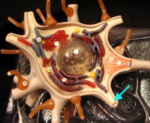
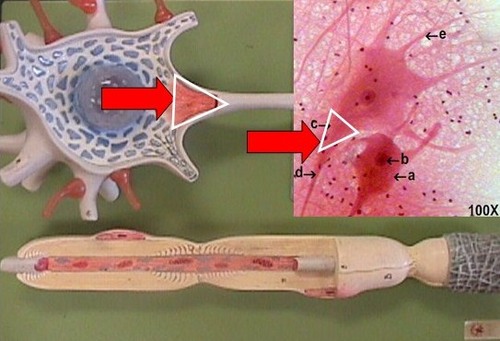
Perikaryon
Cell body of a neuron
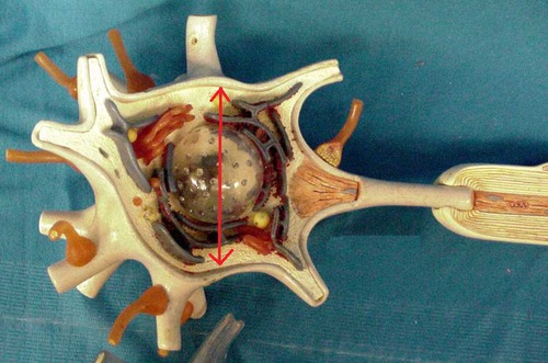
Nucleus
Inside the cell body
Neurofibrils
Protein filaments that form the core of the nerve fiber (red stuff)
Dendrites
Receive signals (the thick sticks sticking out of the cell body)
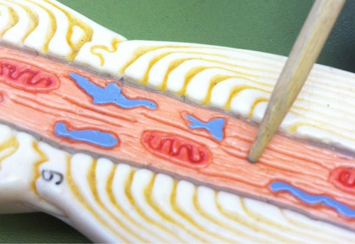
Axon
Transmits signals away from the dendritic zone (grey thing inside the neurolemmocytes)
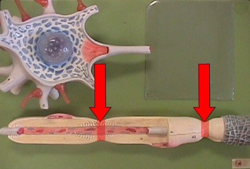
Neurolemmocytes (Schwann cells)
Covering over neural fibers of all peripheral neurons. Wrapped around axon to form a Myelin sheath, Schwann cells (only found in the peripheral neural system, wrapped around axon, it's a cell, and the layers are the cell membrane, facilitate the speed of action potential, will help regeneration of any fibers.
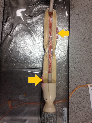
Neurofibril Node (Node of Ranvier)
Points of discontinuity between neurolemmocytes. Facilitates speed of nerve impulse transmission
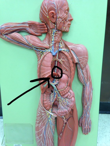
Sensory Neuron (afferent)
Yellow color on model, in the dorsal side, takes stimulus to CNS
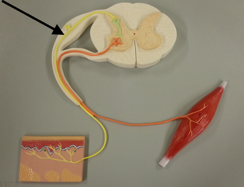
Motor Neuron (efferent)
Orange color on model, takes CNS to PNS, go to the spinal nerve into muscle
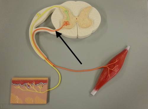
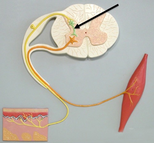
Interneuron (integrator cells between sensory and motor)
Green color on the model, within the central nervous system, located in grey matter, communterm-7icates between sensory and motor
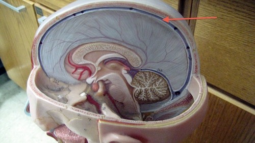
Meninges
3 layers of protective connective tissue that surrounds brain and spinal cord. Contains and controls the flow of CSF.
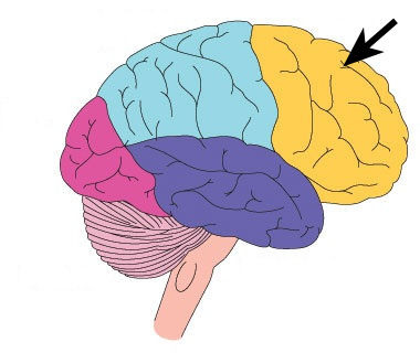
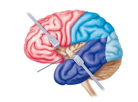
Frontal lobe
Includes the motor cortex and areas for thought and memory
Occipital
Visual cortex
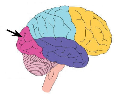
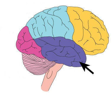
Temporal lobe
Processing center for hearing and emotional behavior
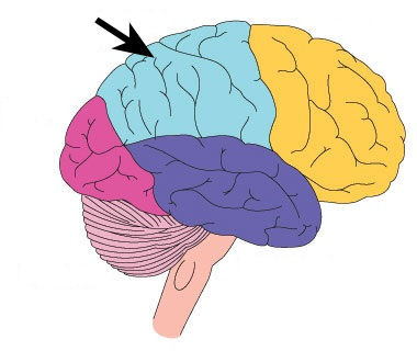
Parietal lobe
Includes the sensory cortex, main receiving area for skin and joint sensation
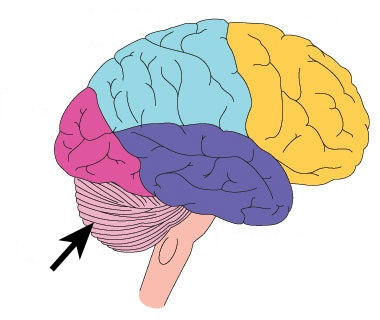
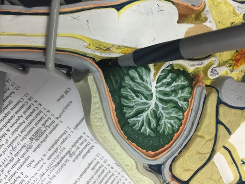
Cerebellum
Reflex center for balance, maintenance of posture, fine/complex muscle movements, remember with cerebalance
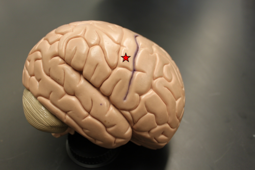
Central Sulcus
The groove that separates frontal and parietal lobe

Precentral Gyrus
Bumps in front of central sulcus, motor
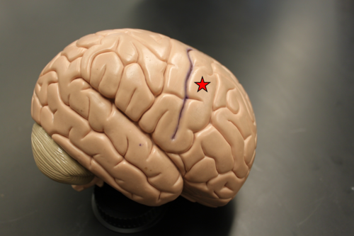
Postcentral Gyrus
Bumps behind the central sulcus, sensory
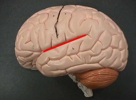
Lateral Cerebral Fissure
Separates the temporal lobe from the parietal lobe
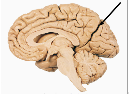
Parieto-occipital Fissure
Separates parietal lobe from occipital lobe
Insula
Little mini brain looking one inside the brain, deals with emotion
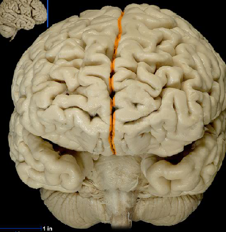
Longitudinal Cerebral Fissure
Splits the hemispheres, runs front to back
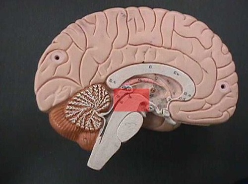
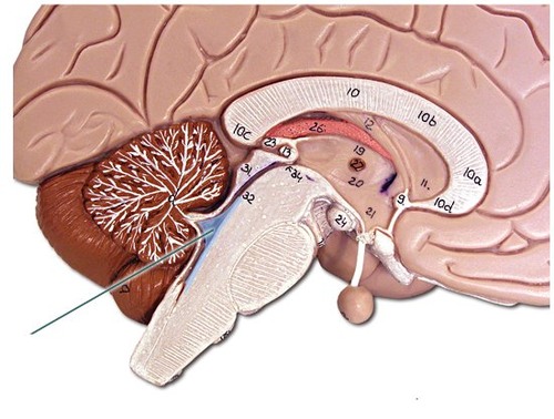
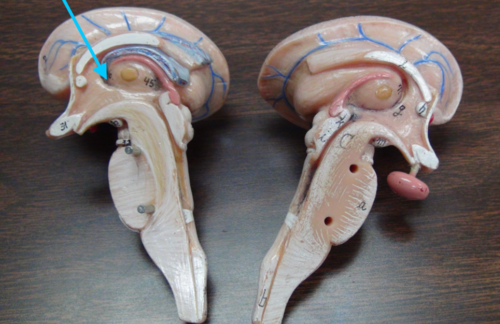
Midbrain
Part of the brain stem, second part, underneath diencephalon, cerebral peduncle, corpora quadrigemina
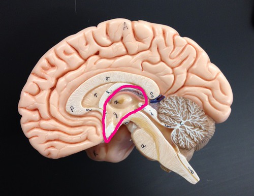
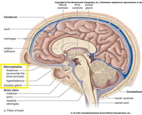
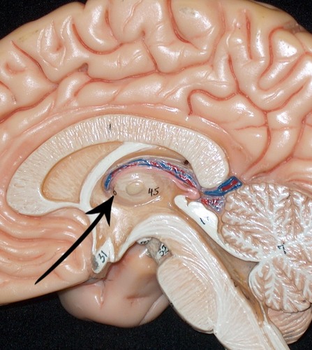
Diencephalon
Part of the brain stem, first part, thalamus. hypothalamus, pineal gland
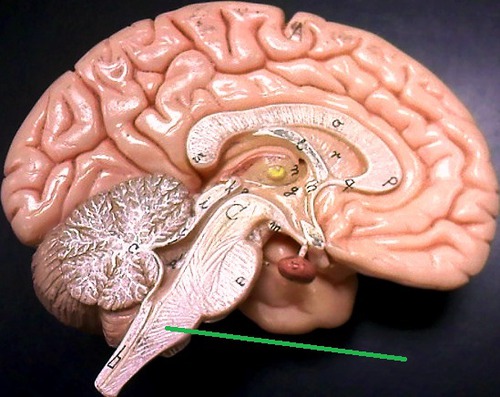
Pons
Part of the brain stem, third part, the little belly sticking out, a bridge between your spinal cord and higher brain functions

Medulla Oblongata
Part of the brain stem, last part, underneath pons
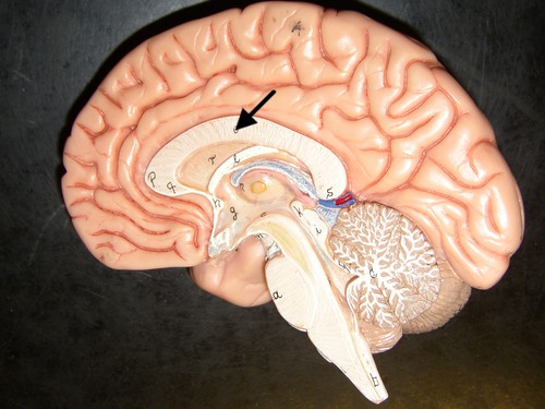
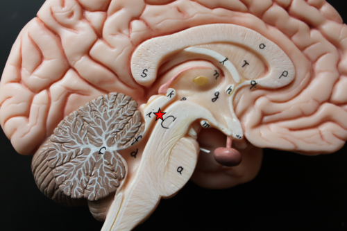
Corpus Callosum
Connects right and left brain, mass above the diencephalon
Parts of the Diencephalon
a. Fornix right at the top of the diencephalon b. intermediate mass of the thalamus c. hypothalamus, mammillary body d. hypophysis (pituitary) e. corpora quadrigemina
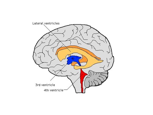
Septum pellucidum
Separates lateral ventricles, between fornix and corpus colosum

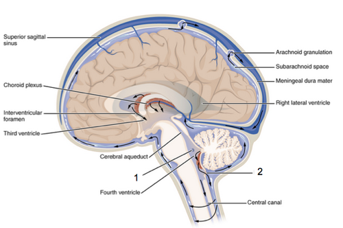
Third ventricle
Choroids plexus of 3rd vent
Fourth ventricle
Choroids plexus of 4th ventricle behind pons medullum
2 lateral ventricles
Choroid plexus produces CSF, behind septum pellucidum
Interventricular Foramen (of monro)
3rd ventricle, the little hole
Cerebral Aqueduct
4th ventricle
Lateral Aperture
Magendie, through median aperture
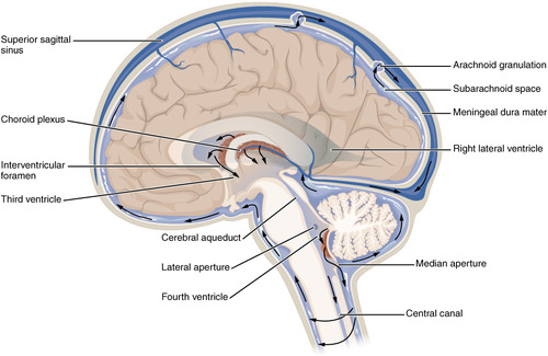
Median Aperture
Cisterna cerebellomedularis of (subarachnoid space)
Cisterna superior
Above cerebellum & down Subarachnoid space of Spinal Cord (posterior) & up along the anterior
Subarachnoid Space
Around cerebral hemispheres
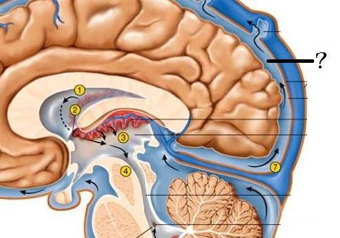
Arachnoid Granulations
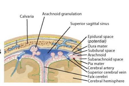
Sagital Sinus