Microbiology Exam 1
1/144
There's no tags or description
Looks like no tags are added yet.
Name | Mastery | Learn | Test | Matching | Spaced | Call with Kai |
|---|
No analytics yet
Send a link to your students to track their progress
145 Terms
Cellular
Archaea, bacteria, fungi, protozoans
Acellular
Viruses, Prions
Prokaryotes
No membrane bound organelles, contains nucleoid region
Eukaryotes
Membrane-bound organelles, has nucleus
Ubiquitous
Microbes are found everywhere
Prions
No nucleic acid, only protein, acts as infectious organism
Virus
Small amount of DNA or RNA, protein coat and sometimes a membrane
Louis Pasteur
Disproved abiogenesis
Louis Pasteur
Invented pasteurization
Louis Pasteur
Created Germ Theory of Disease
Robert Hooke
Studied objects, plants, and trees. Drew sketches of cell-like structures
Antonie von Leeuwenhock
Invented simple microscope to study fabric. Observed “animals in a drop of water”
Oliver Wendell Holmes & Ignaz Semmelweis
Pushed for handwashing in hospitalsJo
Joseph Lister
Used aseptic techniques & phenol during surgery
Robert Koch
Created Koch’s postulates
Koch’s Postulates
Steps to determine if an organism is pathogenic, and what disease it causes
Koch's Postulates
Showed that bacillus anthracis caused Anthrax
Carbohydrate
Made of monosaccharides
Protein
Made of amino acids
Nucleic acid
Made of nucleotides
Lipid
Made of fatty acids
Pentose Sugar, Nitrogenous Base, Phosphate
3 parts of a nucleotide
Pentose Sugar
Ribose in RNA, Deoxyribose in DNA
Adenosine Triphosphate
Stores energy for later inside cell
Adenosine Diphosphate + Energy
Created when ATP is broken down
CHO(N)(P)
What are organic macromolecules composed of?
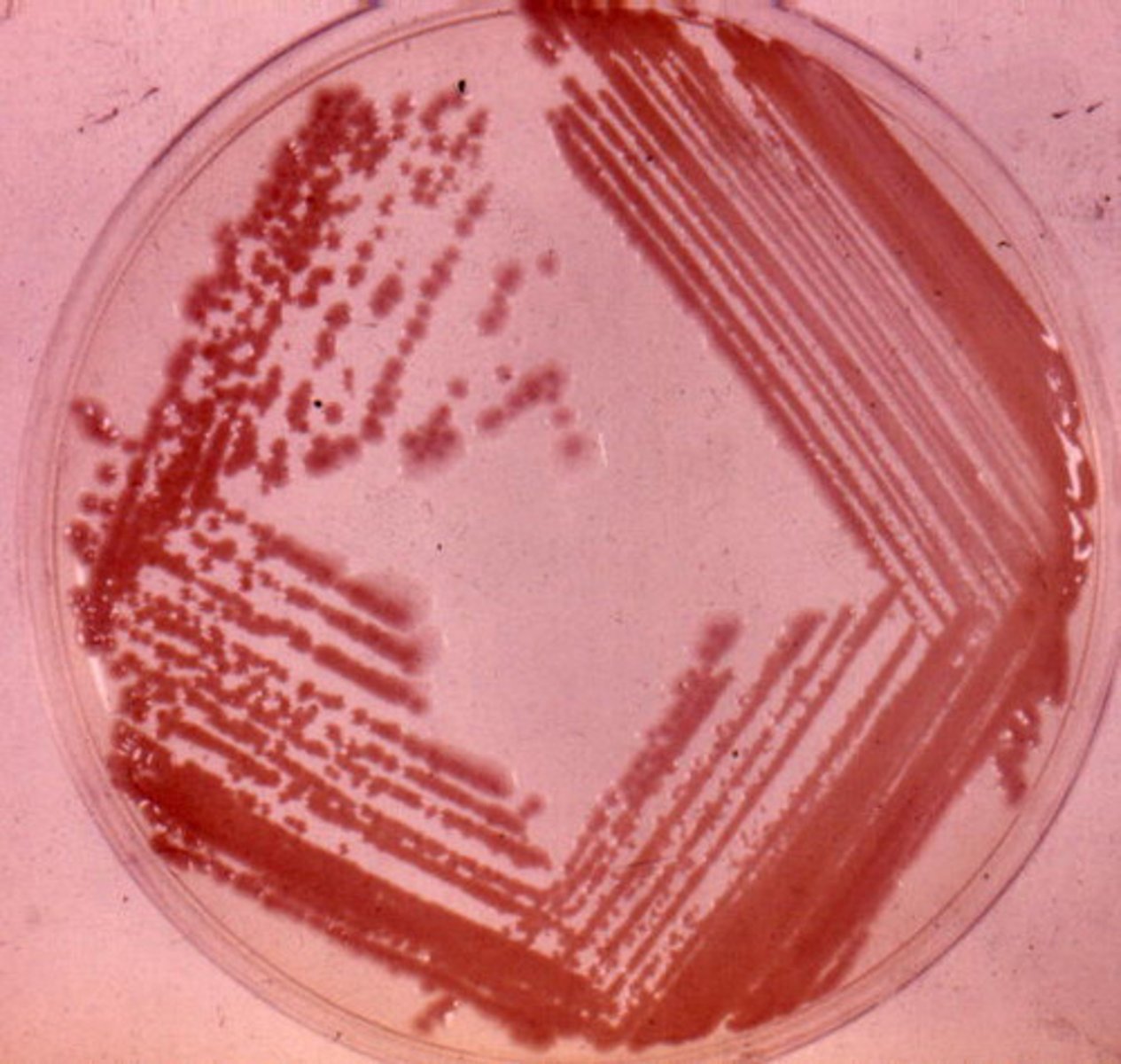
Inoculation
introduction of a sample into a container of media to produce a culture of observable growth
Incubation
inoculated media are placed in a temperature and atmosphere controlled environment (incubator) to promote growth
(****during the hours or days of this process, a culture develops as visible growth of microbes in the container of the medium

Isolation
if an individual bactria cell is separate from other cells and has space on a nutrient surface, it will grow into a mound of cells (= a colony ---> consists of ONE species)
Inspection
cultures are observed for the macroscopic appearance of growth characteristics
*cultures are examined under the microscope for basic details such as cell TYPE AND SHAPE
pure culture
grows only single known species of microorganisms
mixed culture
two or more identified species/microbes growing
contaminated culture
pure or mixed culture that has unwanted microbes growing
*this is important because it BAD and can induce FALSE DIAGNOSIS
ex) fungi.
information gathering
testing of cultures with procedures that analyze biochemical and enzyme characteristics, immunologic reactions, drug sensitivity, and genetic makeup
**IMPORTANT to gather the most information about your identification in order to rule out other microorganisms.
Identification
the goal of these procedures is to attach a name to the microbe, usually to the level of species.
-appearance
-biochemical tests
-genetic characteristics
-immunological testing
Magnification
the ability to make things look larger than they are
Resolving Power
ability to show detail
Refraction
the bending of light passing through convex surface of glass
total magnification
objective lens x ocular lens
Resolution (resolving power )
the capacity to distinguish or separate 2 adjacent objects
What is the purpose of oil?
to prevent light scattering for an overall clearer image
*used only 100X objective
Purpose of Staining
-increase contrast
- distinguish between gram-positive and gram-negative bacteria.
Positive staining
surfaces of microbes are negatively/positively charged and attract basic/acidic dyes
Negative Staining
microbe repels dye, the dye stains the background
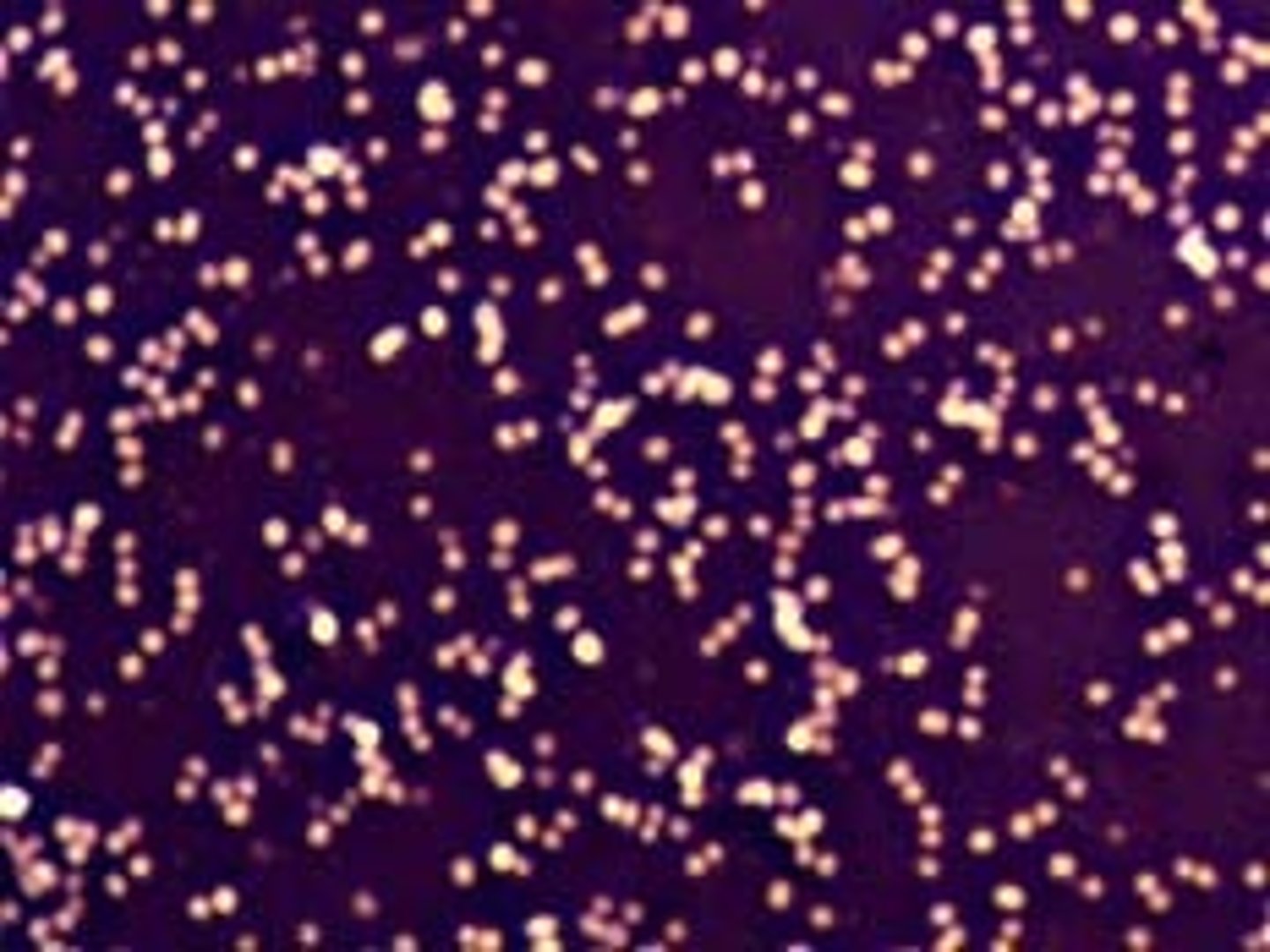
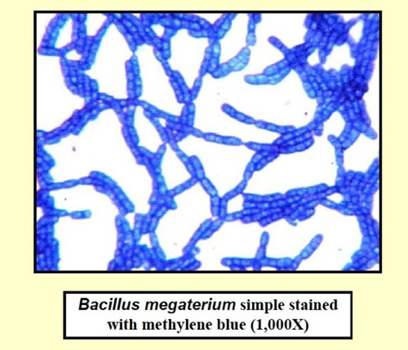
Simple Stains
one dye is used; reveals shape, size, and arrangement
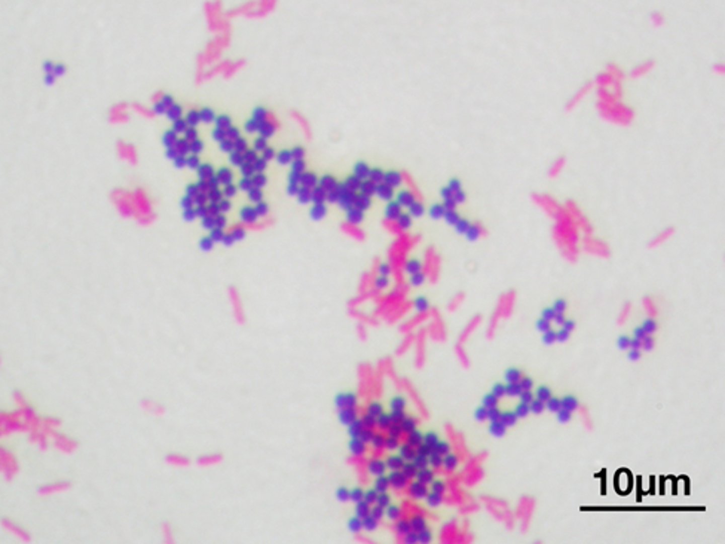
Differential Staining
use a primary stain and a counter stain to distinguish cell types
Example: gram-staim, acid-fast stain, endospore
Gram stain
-its a differential stain used to classify bacteria as gram-negative or gram-positive
-most significant technique in Microbiology
Physical states of media
1. liquid: nutrient broth; does not solidify
2. solid (agar): a firm surface for colony formation
3. semi-solid: clot-like consistency, contains agar solidifying agent
Types of Media
general, enrichment, selective, and differential media
General Media
used to grow a variety of bacteria, no special growth factors
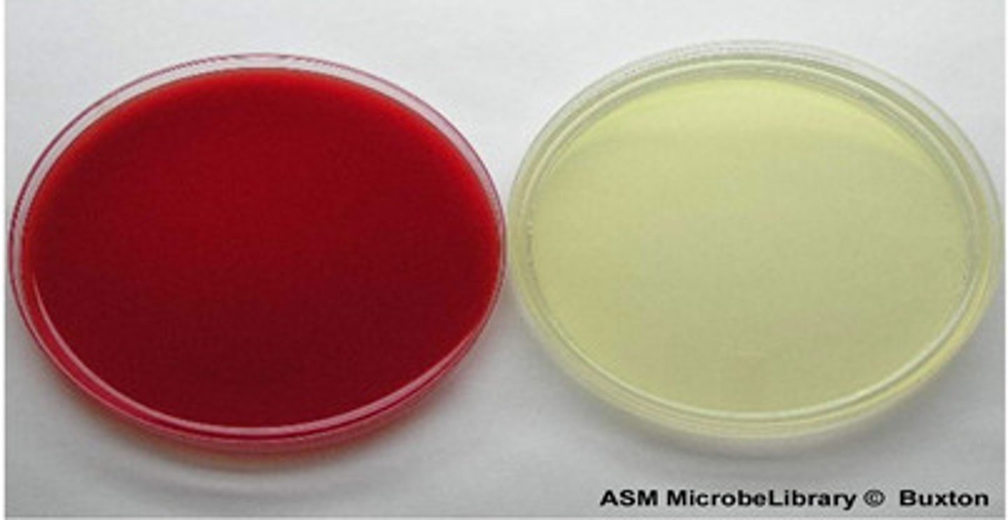
Enriched Media
contains complex organic substances such as blood, serum, hemoglobin, or special growth factors required by microbes
examples:
- blood agar (microbes lice through)
- chocolate agar (already liced)
Selective Media
contains one or more agents that inhibit growth of some microbes and encourage growth of the desired microbes
Differential media
allows growth of several types of microbes and displays visible differences among desired and undesired microbes
Endospore Stain
Used on clostridium and bacillus species
Green/Pink
What colors are endospores and vegetative cells after an endospore stain?
Flagellar stain
Binds to flagella, thickening & changing color
Pink/Blue
What colors are acid fast cells and non acid fast cells after an Acid fast stain?
Transmission electron microscope
Beam of electrons passes through specimen, excellent resolution of cellular details
Scanning electron microscope
Electrons pass over surface, produces 3-D images with great surface detail
2000x
Max magnification of light microscope
Magnification
Results from light or electron beam refracting as it passes through lens or magnetic field
Resolution
Ability to distinguish objects that are close together clearly
Contrast
Differences in intensity between 2 objects or an object and its background
1 um to 4+ mm
Protozoan size
3 to 10 um
Yeast size
200 nm to 750 um
Bacteria Size
20 to 400nm
Viruses
Synthetic/Defined Media
Same formula every time, every ingredient is known
Non-Synthetic/Complex Media
Contains extracts of animal, plant, or yeast products. Composition varies from batch-to-batch
Agar
Polysaccharide derived from Gelidium
Monotrichous
Describing a microorganism that bears a single flagellum.
Lophotrichous
Having a tuft of flagella at one or both poles
Peritrichous
having flagella distributed over the entire cell.
Fimbria
A short, numerous surface appendage on some bacteria that provides adhesion but not locomotion.
Conjugation
In bacteria, the contact between donor and recipient cells associated with the transfer of genetic material such as plasmids. Can involve special (sex) pili.
Nanotubes
Extensions of bacterial membranes that are channels for nutrient or energy exchange.
Capsule
In bacteria, the loose, gel-like covering or slime made chiefly of polysaccharides. This layer is protective and can be associated with virulence.
L form
A stage in the lives of some bacteria in which they have no peptidoglycan.
Bacterial chromosome
A circular body in bacteria that contains the primary genetic material. Also called nucleoid.
Endospore
A small, dormant, resistant derivative of a bacterial cell that germinates under favorable growth conditions into a vegetative cell.
Glycocalyx
A coating or layer of molecules external to the cell wall. It serves protective, adhesive, and receptor functions. It may fit tightly (capsule) or be very loose and diffuse (slime layer).
Nanotubes
Membrane extensions that allow bacteria to transmit electrons or nutrients to other bacteria or onto environmental surfaces
Plasmid
Double-stranded DNA circle containing extra genes
Lophotrichous
With small bunches or tufts of flagella emerging from the same site.
Amphitrichous
With flagella at both poles of the cell.
Peritrichous
Flagella are dispersed randomly over the surface of the cell.
Bacillus
Bacterial cell shape that is cylindrical (longer than it is wide).
Spirillum
A type of bacterial cell with a rigid spiral shape and external flagella.
Flagellum
A structure that is used to propel the organism through a fluid environment.
Palisades
The characteristic arrangement of Corynebacterium cells resembling a row of fence posts and created by snapping.
Appendages
Accessory structures that sprout from the surface of bacteria. They can be divided into two major groups: those that provide motility and those that enable adhesion.
Motility
Self-propulsion.
Pili
Long, tubular structures made of pilin protein produced by gram-negative bacteria and used for conjugation.
S layer
Single layer of thousands of copies of a single type of protein linked together on the surface of a bacterial cell that is produced when the cell is in a hostile environment.
Glycocalyx
A filamentous network of carbohydrate-rich molecules that coats cells.
Slime layer
A diffuse, unorganized layer of polysaccharides and/or proteins on the outside of some bacteria.
Biofilm
A complex association that arises from a mixture of microorganisms growing together on the surface of a habitat.
Cell wall
In bacteria, a rigid structure made of peptidoglycan that lies just outside the cytoplasmic membrane.
Peptidoglycan
A network of polysaccharide chains cross-linked by short peptides that forms the rigid part of bacterial cell walls. Gram-negative bacteria have a smaller amount of this rigid structure than do gram-positive bacteria.
Gram stain
A differential stain for bacterial useful in identification and taxonomy. Gram-positive organisms appear purple from crystal violet mordant retention, whereas gram-negative organisms appear red after loss of crystal violet and absorbance of the safranin counterstain.