CV pathology 3
1/46
There's no tags or description
Looks like no tags are added yet.
Name | Mastery | Learn | Test | Matching | Spaced |
|---|
No study sessions yet.
47 Terms
What is the meaning of cardiomegaly and what are the different types?
Enlargement of the heart
Hypertrophy
Dilation
Cardiomyopathy
What is ventricular hypertrophy?
“Compensatory mechanisms” – physiological response, maintains adequate cardiac output e.g. response to exercise
Represents a reversible increase in muscle mass (increase in size of muscle cells, NOT hyperplasia).
What is the difference between primary and secondary ventricular hypertrophy?
Secondary - compensatory response to increased workload
Physiological response (athletic animal) - maintain adequate cardiac output
Pathological - volume overload
Primary - irreversible idiopathic hypertrophic cardiomyopathy
What are the features of eccentric ventricular hypertrophy?
Heart with normal / enlarged ventricular chambers
Walls of normal / decreased thickness
Produced by volume overload (valve insufficiencies, septal defects)
What are the features of concentric ventricular hypertrophy?
Heart with small ventricular chambers that have thick walls which compress the chamber lumens
Produced by pressure overload (e.g. valvular stenosis, systemic hypertension, pulmonary disease (hypertension))
What could cause a pressure overload?
valvular stenosis
systemic hypertension
pulmonary disease (hypertension)
What is the meaning of cor pulmonale?
An alteration in the structure and function of the right ventricle (RV) of the heart caused by a primary disorder of the respiratory system
Which is concentric/eccentric ventricular hypertrophy?
Concentric - left (thicker walls)
Eccentric - right (dilated chamber lumen and normal walls)
What causes right ventricular hypertrophy?
Cor pulmonale due to increase flow resistance in pulmonary circulation.
Dirofilariosis (heart worm) and congenital pulmonic stenosis in dogs
High altitude disease (pulmonary hypertension) in cattle
Chronic alveolar enphysema in horses (heaves)
What can cause left sided hypertrophy?
Systemic hypertension (chronic renal failure)
Congenital subaortic stenosis
When does ventricular dilation occur?
Myocardium cannot undergo hypertrophy because of insufficient time, inadequate nutrition or diseases
Same reasons as hypertrophy - compensatory response to inc CO
Dilation allows stretching of cardiac muscle cells to increase contractile force and increases stroke volume is the result
(terminal lesion in many cardiac diseases)
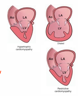
What are examples of primary cardiomyopathies
Dilated cardiomyopathy
Hypertrophic cardiomyopathy
Different to hypertrophies because they are secondary to other disease (these happen spontaneously)
Restrictive cardiomyopathy
What cardiomyopathies can you have with a known pathogenesis?
Toxic
Nutritional
Genetic causes
What species is hypertrophic cardiomyopathy common in and what occurs secondary to this?
Young adult to middle aged cats
Cats die from left atrial thrombosis due to pooling of blood and caudal aorta thromboembolism (from the atrial thrombi -one of top differentials for cat with sudden death)
(Uncommon in dogs, may occur in large breeds)
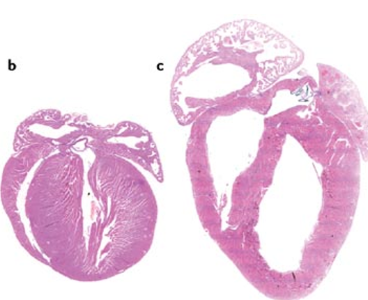
How does Hypertrophic cardiomyopathy present grossly?
Hearts are enlarged (increased cardiac silhouette)
Prominent concentric hypertrophy of the left ventricle, interventricular septum
Dilation of the left atrium
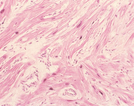
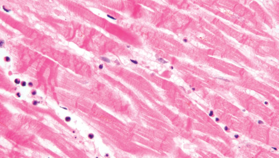
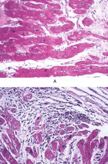
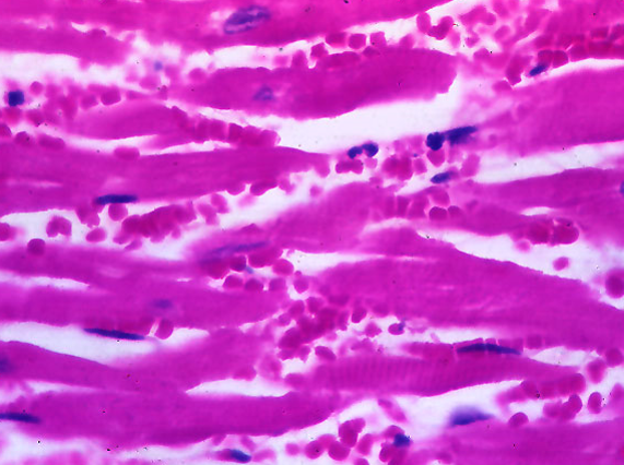
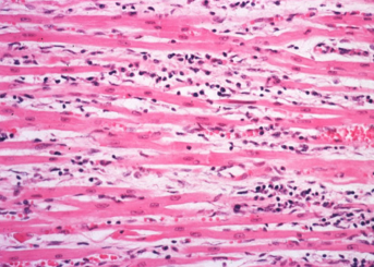
How does Hypertrophic cardiomyopathy present histologically?
Disarrays of hypertrophic degenerated myocytes (unusual angles)
Interweaving arrangement of fibres and interstitial fibrosis
(normal below for reference)
What species is dilated cardiomyopathy common in and what causes it?
Middle aged dogs – idiopathic or autosomal recessive or X-linked mode of inheritance.
Cats with low tissue concentrations of taurine. Taurine supplementation in commercial diets has resulted in dramatic reduction of cases
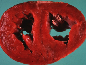
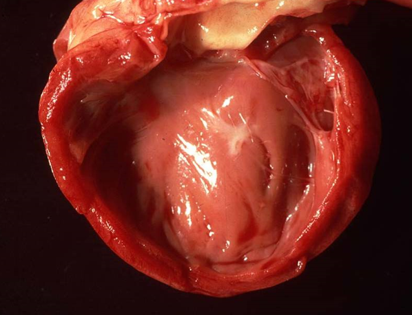
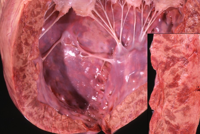
How does dilated cardiomyopathy present grossly?
Biventricular dilation
White thickened endocardium
Walls are normal, may be thinner
Increased heart weight (more than 1% of BW)
How does dilated cardiomyopathy present histologically?
Interstitial fibrosis
Fatty infiltration and myocyte degeneration
Attenuated wavy fibres
Variable
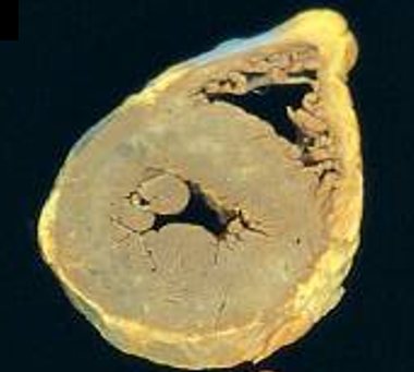
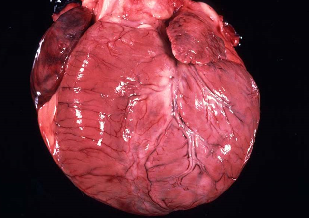
Describe the features of bovine dilalated cardiomyopathy?
well–grown 2-3 year-old Holstein cattle.
peripheral oedema, jugular distension,
fluid accumulations in the body cavities.
enlargement of the heart with a rounded “globose” shape
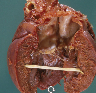
What is restrictive cardiomyopathy and when would this occur?
Walls are rigid and the heart is restricted from stretching and filling with blood properly
Rhythmicity and contractility of the heart may be normal
Stiff walls of the heart chambers keep them from adequately filling.
Cats with endocardial lesions (inflammation, fibrosis, fibroelastosis) that impair the ventricular flow
What will be seen with restrictive cardiomyopathy?
How does myocardial necrosis lead to death?
When cardiac conduction is disrupted
or
Cardiac decompensation, cardiac dilation and congestive heart failure
What causes myocardial necrosis?
Nutritional deficiencies: Vitamin E / Selenium deficiency [calves, lambs, foal] mulberry heart disease [pigs]
Plant intoxication
Ionophore toxicity
Doxirubicin - chemo drug
Secondary to myocarditis
Describe how myocardial necoriss presents grossly
Affected areas are pale, yellow to white and dry
They can become gritty due to dystrophic calcification
Common in the papillary muscles
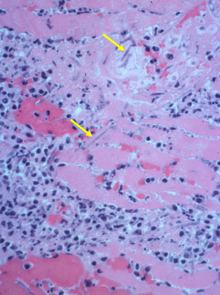
Describe how myocardial necoriss presents histologically
Fibres appear swollen and hypereosinophilic (hyalin necrosis)
Striations are indistinct, and nuclei are pyknotic
Infiltration of inflammatory cells (macrophages and neutrophils)
What are the features of mulberry heart disease in pigs?
Peroxidation of the cell membranes due to Vit E/ selinium deficiency
Necrosis
Hydropericardium
Haemorrhages
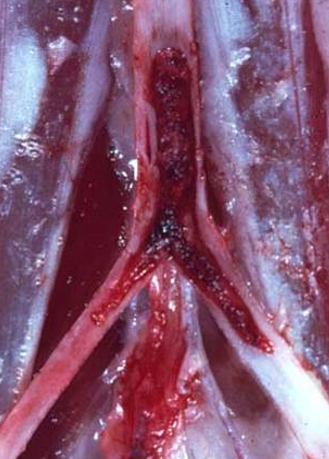
What can Equine myocardial degeneration cause?
arrhythmic heart beat
ventricular fibrillation
petechial pericoronal haemorrhages
necrosis / hyalinosis of myocytes
How does the use of doxorubicin make the heart appear grossly?
Pale myocardium
Hyaline necrosis
Hydropic degeneration of the myocytes
(chemo drug)
What is the portal of entry for myocarditis?
Haematogenous dissemination
Embolic dissemination from vegetative endocarditis into the coronary arterial tree
What are the types of myocarditis?
purulent (from vegetative endocarditis)
necrotising (toxoplasmosis in dogs and cats)
haemorrhagic (black leg)
lymphocytic ( parvoviral myocarditis)
eosinophilic (sarcocystosis)
granulomatous (fungi)
When can canine parvovirus cause myocarditis?
Puppies less than 10 weeks of age - cardiomyocytes are not post mitotic
Acute necrosis with little or moderate lymphocytic inflammatory response
Describe this histological image of purulent myocarditis?

Increase in neutrophils
Degeneration of surrounding cells
What can cause purulent myocaditis?
Direct extension of pericarditis (eg. "hardware disease" wire in cattle)
Direct extension of endocarditis (eg. erysipelas in pigs)
What myocarditis is being shown here?
Equine disseminated granulomatous myocarditis - Mycosis / aspergillosis
Macrophages and fungal hyphae present
What protozoan parasites are found in the heart?
Sarcocystis sp.
Toxoplasma sp.
Neospora sp.
What metazoan parasites are found in the heart?
Cestodes
Cysticercus sp.
Hydatid cyst.
Nematodes
Dirofilaria immitis (dogs and cats)
Angiostrongylus vasorum (dogs)
What does toxoplasma gondii cause in the heart?
Necrosis and chronic pyogranulomatous myocarditis

What does neospora cause?
CNS /neuronal symptom
dermatitis
hepatitis
pneumonia
myocarditis can occur - adult dogs sporadically develop neosporosis
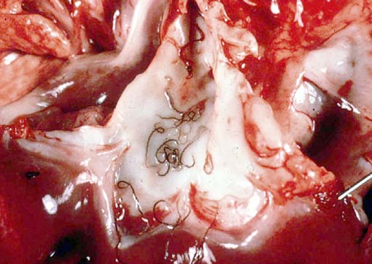
What causes lungworm in dogs and where are the worms found?
Angiostrongylus vasorum
Adults live in pulmonary artery and RV
Larvae in the lung parenchyma
What does Angiostrongylus vasorum cause?
Enlarged RV, cardiac silhouette rounded
Right sided heart failure
Eosinophilic vasculitis
Multifocal granulomatous and eosinophilic pneumonia
Bleeding disorder
What is the most prevalent tumour of the heart and pericardium?
Endothelial cell neoplasia
Haemangio-
Where do haemangiomas originate?
Tumour arising from endothelial cells in vessels (e.g. of the skin)
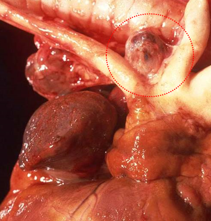
How does a haemangioma present grossly and histologically?
Gross - Red-black blood containing masses that protrude into the lumen or epicardial surface.
Histologically – well differentiated vascular spaces lined by endothelial cells
Where will you often find a haemangiosarcoma?
Right atrium in dogs, spleen in dogs
How does a haemangiosarcoma present grossly and histologically?
Gross – similar to haemangioma
Histologically - scattered, elongated plump neoplastic endothelial cells
What are examples of heart base tumours?
Aortic body tumours - chemodectoma (could occlude great vessels - pressure overload)
Ectopic thyroid
Metastatic (bovine lymphoma)