A225 - IU - Exam 5
 Call Kai
 Learn
Learn Practice Test
 Spaced Repetition
 Match
Match Flashcards
 Knowt Play
1/198

There's no tags or description
Looks like no tags are added yet.
Name | Mastery | Learn | Test | Matching | Spaced | Call with Kai |
|---|
No analytics yet
Send a link to your students to track their progress
199 Terms
What is the gastrointestinal tract?
A tube that extends from the oral cavity to the anal canal, including the oral cavity, pharynx, esophagus, stomach, small intestine, large intestine, and anal canal.
What is peristalsis?
Muscular contractions that occur in waves to push material further along the gastrointestinal tract.
What are the two types of digestion?
Mechanical digestion (chewing) and chemical digestion (separating chemical bonds to receive nutrients).
What is absorption in the digestive system?
The process where water and food molecules pass through to the GI lymphatics and blood vessels.
What are accessory digestive organs?
Organs that assist with digestion but are not part of the GI tract, including the tongue, teeth, salivary glands, liver, gallbladder, and pancreas.
What is the role of the autonomic nervous system in digestion?
The parasympathetic system stimulates or increases activity of the GI tract, while the sympathetic system inhibits digestion.
What is the structure of the oral cavity?
Lined by non-keratinized stratified squamous epithelium and continuous posteriorly with the oropharynx.
What are the types of teeth and their counts in adults?
Adults have 2 incisors, 1 canine, 2 premolars, and 3 molars per quadrant.
What is the function of the tongue?
Composed of skeletal muscle, it helps in manipulation of food and is covered in non-keratinized stratified squamous epithelium.
What is the function of the uvula?
It rises to close off the nasopharynx during swallowing.
What are the three pairs of salivary glands?
Parotid, submandibular, and sublingual glands.
What is the main function of the stomach?
Digestion through both chemical (mixing food with gastric juice) and mechanical (churning food) processes.
What are the parts of the small intestine?
Duodenum (first part), jejunum (second part), and ileum (third part).
What is the role of the large intestine?
Absorbs water and electrolytes, stores undigested materials, and expels waste from the body.
What is the histology of the esophagus?
Mucosa is lined with non-keratinized stratified squamous epithelium, with varying muscle types in the muscularis layer.
What is the liver's primary function?
Produces and secretes bile, processes products of digestion, stores nutrients, and produces plasma proteins.
What is the structure of the liver?
Divided into four lobes: right lobe, left lobe, quadrate lobe, and caudate lobe.
What is the function of the gallbladder?
Stores bile received from the liver.
What is the role of the pancreas?
Secretes pancreatic juice containing digestive enzymes and bicarbonate.
What is the blood supply to the GI tract?
Arterial supply includes the celiac trunk, superior mesenteric artery, and inferior mesenteric artery.
What is the function of the kidneys in the urinary system?
Filter blood and convert filtrate into urine.
What is the pathway of urine from the kidneys?
Urine travels from the kidneys through the ureters to the urinary bladder and then out through the urethra.
What is glomerular filtration?
The process of removing waste from the blood in the kidneys.
What are the components of a nephron?
Each nephron consists of a renal corpuscle, glomerular capsule, proximal convoluted tubule, nephron loop, and distal convoluted tubule.
What is the histology of the ureters?
Mucosa lined with transitional epithelium, which allows for stretching.
What is the significance of the renal papilla?
It collects urine from the collecting duct before it moves to the minor calyx.
What is the function of the collecting duct in the nephron?
It is the target of ADH and where filtrate becomes urine.
What is the structure of the large intestine?
Includes the cecum, ascending colon, transverse colon, descending colon, sigmoid colon, rectum, and anal cavity.
What is the role of the peritoneum in the digestive system?
It lines the abdominal cavity and covers the organs, providing support and protection.
What is the function of the urinary bladder?
It serves as a urine reservoir and muscular storage sac for urine.
What shape does the urinary bladder take when empty?
An upside-down pyramid in the true pelvis.
What are renal calculi?
Calcifications or crystals in the filtrate that can get stuck in the calyx, bladder opening, or ureter.
What type of muscle is found in the muscularis layer of the urinary bladder?
Two layers of smooth muscle.
What is the histological structure of the urinary bladder's mucosa?
Transitional epithelium.
What is the role of the internal urethral sphincter?
It is under the control of the autonomic nervous system and is an involuntary response.
What is the urethra?
A singular muscular tube running from the urinary bladder to the exterior of the body.
What is the difference between the male and female urethra?
The female urethra carries only urine and is shorter, while the male urethra carries both urine and semen and is longer.
What is meiosis?
A type of cell division that produces gametes, resulting in cells with half the DNA of a normal cell.
What is the difference between diploid and haploid cells?
Diploid cells have two copies of each chromosome, while haploid cells have a single copy.
What happens during meiosis 1?
Homologous chromosomes are separated to reduce the chromosome number from diploid to haploid.
What occurs during crossing over in meiosis?
Genetic material is exchanged between homologous chromosomes.
What is the outcome of meiosis 2?
It results in four daughter cells, each containing 23 single-stranded chromosomes.
What is nondisjunction?
The failure of chromatids to separate during meiosis, leading to an abnormal number of chromosomes.
What is trisomy?
A condition where there are three sets of one chromosome.
What are spermatogonia?
The most immature sex cells in men that undergo mitosis to produce primary spermatocytes.
What is the function of sustentacular cells?
They support, protect, and nourish developing sperm.
What is the role of the epididymis?
It receives sperm from the testis and is the site of sperm maturation.
What is the ductus deferens?
An 18-inch long tube that transports sperm from the testis to the prostate gland.
What do the seminal vesicles produce?
A nutritive alkaline fluid that combines with sperm to form semen.
What is the function of the prostate gland?
It produces prostatic secretions and is located inferior to the bladder.
What are the functions of the ovaries?
To produce oocytes and sex hormones (estrogen and progesterone).
What is the role of the uterine tubes (fallopian tubes)?
To pick up secondary oocytes and transport them to the uterus.
What is the myometrium?
The thickest layer of the uterus, composed of three layers of smooth muscle.
What is the endometrium?
The innermost layer of the uterus, highly vascular, where a fertilized oocyte implants.
What are the two layers of the endometrium?
The basal layer (permanent) and the functional layer (grows during the ovarian cycle).
Vestibule
Space between lips and teeth or cheeks and teeth
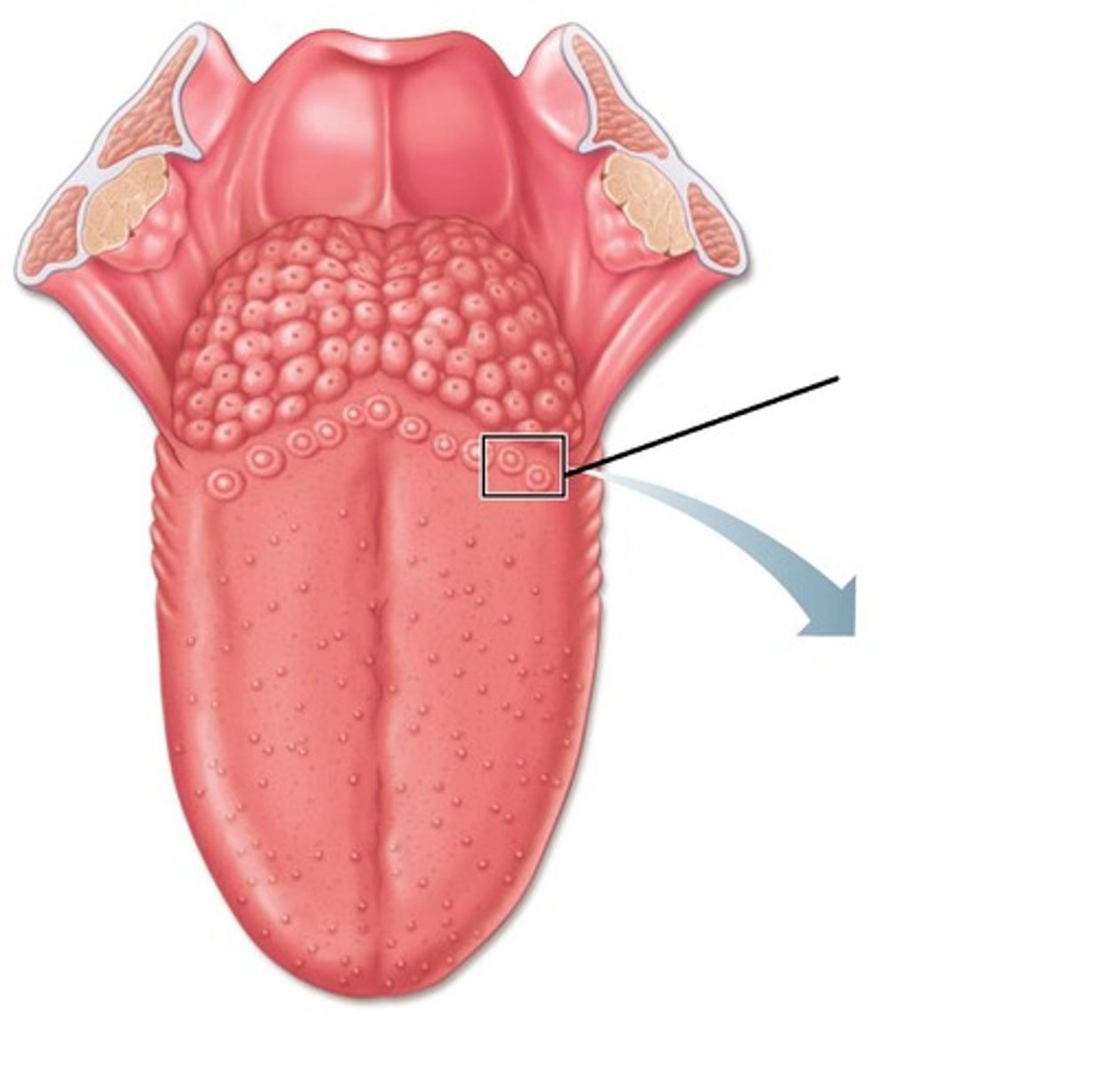
vallate papillae
largest taste buds with 8-12 forming "V" at back of tongue
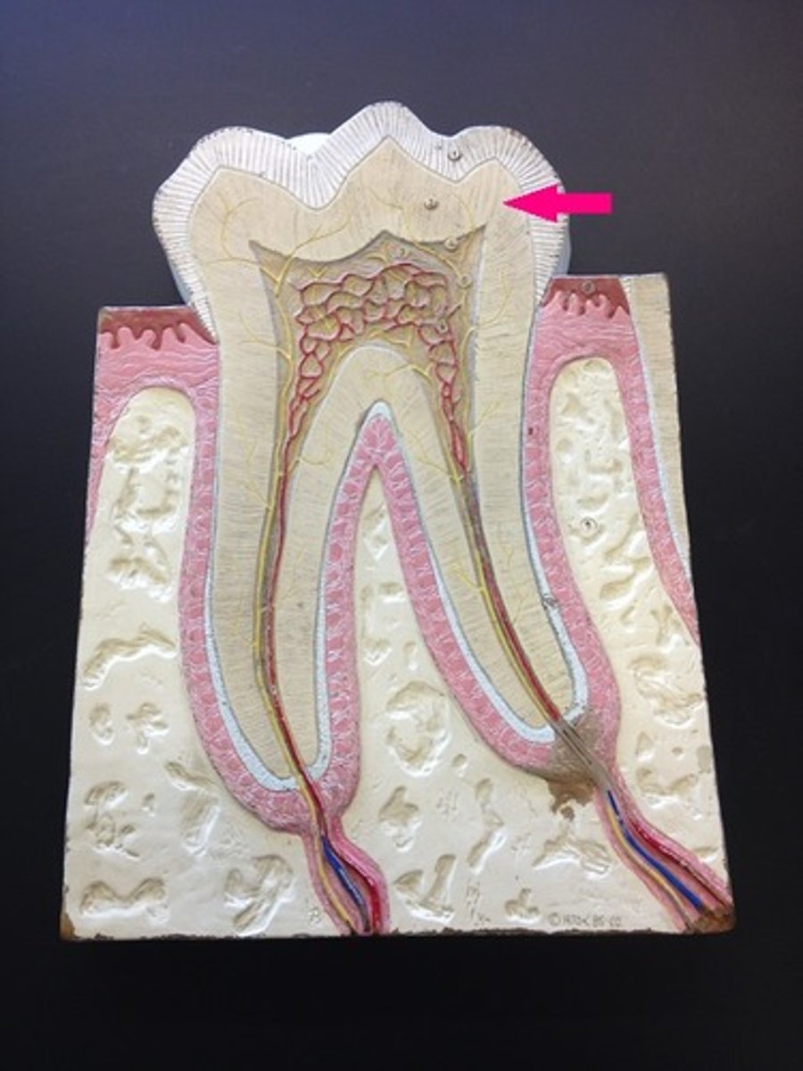
Dentin
forms the bulk of the tooth; it is hard, yellowish, calcified substance
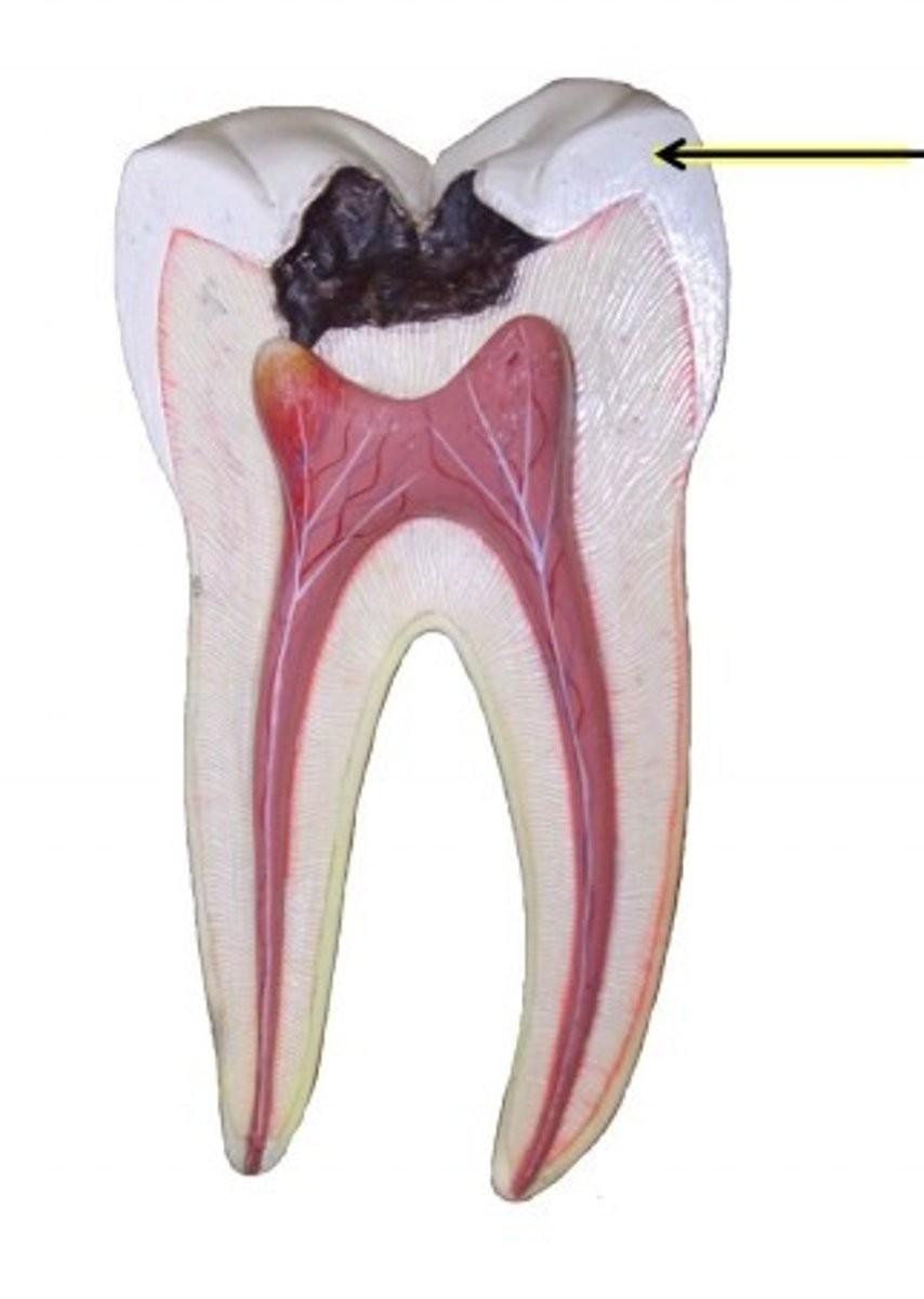
Enamel
Is the hardest substance in the body and is composed largely of inorganic salts, covers dentin of the crown
Cementum
is a bonelike substance which surrounds the dentin of the root and helps anchor the tooth
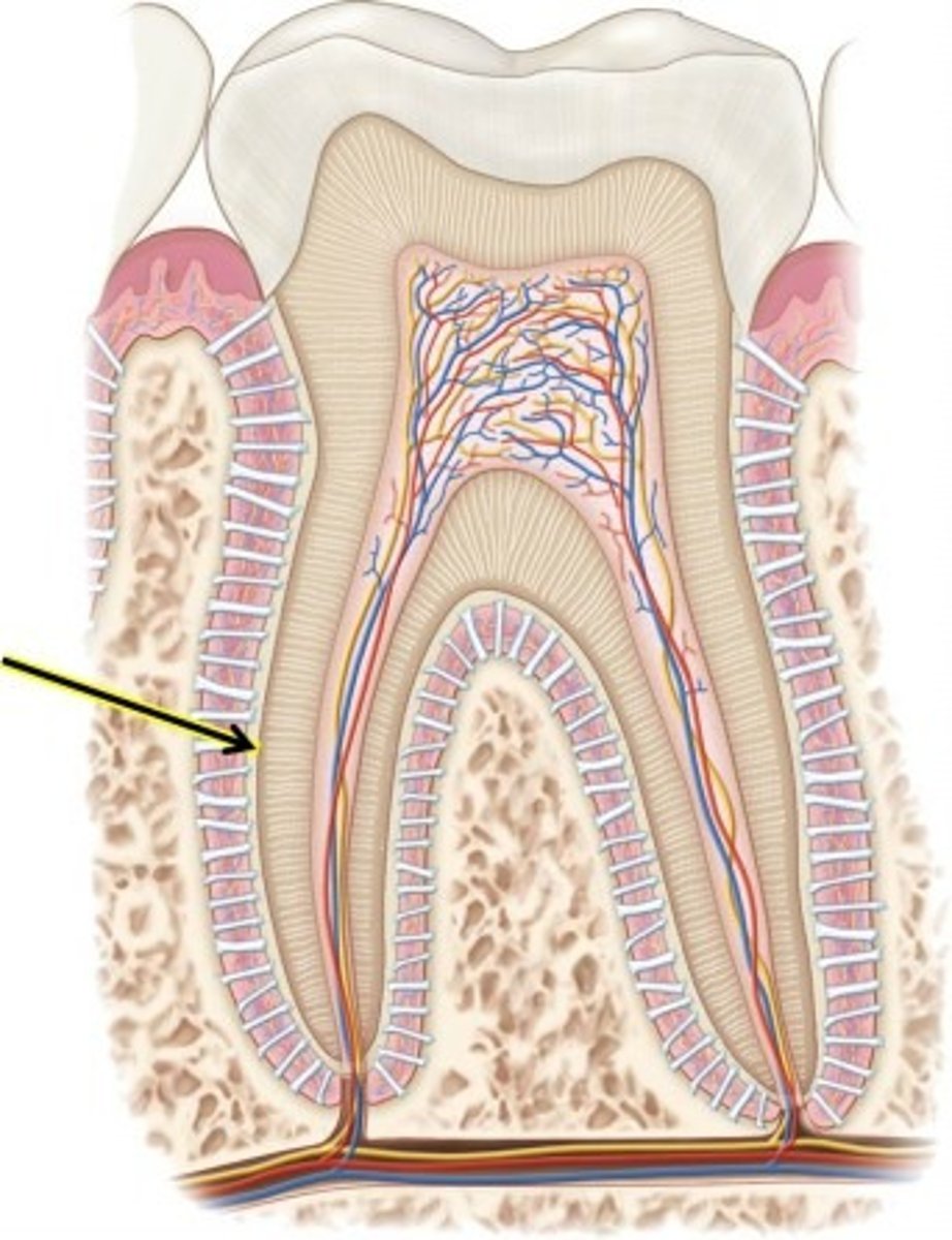
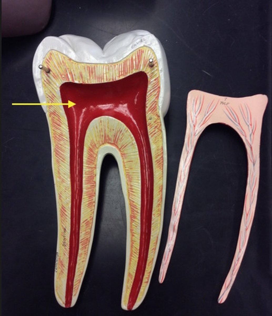
pulp cavity
receives blood vessels and nerves through the root canal
periodontal ligaments
hold tooth in socket
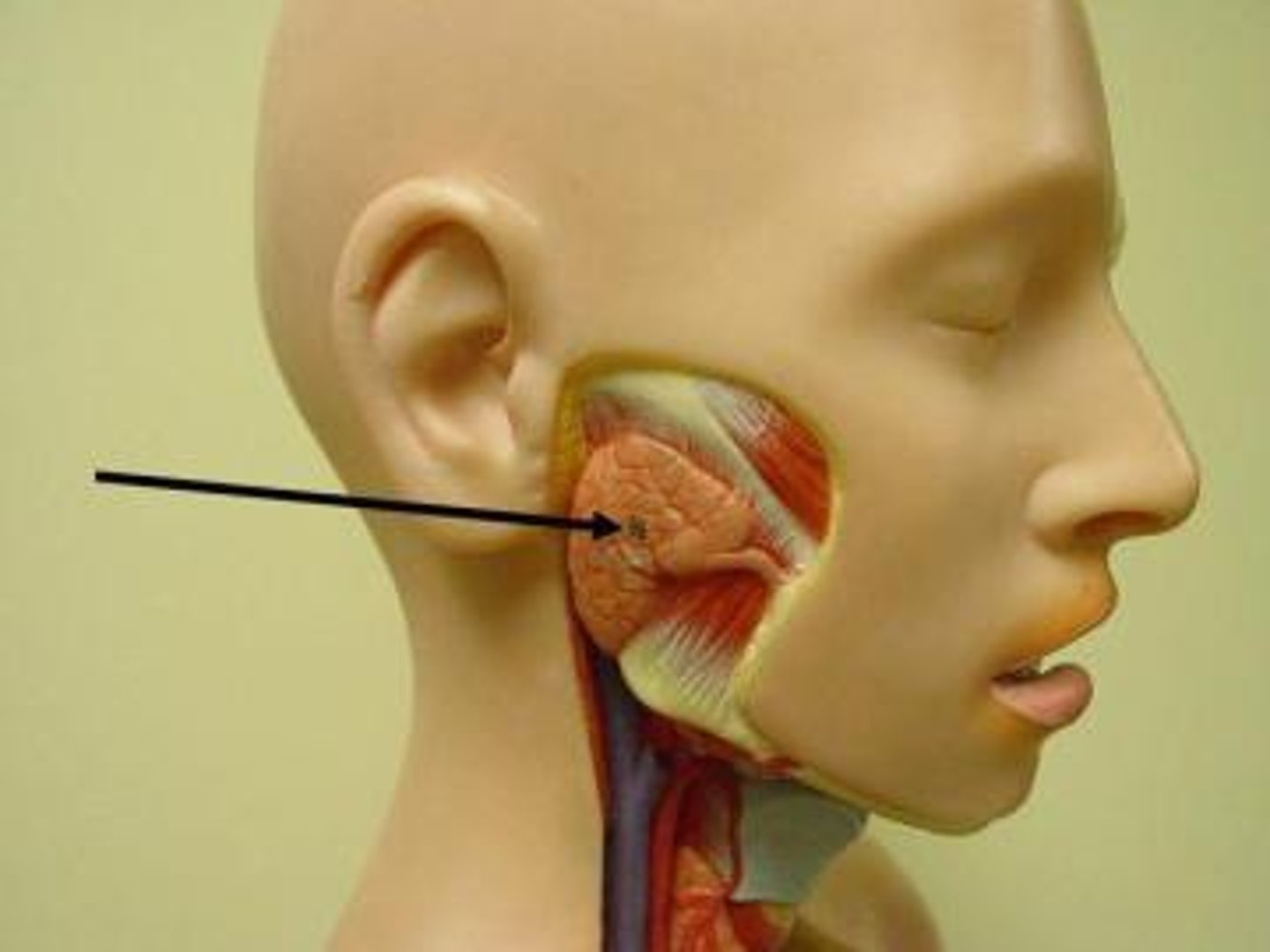
parotid glands
pair of salivary glands in the cheeks in front of the ears
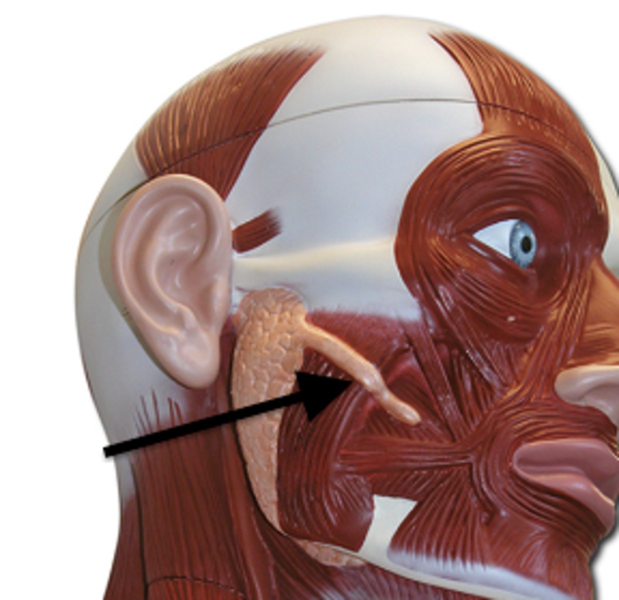
parotid ducts
passageways for saliva from parotid glands to oral cavity
submandibular glands
beneath the mandible at the angle of the jaw
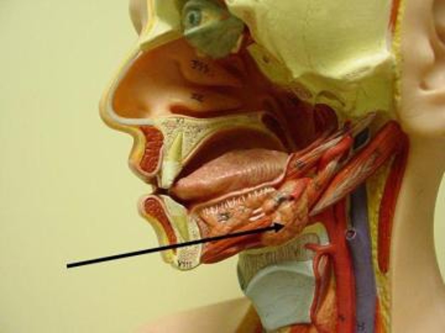
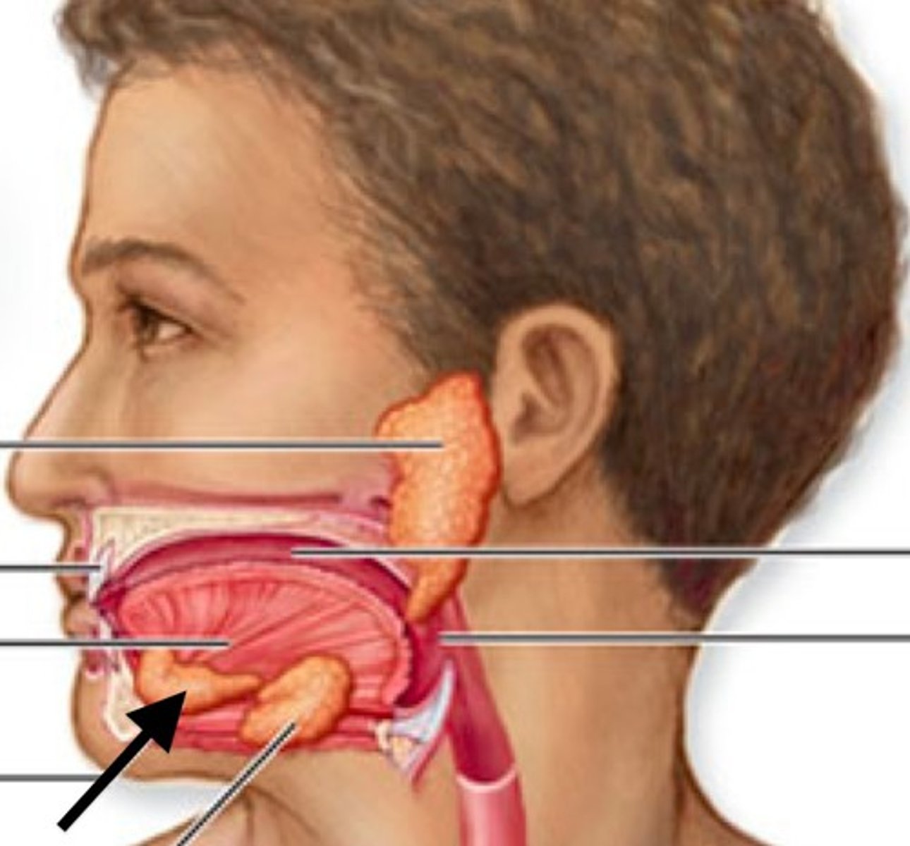
sublingual glands
under the tongue
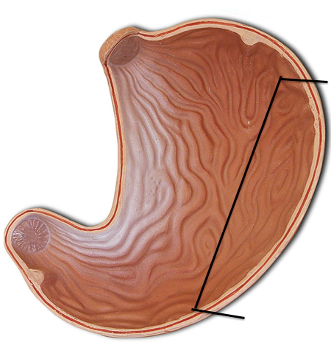
greater curvature
convex lateral surface
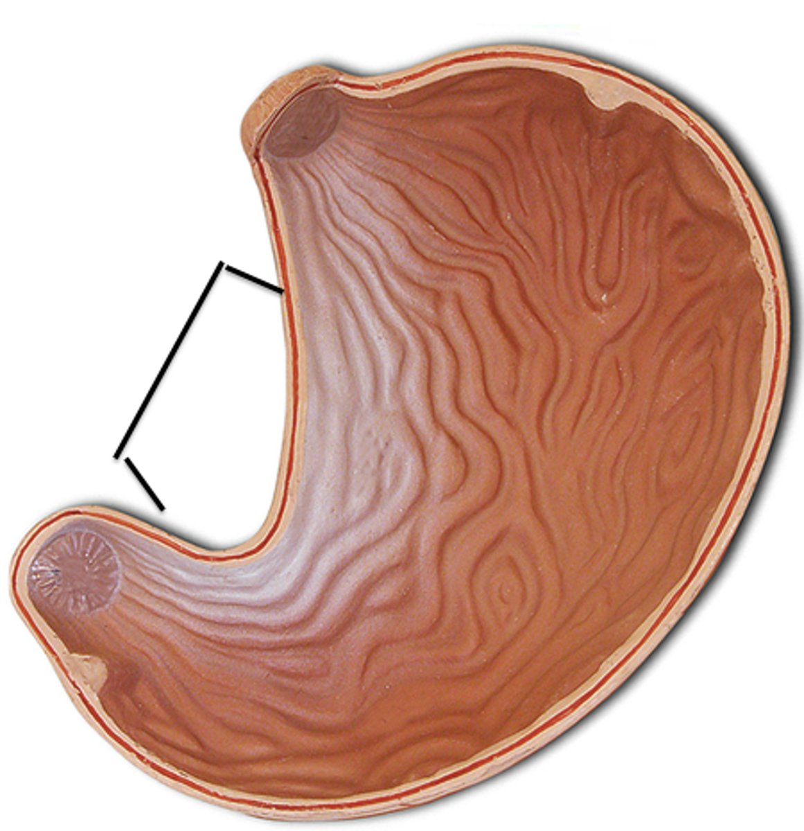
lesser curvature
concave medial surface of the stomach
pyloric sphincter
Controls passage of food from stomach to small intestine
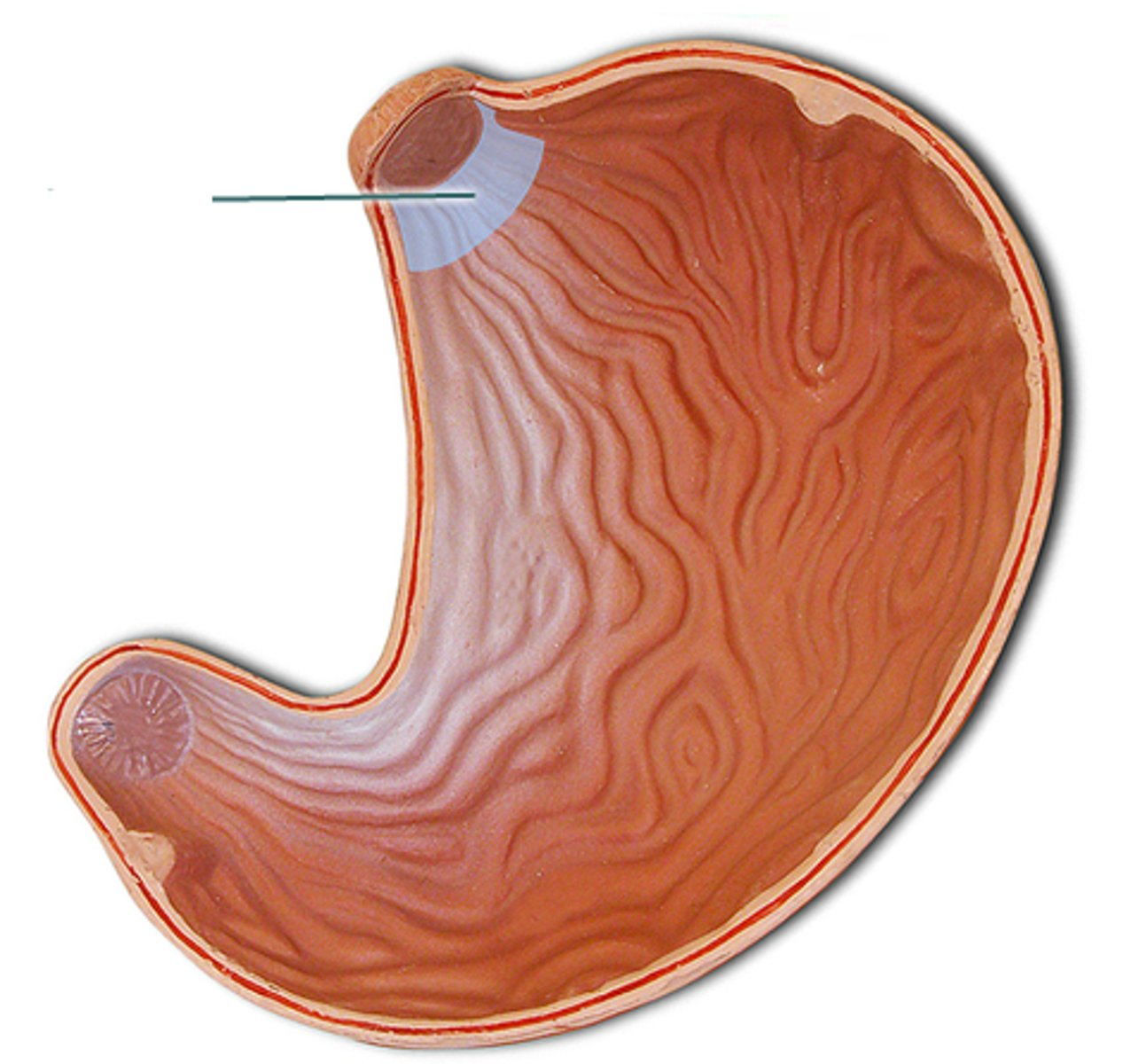
cardiac region of stomach
near the heart
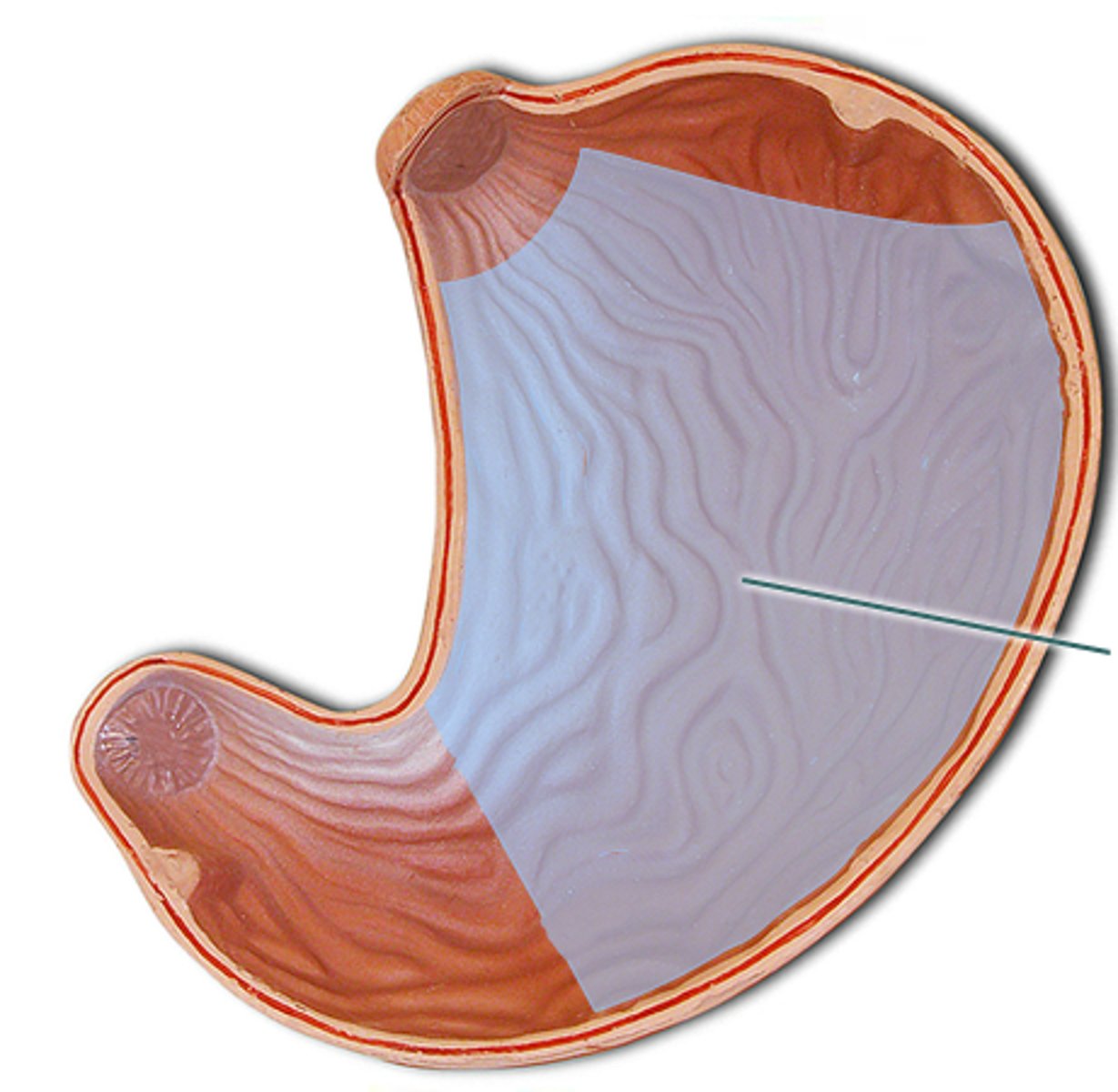
body region of stomach
mid portion of the stomach
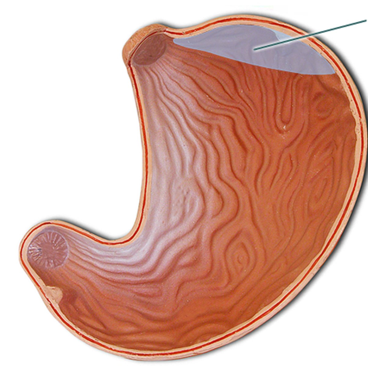
fundus region of stomach
bulges superior to and to the left of the cardiac orifice
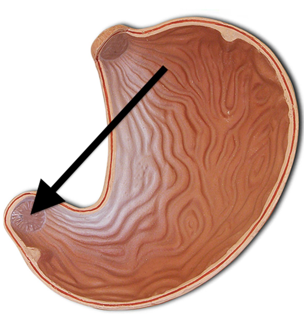
pylorus
distal region of the stomach, opening to the duodenum
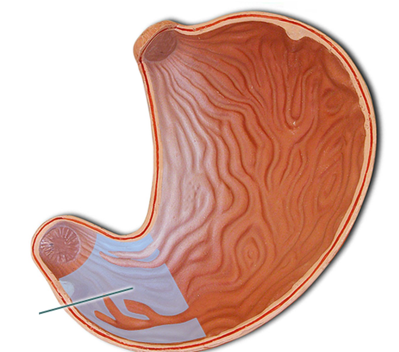
gastric folds
allow stomach to expand
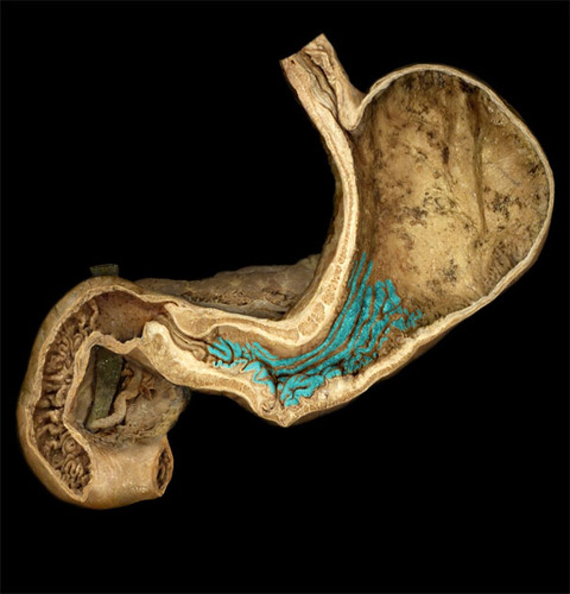
duodenum
first part of the small intestine
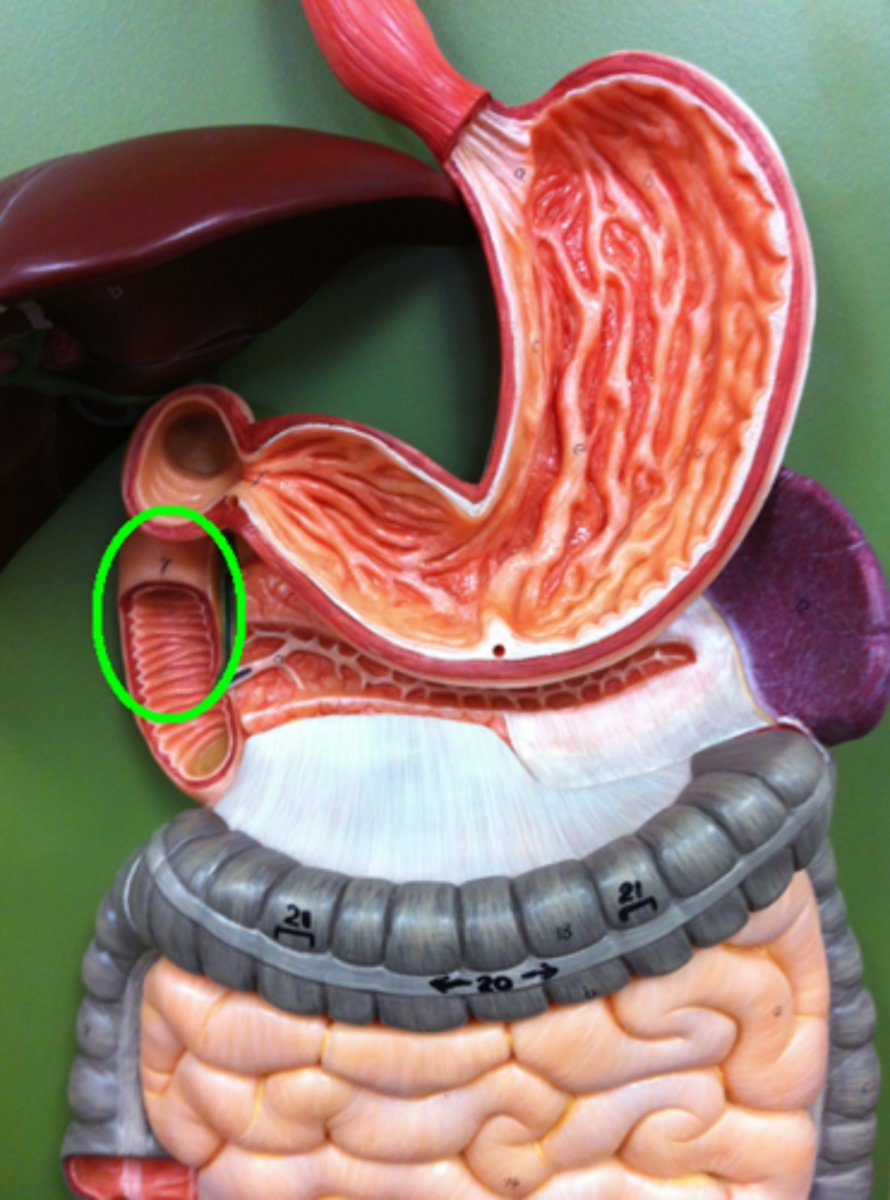
Jejunum
second part of the small intestine
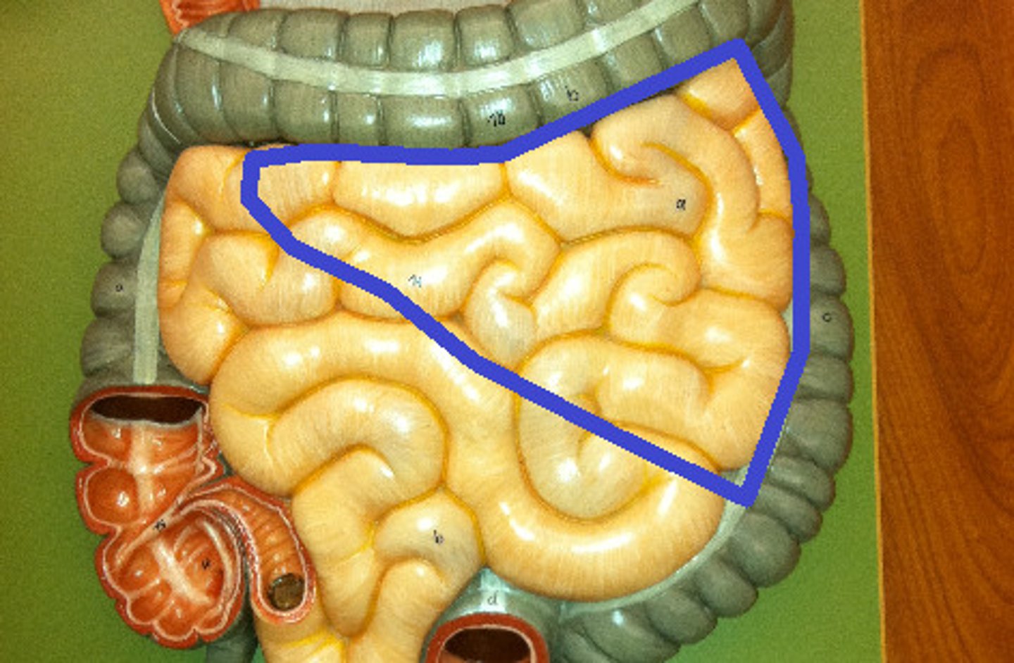
ileum
third part of the small intestine
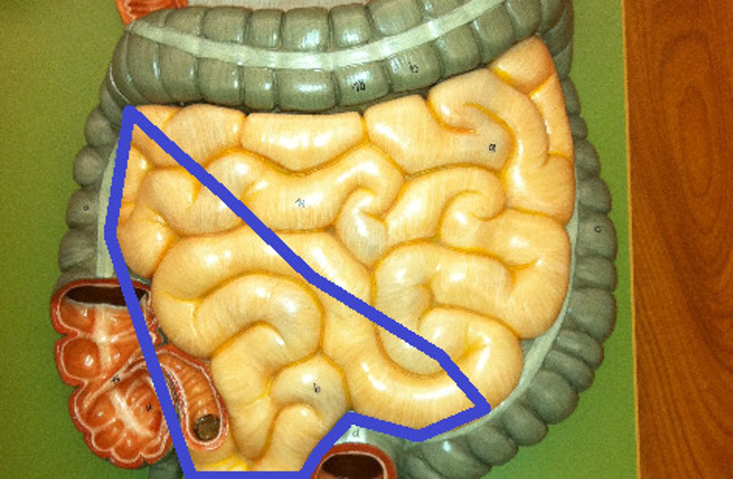
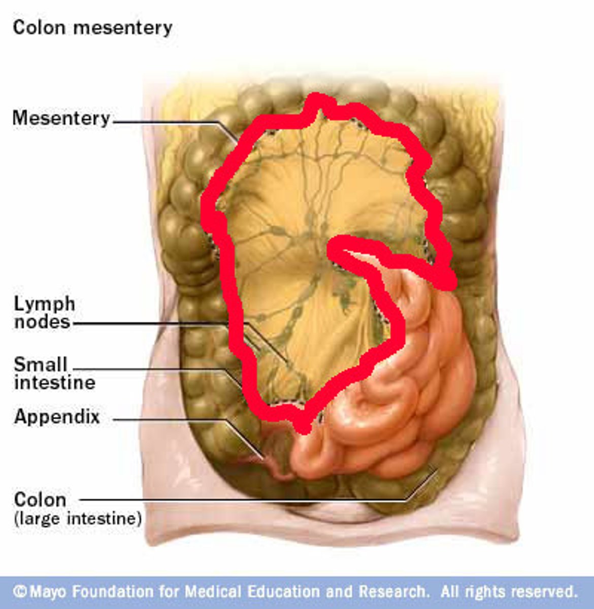
mesentary of small intestine
Location:
Abdomen
Between posterior abdominal wall (root) and small intestine (jejunum and ileum)
Description:
Double layer of peritoneum
Function:
Support for ---------
Contains blood, nerve, and lymphatic supply for -----------
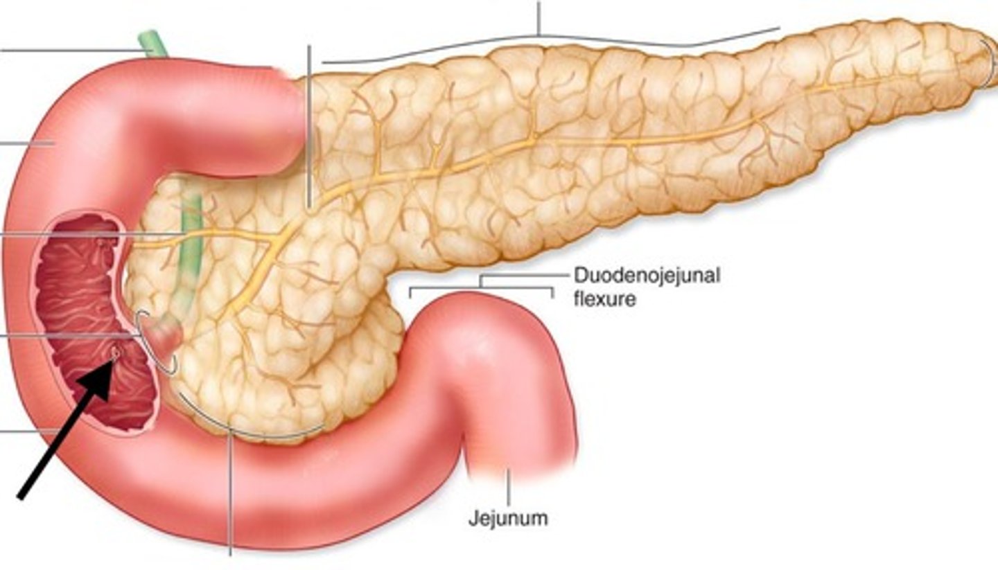
duodenal papilla
the raised area where the common bile duct and pancreatic duct enter the duodenum
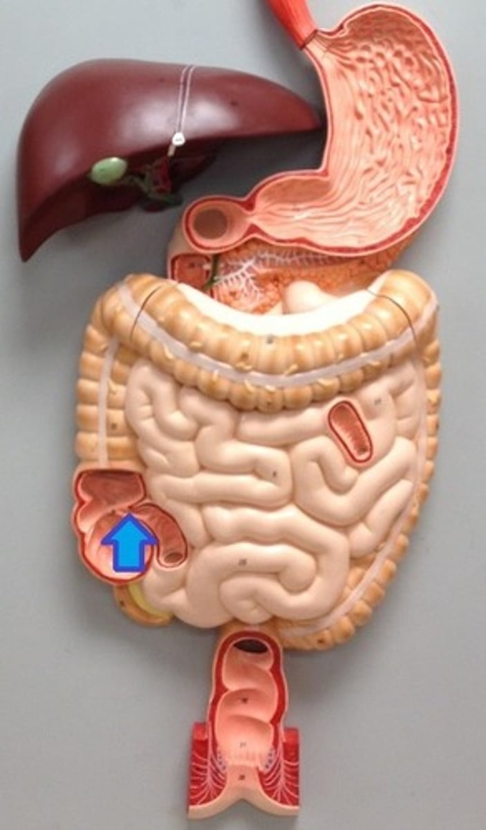
ileocecal valve
Valve at the junction of the small and large intestines
tenaie coli
three bands of longitudinal smooth muscle in muscularis
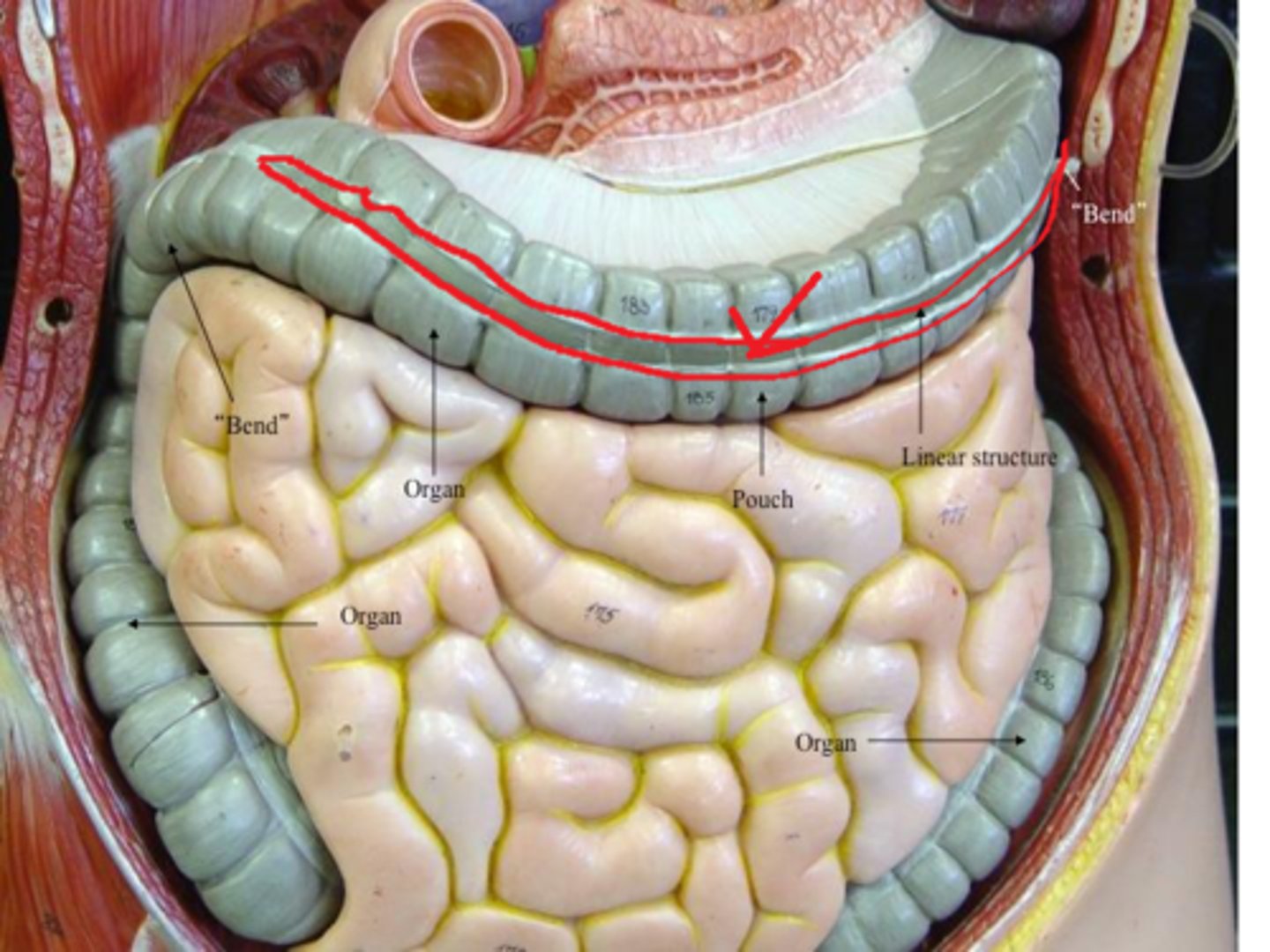
haustra
pouches that form in the large intestine when the longitudinal muscles are shorter than the colon
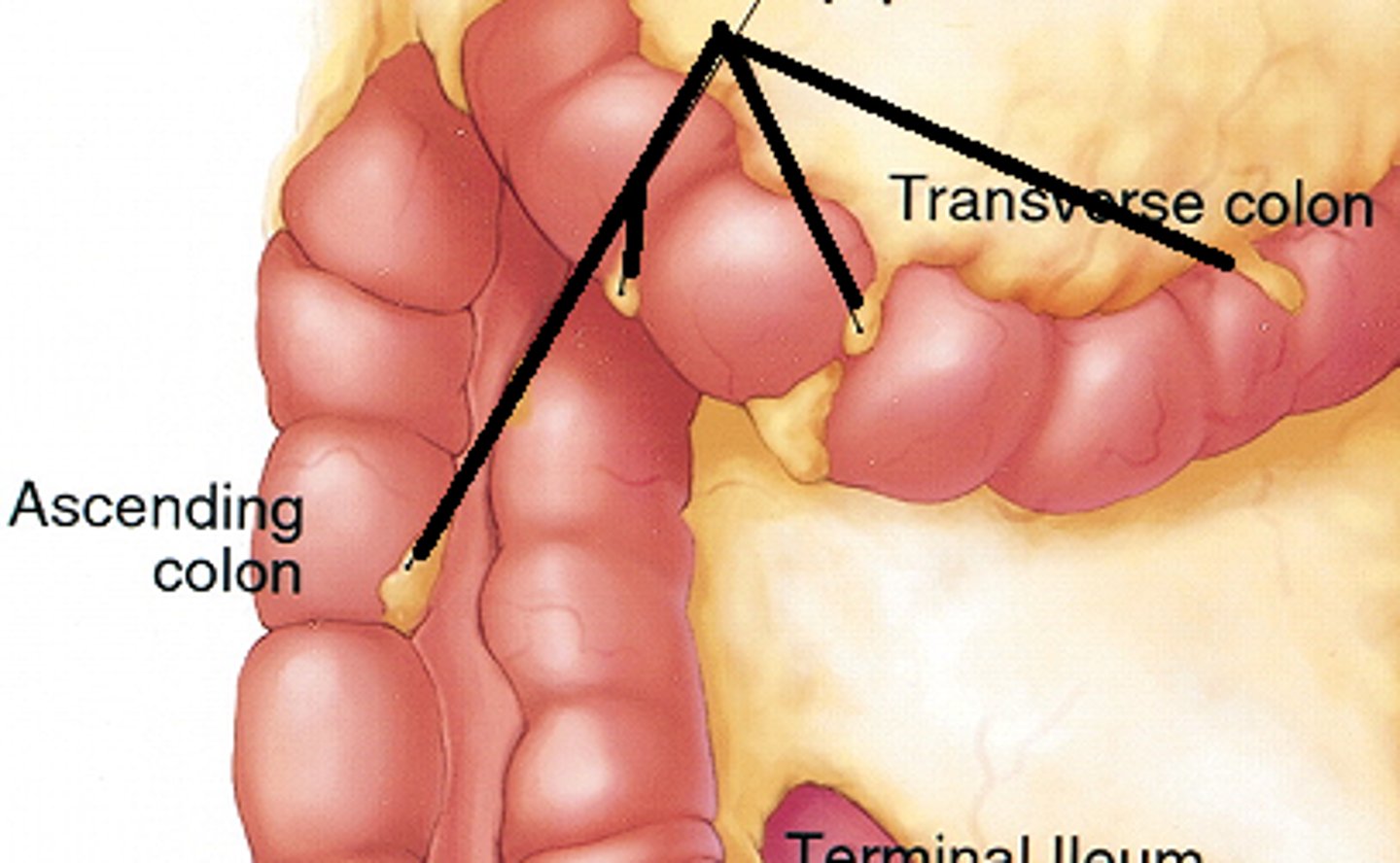
omental appendices
Lobules of fat hanging off external surface of haustra
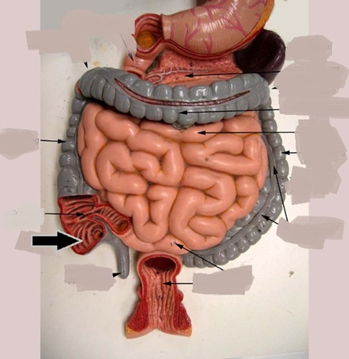
cecum
first part of the large intestine
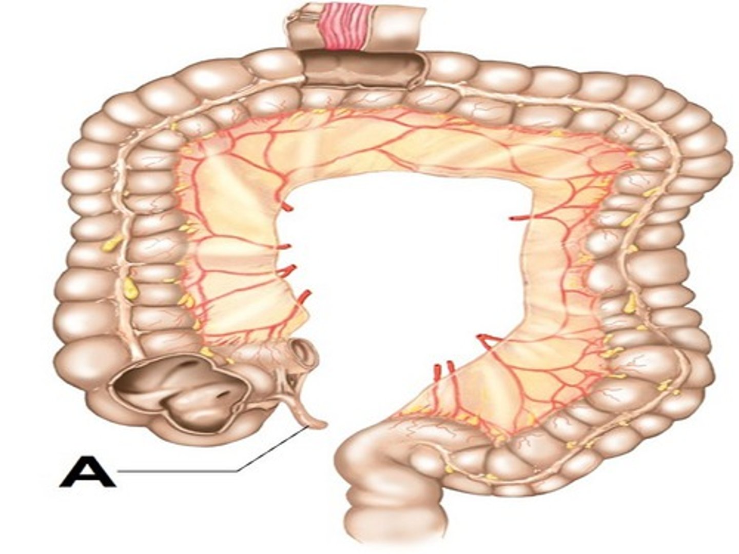
vermiform apendix
a long, narrow, worm-shaped tube connected to the cecum
ascending colon
travels upward from the cecum to the undersurface of the liver
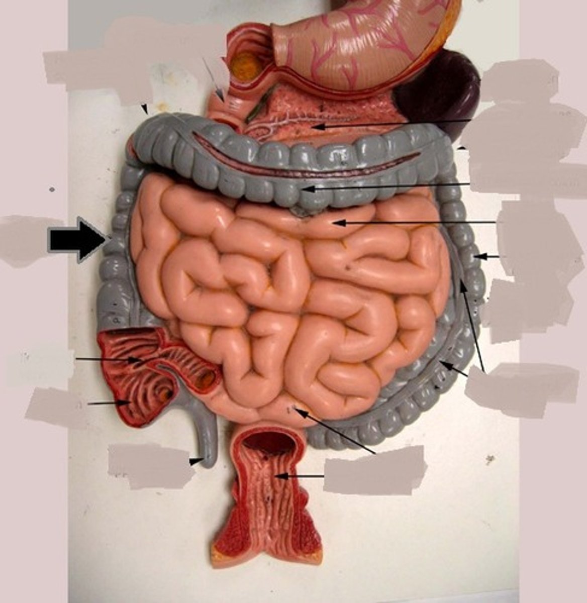
transverse colon
passes horizontally from right to left toward the spleen
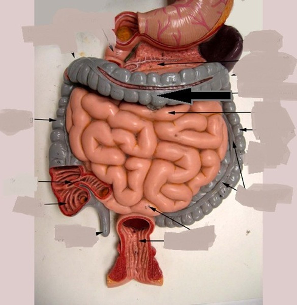
greater omentum
part of the peritoneum attached to the stomach and to the colon and covering the intestines
descending colon
portion of the colon that extends downward from the transverse colon
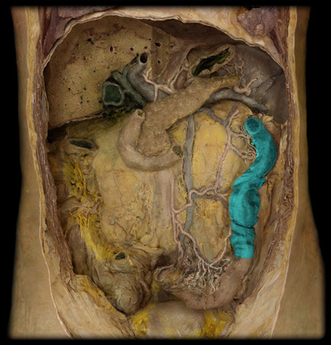
sigmoid colon
an S-shaped structure that continues from the descending colon above and joins with the rectum below
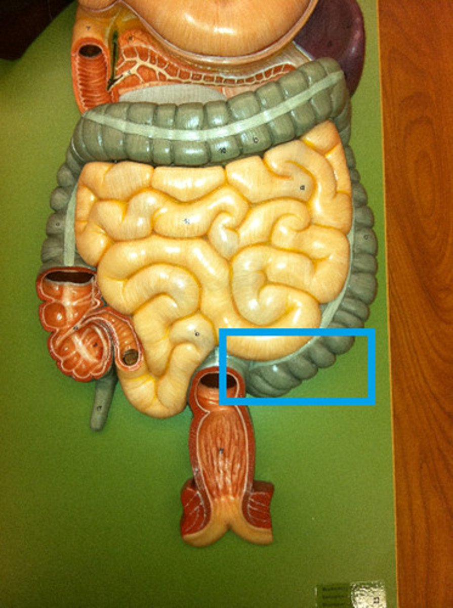
rectum
A short tube at the end of the large intestine where waste material is compressed into a solid form before being eliminated
anal canal
region, containing two sphincters, through which feces are expelled from the body
anal sphincter
The valve that controls the release of feces from the recturm. It has an internal part made of smooth muscle (thus involuntary) and an external part made of skeletal muscle (thus voluntary).
common hepatic duct
large bile duct leading from liver; joins with the cystic duct to form the common bile duct
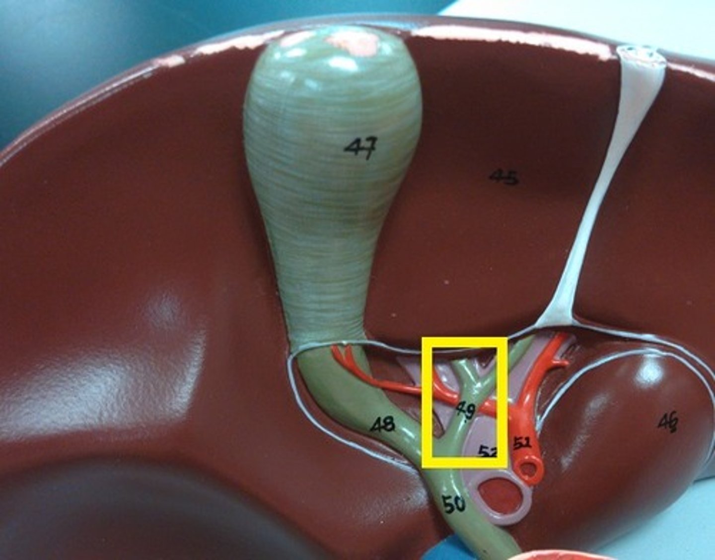
cystic duct
Duct leading from the gallbladder to the common bile duct; carries bile
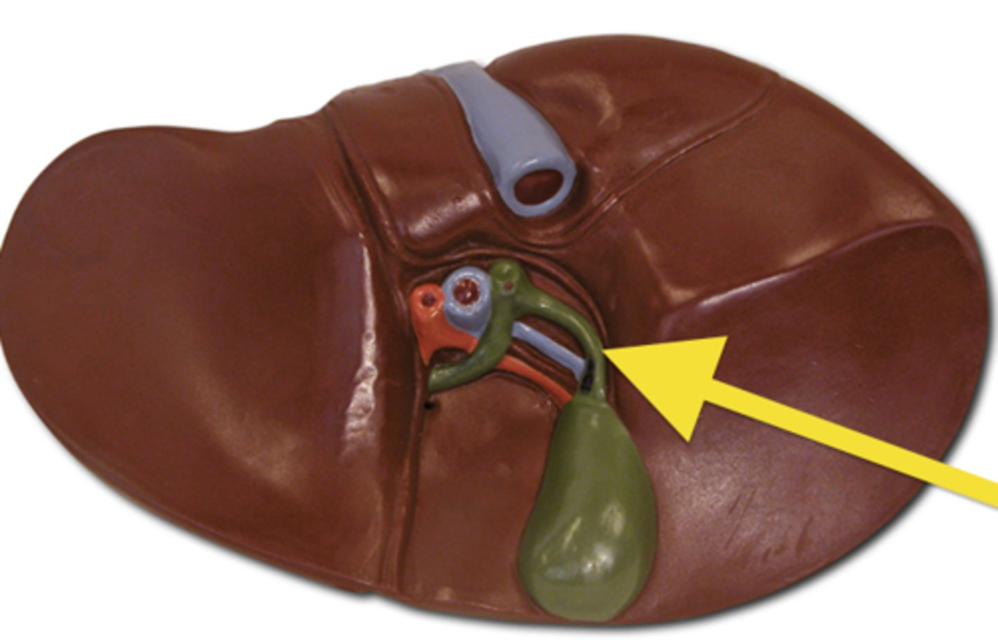
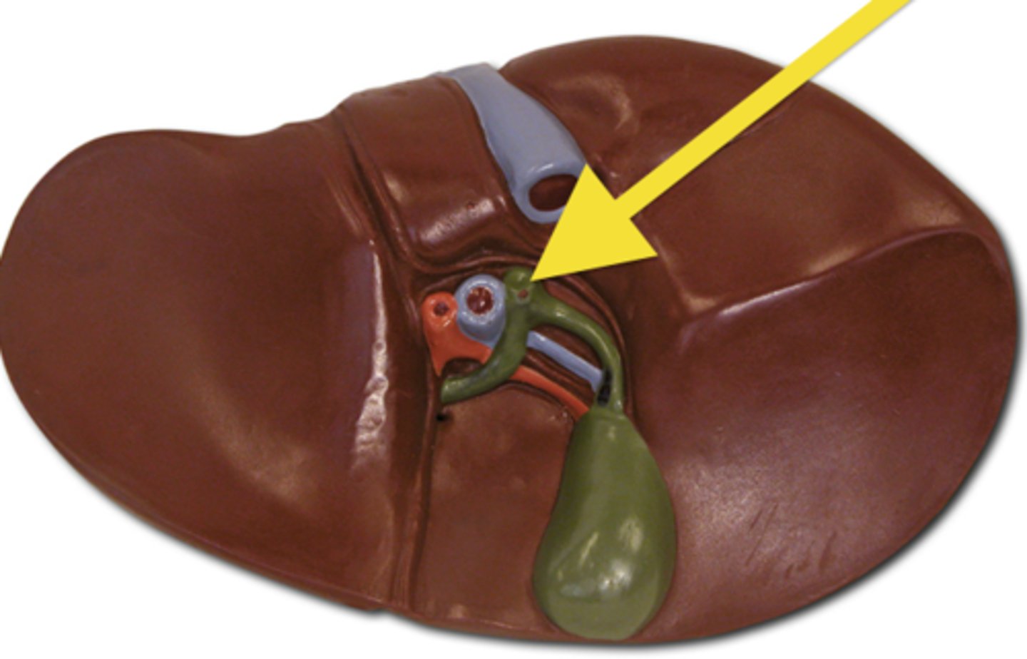
common bile duct
carries bile from the liver and gallbladder to the duodenum
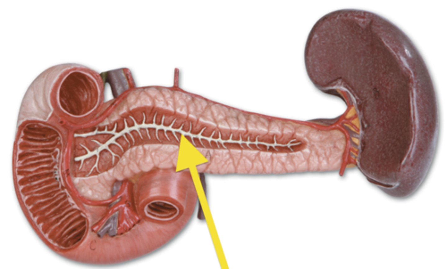
pancreatic duct
conducts pancreatic juice from the pancreas to the small intestine
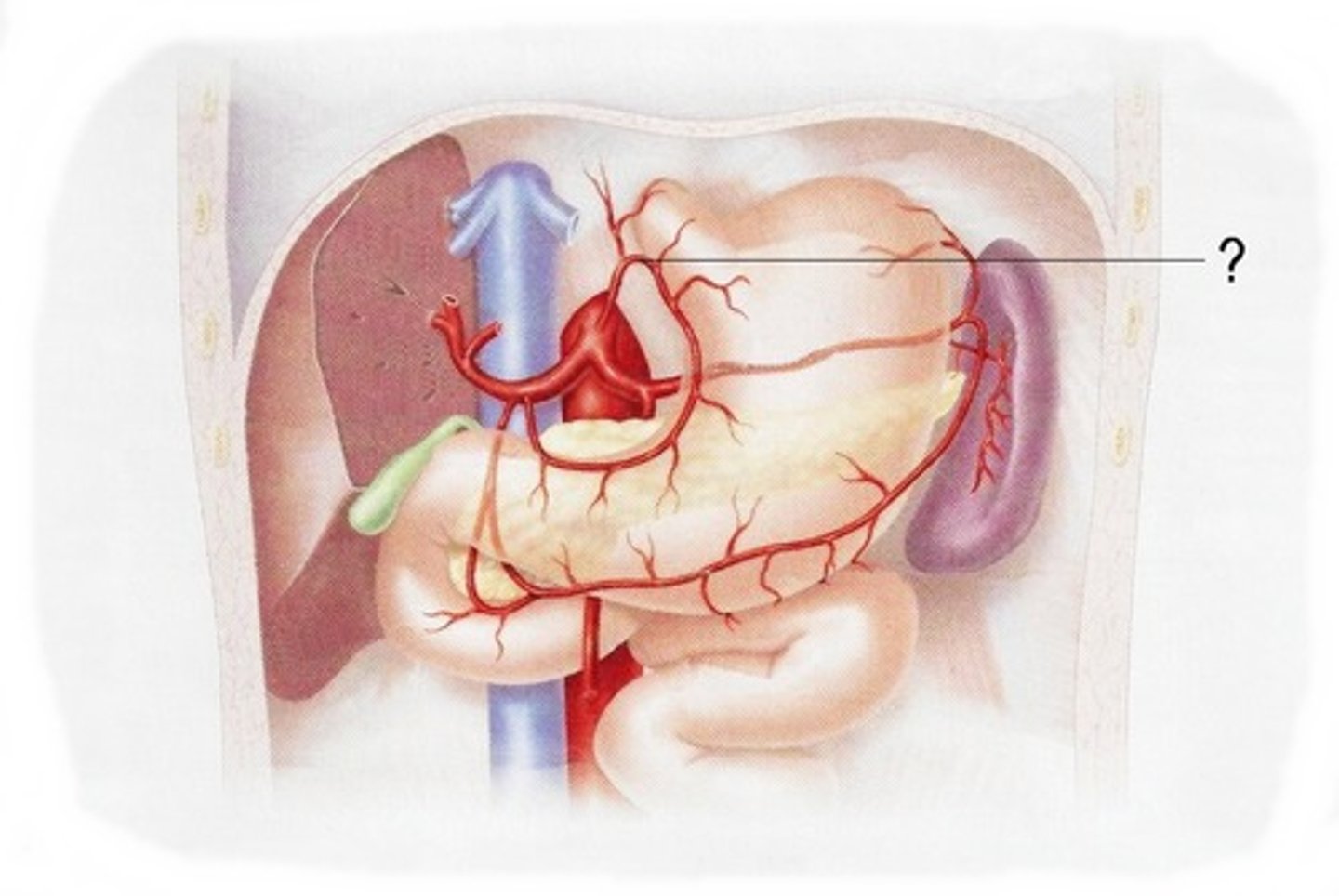
left gastric artery
artery that leads from the celiac trunk to the stomach (on the left)
common hepatic artery
arises from the celiac trunk to supply the liver
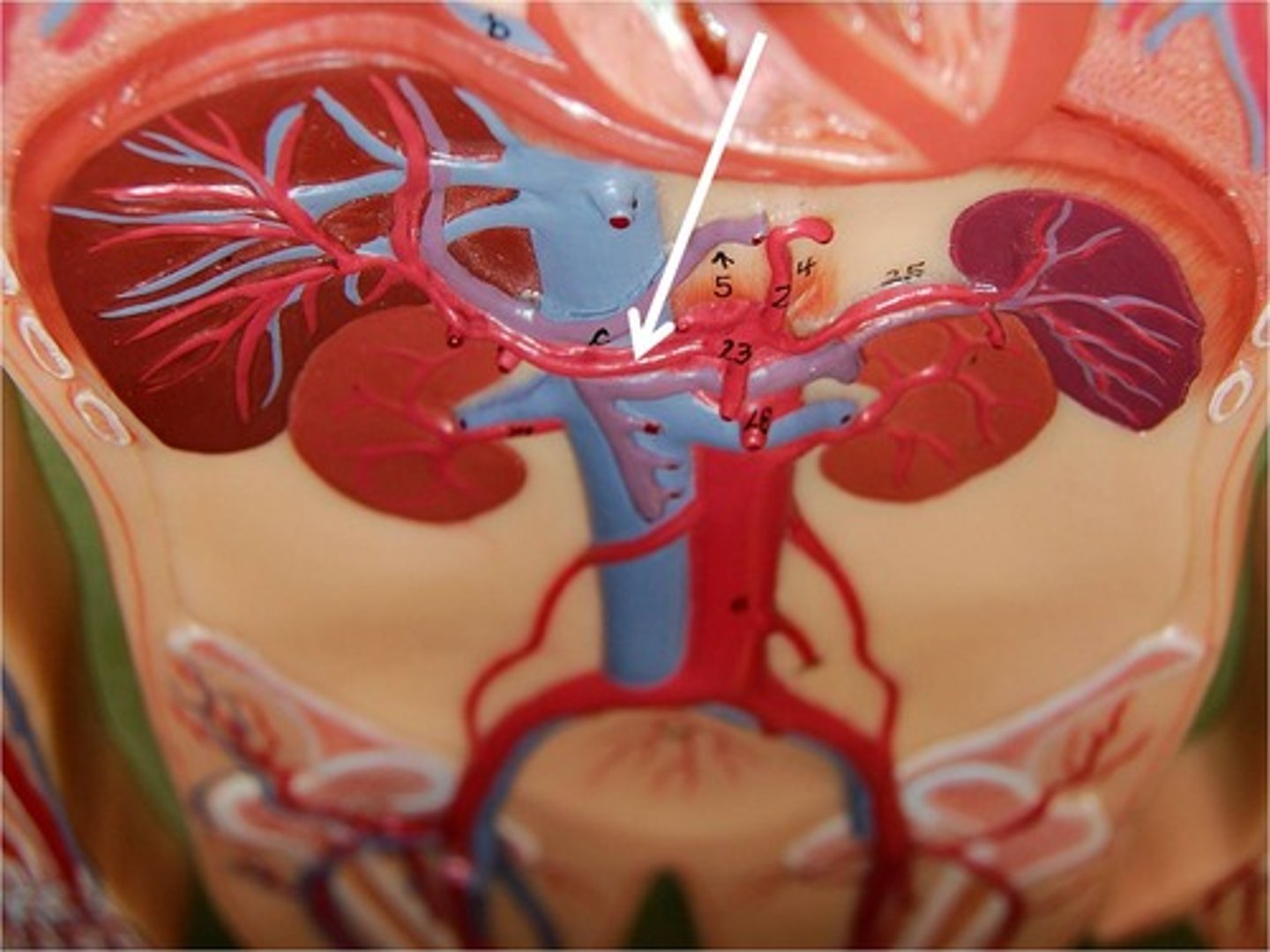
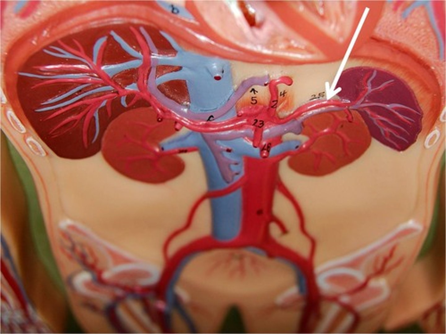
splenic artery
Supplies blood to the spleen.