Biology EXAM
1/31
There's no tags or description
Looks like no tags are added yet.
Name | Mastery | Learn | Test | Matching | Spaced | Call with Kai |
|---|
No analytics yet
Send a link to your students to track their progress
32 Terms
Lymphatic system
Network of vessels and organs that help return fluid from tissue back into the bloodstream and defend the body against infection (lymph nodes, spleen, etc.)
Fluid Balance (lymphatic system)
blood → tissue fluid → lymph
Helps transport fat and immune cells
Blood plasma leaks out of capillaries to form tissue fluid, which bathes cells
Most fluid returns to the blood stream, but some enters lymphatic capillaries becoming lymph
Flow of lymph
Tissue fluid enters lymph capillaries
Moves through lymph vessels and lymph nodes (filtered for pathogens)
Returns to the circulatory system via large veins near the heart
Arteries
Thick muscle layer; small lumen
Function: Carry blood away from the heart under high pressure
Veins
Thin walls; larger lumen; valves present
Function: Carry blood to the heart under low pressure
Capillaries
One cell thick
Fucntion: site of gas and nutrient exchange between blood and tissues
Universal donor
O-
Universal receiver
AB-
Agglutination
Clotting of red blood cells, often caused by an antigen-antibody reaction when blood transfusions
Systemic Circulation (Heart -> body -> heart)
Main role: Delivers oxygen and nutrients to tissues
1. Oxygenated blood leaves the left side of the heart and takes oxygen to cells via the arteries
2. De-oxygenated blood leaves the cells and travels back to the heart via the veins/venules
3. Blood enters from the body to the right side of the heart and then leaves to go to the lungs via pulmonary circulation
Pulmonary Circulation (heart -> lungs -> heart)
1. Deoxygenated blood goes from right side of the body to the lungs via pulmonary arteries
2. In lungs gas exchange allows fresh oxygen to enter the lungs for exhalation
3. Oxygenated blood flows from the lungs to the left side of the heart via pulmonary veins
Coronary Circulation (Within the heart muscle itself)
Supplies the heart with its own oxygen and nutrients
1. The heart muscle is very thick and requires its own blood supply
2. A branch off the aorta supplies the heart muscle with oxygenated blood
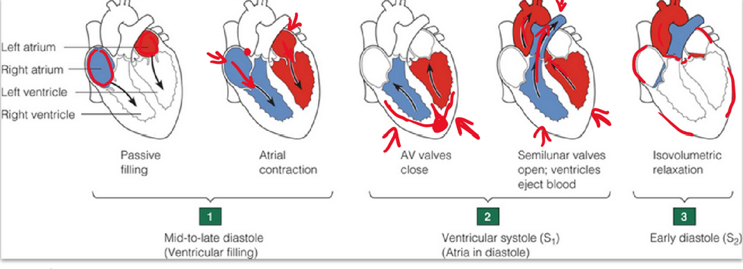
Flow of blood in heart
1. Deoxygenated blood from the body returns to the heart via the superior and inferior vena cava, entering the right atrium
2. As ventricles relax, blood flows through the tricuspid valve into the right ventricle
3. When the right ventricle contracts, blood is forced through the pulmonary semilunar valve into the pulmonary artery
4. Pulmonary arteries carry deoxygenated blood to the lungs
5. In the alveoli of the lungs, gas exchange occurs – oxygen enters the blood and carbon dioxide diffuses out. The blood is now oxygenated
6. The pulmonary veins transport this oxygenated blood back to the heart, entering the left atrium
7. As the left ventricle relaxes, blood passes through the bicuspid (mitral) valve into the left ventricle
8. When the left ventricle contracts, blood is forced through the aortic semilunar valve into the aorta
9. The aorta branches into systemic arteries, delivering oxygen-rich body to all body tissues
10. In the capillary network of tissues, cells take up oxygen and nutrients and release carbon dioxide into the blood
11. This deoxygenated blood then returns to the heart through systemic veins, re-entering the right atrium
Electrical conduction of heart
1. Heartbeat starts with an electrical signal from the SA node (sinoatrial node), also called the heart’s pacemaker
2. Signal spreads through the atria, causing them to contract
3. Passes to the AV node, down the Bundle of His, and into Purkinje fibres, which trigger the ventricles to contract
Factors affecting HR
· Autonomic innervation (Nervous system)
· Hormones (adrenaline, nonadrenaline)
· Fitness levels (more fit, lower HR)
Factors affecting SV
· Heart size (bigger = Higher SV)
· Fitness levels (fitter = Higher SV)
· Gender (males = higher SV)
Hemoglobin binds O2 and helps transport CO2 in three forms:
1. Dissolved in plasma
2. As bicarbonate ions (HCO3-)
3. Bound to hemoglobin as carbaminohemoglobin
Cellular Respiration
1. Glycosis
2. Kreb Cycle
3. Electron transport chain
Tidal volume (TV)
amount of air moved in or out in a NORMAL breath
Inspiratory Research Volume (IRV)
Extra air inhaled during a deep breath
Expiratory Reserve Volume (ERV)
Extra air exhaled after a normal exhalation
Residual Volume (RV)
air remaining after maximal exhalation; prevents lung collapse
Vital capacity (VC)
max air moved in and out
Total lung capacity (TLC)
Total air the lungs can hold; VC + RV
Functional Residual Capacity (FRC)
Air left in lungs after a normal exhalation
Herbivores
Broad flat molars grind tough plant material helping break down CELLULOSE for digestion
Omnivore:
Mix of cutting incisors, tearing canines, and grinding molars allow them to eat and process both plant and animal foods
Carnivore
Sharp canines and slicing teeth allow them to tear meat and crush small bones needed for a high-protein, animal based diet
What enzymes are produced by salivary glands and what do they break down?
Enzymes: Salivary Amylase
Works in: Mouth
pH: 7
Breaks down: carbohydrates (starch)
End product: maltose
What enzymes are produced by the stomach and what do they break down?
Enzymes: Protaese (pepsin)
Works in: Stomach
pH: 2
Breaks down: proteins
End product: peptides
What enzymes are produced by pancreas and what do they break down?
Enzymes: Pancreatic amylase, tripsin, and lipase
Works in: SI
pH: 7-8
Breaks down: Carbohydrates, Proteins, lipids
End product: Maltose, amino acids, fatty acids and glycerol
What enzymes are produced by SI lining and what do they break down?
Enzymes: Disaccharidases (maltase, lactase, sucrase) and Peptidases
Works in: SI
pH: 7-8
Breaks down: Disaccharides (maltose, lactose, sucrose), Peptides
End Product: Monosacharides (glucose), amino acids