UNIT 3
1/77
There's no tags or description
Looks like no tags are added yet.
Name | Mastery | Learn | Test | Matching | Spaced | Call with Kai |
|---|
No analytics yet
Send a link to your students to track their progress
78 Terms
What are qualitative tests used for?
Qualitative tests used to show if certain substances are present or not. DO NOT give an indication of amount of particular substance.
Describe Benedict’s test for reducing sugars.
Add an equal volume of Benedict’s reagent to the test solution and heat to at least 80°C in a water bath. Blue – Brick Red Precipitate.
Describe Benedict’s test for non-reducing sugars.
Initially test for presence of reducing sugar using details above. Hydrolyse sample by heating with dilute hydrochloric acid in a water bath. Once cooled, neutralise by adding sodium hydrogen carbonate. Test with Benedict’s solution as stated above. Blue – Brick Red Precipitate.
Describe the iodine test for starch.
Add iodine solution. Yellow Brown – Blue Black.
Describe the Biuret test for protein.
Add potassium hydroxide to the sample and add a few drops of copper sulfate solution. Blue – Lilac/Mauve.
What is important about Benedict’s test being partially quantitative?
The Benedict’s reagent will change through the sequence blue-green-yellow-orange-brick red, depending on how much reducing sugar is present in the sample.
What is Clinistix used for?
Clinistix is a glucose specific test.
What is the principle of paper chromatography?
Some solutes are more soluble than others in the same solution. The more soluble substances will travel further than others in a solvent that is moving through chromatography paper. Separation of the solutes allows them to be identified. This is useful in identifying which amino acids are present in a mixture.
What are the stages of paper chromatography?
Preparing the chromatogram. Running the chromatogram. Developing the chromatogram.
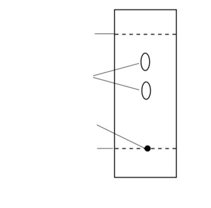
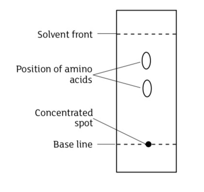
How is a chromatogram prepared?
Chromatography paper cut to fit tank or vessel used to hold it in a vertical position., Chromatogram should be long enough to be attached to the lid of the container and drop just BELOW the volume of solvent at the bottom of the tank. , A horizontal line should be drawn in pencil a set distance from the base. The line should be above the solvent level. , The atmosphere of the tank should be saturated with the solvent prior to placing the chromatogram inside. , The solution containing amino acids needs to be spotted onto the origin line.This is achieved using a micro-pipette, capillary tube or point of pin add extract from the sample. After adding the drop of solution, it is then dried before repeating the process and adding the next drop. This allows the spot to become concentrated and ensures it occupies a small area.
What must be ensured when running a chromatogram?
The line where the spots have been placed does not make contact with the solvent. The chromatogram is securely attached. Chromatogram is not suspended at an angle. The solvent run should be stopped before reaching the top and marked.
How to prevent contamination
only holding it at edges of paper and avoid setting it on laboratory benches that could be contaminated with chemicals. Gloves can also be worn to prevent contamination
How is a chromatogram developed?
The chromatogram should be dried and the solvent front marked. Amino acids are colourless and so the chromatogram should be sprayed with ninhydrin in a fume cupboard. The chromatogram should be re-dried and amino acids will appear as purple spots. Proline appears yellow.
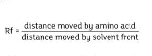
What is the Rf value formula?
Rf = distance moved by the solute / distance moved by the solvent.
Why must the solvent front be marked quickly?
It evaporates and becomes invisible.
Why must the origin line be drawn in pencil in chromatography?
Ink would dissolve in the solvent.
What are key rules about Rf values?
A consistent approach must be taken when measuring from origin. Pigments closest to origin are least soluble and pigments furthest away are most soluble. A Rf is always less than 1.
Why must the chromatography tank be saturated with solvent?
Prevents evaporation and ensures consistent solvent movement.
Example of reactions; Enzymes
Amylase breaking down starch
Protease breaking down protein
Catalase breaking down hydrogen peroxide
What must be considered when investigating temperature on enzyme activity?
A range of temperatures should be used. Thermostatically controlled waterbaths should be used and monitored. Other variables must be controlled including pH, volume and concentrations. Enzyme and substrate should be brought to temperature before mixing.
Why must enzyme and substrate be pre-heated before mixing?
Ensures the reaction starts at the correct temperature.
What must be considered when investigating substrate concentration?
A suitable number and range of substrate concentrations should be used. Controlled variables include temperature, pH, volume and enzyme concentration. Rate may level off due to enzyme saturation.
What must be considered when investigating enzyme concentration?
A suitable number and range of enzyme concentrations should be used. Controlled variables include temperature, pH, volume and substrate concentration. Rate increases with more enzyme substrate complexes.
What must be considered when investigating pH?
A range of pH values should be used. Controlled variables include temperature, volume and concentrations. Buffers are used to maintain pH. Buffer must be added before mixing enzyme and substrate.
Why must buffer be added before mixing enzyme and substrate?
Ensures the correct pH conditions before the reaction starts.
What do results of pH investigations show?
Enzyme has a higher rate of activity at a particular pH. The optimum pH corresponds with the pH in which the enzyme is normally active. Either side of the optimum activity falls.
Why does enzyme activity decrease at extreme pH?
Ionic bonds in the tertiary structure break and the active site changes shape so it is no longer complementary.
Effect of ph on enzyme activity method
Label six boiling tubes 1-6.
Cut six 1 cm3 cubes of jelly.
Add 10 cm3 of 2% trypsin to each boiling tube.
Add 10 cm3 of the appropriate buffer to five of the boiling tubes and distilled water to the remaining one.
Place the boiling tubes in a water bath at 25oC.
After 5 minutes add a cube of jelly to each of the boiling tubes 1 to 5.
After 24 hours remove a sample from tube 6 to act as a blank for the colorimeter. Using the appropriate filter, set the percentage transmission on the colorimeter to 100%.
Shake the contents of boiling tube 1 and remove a sample, place in the colorimeter and record the percentage transmission.
Repeat for the other boiling tubes 1 to 5.
Tabulate the results and use an appropriate graphical technique to present the results.
Demonstrate enzyme immobilisation method
Add 8 cm3 of sodium alginate solution (2%) to a small beaker.
Add 2 cm3 of beta-galactosidase (lactase) to the beaker.
Add one drop of food colouring – this allows the reaction to be seen more clearly.
Mix thoroughly but keep bubbles to a minimum.
Draw this mixture into the barrel of a 10 cm3 plastic syringe.
Add 1.5g of calcium chloride to 100 cm3 of distilled water in 250 cm3 beaker.
Add the enzyme mixture dropwise from the syringe to the calcium chloride solution. Allow the immobilised enzyme beads that start to form to harden for about 10 minutes. Remove the beads and rinse thoroughly with water.
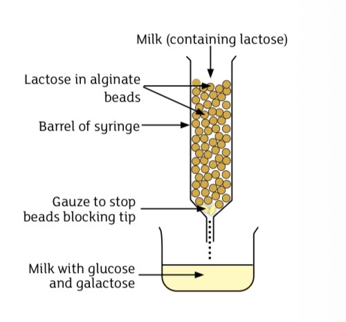
Rinse the syringe and remove the plunger and fix the barrel to a retort stand.
Place a small piece of gauze near the tip of the syringe to prevent the beads from blocking the syringe nozzle.
Add the beads to the syringe.
Test the milk after it has been filtered through the beads with Clinistix or a similar specific test for glucose.
Enzyme immobilisation diagram,
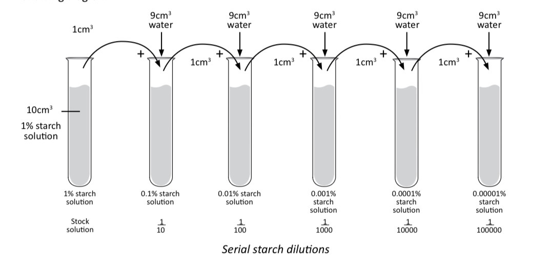
What are serial dilutions?
Each solution is 10 times less concentrated than the previous one by adding 1 cm³ of solution to 9 cm³ of water.
Why must fresh pipettes be used?
To prevent contamination.
What does a colorimeter measure?
A colorimeter measures the change of light intensity as it passes through a solution.
What can a colorimeter record?
The amount of light absorbed or transmitted.
Why is a red filter used in amylase starch reactions?
“It is at the opposite end of the spectrum to the colour change observed
How can accuracy be improved in colorimetry?
Cuvettes must be clean, rinsed and dried between samples, filled correctly, Orientation of cuvette in the colorimeter is correct and recalibrated when necessary.
What is the purpose of calibration in colorimetry?
To set a blank baseline for accurate measurement.
What is a calibration curve used for in colorimetry?
To convert transmission or absorbance values into concentrations.
colorimeter effect of factor, e.g temperature, on permeability of cell-surface membranes in beetroot: procedure:
cut several small sections of beetroot of equal size using a cork borer.
Rinse the beetroot in water until the water remains clear.
Set up a number of water baths at, e.g. 20oC, 40oC, 60oC, 80oC.
Add 10 cm3 of water to each of 5 test tubes and place one in each water bath for 5 minutes to allow the temperature of the water to equilibrate.
Place a section of surface-dried beetroot in each of the test tubes.
Leave in the water bath for 10 minutes.
Set up a colorimeter using a blue/blue-green filter. Calibrate distilled water as 0 absorbance (or 100% transmission).
After the 10 minutes, sample the water surrounding the beetroot sections and check its absorbance (or transmission) for each temperature.
Add the results to an appropriate results table and draw a graph of % absorbance (or transmission) against temperature.
What is homogenisation?
Cell fractionation which is the breaking up and mixing of the material to give a uniform preparation. This can be carried out using a mortar and pestle or blender.
What is centrifugation?
Spinning samples at high speeds so forces cause larger, heavier particles to sediment at bottom of centrifuge tube at smaller, lighter particles to remain at top of supernatant in centrifuge tube.
What determines position of organelles after centrifugation?
Their size and density.
Why must a centrifuge be balanced?
To prevent damage and ensure proper operation.
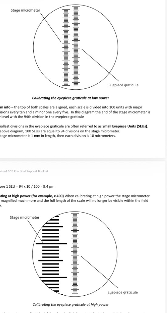
What is the key value of a stage micrometer division?
Each division equals 10 micrometres.
How can you distinguish between the stage micrometer and eyepiece graticule?
Adjusting focus moves the stage micrometer but not the graticule. Rotating the eyepiece rotates the graticule but not the stage micrometer.
Why should high power be used when measuring cells?
Provides greater precision and increases accuracy.
procedure for measuring water potential in potato tissue?
Add a range of sucrose solutions (and water) to separate beakers.
Cut cylinders of potato and weigh.
Add a cylinder to each beaker.
After 24 hours remove the potato and reweigh.
The percentage change in mass should be calculated for the cylinder in each solution.
Plot the percentage change in mass against sucrose solution.
Where the line of best fit crosses the X-axis, the water potential in the potato is equal to the solute potential of the sucrose solution.
The solute potential of the sucrose solution at the point of intercept can be calculated from a conversion table.
What is incipient plasmolysis?
When the cell membrane just touches the cell wall and pressure potential is zero.
How is solute potential measured at incipient plasmolysis?
Add sections of onion epidermal tissue to pure water to make sure all the onion cells are turgid.
Place sections of the onion epidermal cells in beakers, with each beaker containing either water or one of a range of sucrose solutions.
Leave the epidermal tissue in the beakers for 30 minutes.
After 30 minutes remove the onion epidermal tissue and place on a microscope slide.CCEA Revised GCE Practical Support Booklet
Observe the onion tissue under the microscope and calculate the percentage of cells that are plasmolysed for each solution.
Draw a graph of percentage plasmolysis against sucrose solution. Use the graph to identify the point at which 50% of the cells are plasmolysed. At this point the average solute potential of the onion cells is the same as the solute potential of the sucrose. The solute potential of the sucrose at that concentration can be calculated from a conversion table.
Why must potato cylinders be the same size?
Ensures a fair test.
Why must potato be surface dried before weighing?
Removes excess solution for accurate mass.
What is the principle of beetroot permeability experiment?
Damage to membranes allows pigment to leak out causing colour change.
What is the procedure for beetroot permeability experiment?
Cut equal pieces, rinse, heat in water baths, measure absorbance and plot graph.
Why must beetroot be rinsed before the permeability experiment?
Removes pigment released during cutting.
Why is a blue or blue green filter used in beetroot colorimetry?
To obtain maximum differentiation across results.
What is the purpose of staining root tips?
Chromosomes are stained and tissue softened for observation.
Measuring the average solute potential of cells at incipient plasmolysis procedure
Add a small section of root containing lateral roots to a boiling tube containing acetic orcein;
place the boiling tube in a water bath at 60 oC for 30 minutes;
after 30 minutes remove a section of root from boiling tube and use a scalpel to remove last few mm or so from one of the lateral roots. Add this short section to a microscope slide and add more acetic orcein if necessary to stop the root tip from drying out;
add a cover slip and gently tap with a blunt end of a pointed needle. This will ‘squash’ the root tip into a single layer of cells; and
observe under a microscope.
Common features between each staining process
Chromosomes are stained
Procedure softens the root tissue allowing it to be easily squashed into a single layer
Other staining techniques
There are many different techniques used to stain chromosomes and prepare root tip squashes for observing mitosis. Using toluidine blue with garlic tips works well. Common features between each process are that the chromosomes are stained and that part of the procedure used softens / breaks up the root tissue allowing it to be easily ‘squashed’ into a single layer.
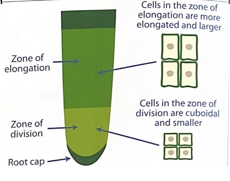
Finding cells undergoing mitosis
Using low / medium power (for example, x100), scan the root tip section and look for the zone of division. Cells in the zone of division are characteristically small and cuboidal in shape with the nucleus appearing relatively large. Once the zone of division is located switch to high power (x400) to observe cells at different stages of mitosis.
Good block diagrams:
•have all the obvious (tissue) layers included;
•have layers added in the correct proportions;
•have continuous (not sketchy) lines; and
•have labels as appropriate.
What is the principle of a respirometer?
Oxygen uptake is measured as carbon dioxide is absorbed causing a pressure change.
How is carbon dioxide production measured in a respirometer?
Replace potassium hydroxide with water and observe movement of liquid.
What does movement of liquid in a respirometer indicate?
Towards organism indicates oxygen uptake and away indicates more carbon dioxide produced.
What must be controlled in respirometer experiments?
Same apparatus, organisms, conditions, time and temperature.
Why must temperature be controlled in respirometer experiments?
It affects respiration rate and gas volume.
What are the main types of sampling?
Density, percentage cover and frequency.
What is random sampling
Quadrats placed randomly to avoid bias
What is systematic sampling
Sampling along a transect
What are abiotic factors?
Non living factors such as temperature, pH and light intensity.
What are biotic factors?
Living factors such as competition, grazing and disease.
How is soil moisture measured?
Weigh, dry at 105°C, reweigh and calculate percentage water content.
How is soil organic content measured?
Burn dried soil and calculate percentage organic content.
How is light intensity measured?
Light at sample divided by light in open multiplied by 100.
What are key rules for block diagrams?
Do not draw individual cells, include layers, keep proportions correct, use continuous lines and label structures.
Why should multiple readings be taken for light intensity?
Light intensity varies due to environmental conditions so multiple readings improve reliability.
Why must soil be stored in plastic bags?
Prevents moisture loss before testing.