Histology Slides
1/40
Earn XP
Description and Tags
FUCKASS HISTOLOGY SLIDES AND THEIR DESCRIPTION FIRST YEAR
Name | Mastery | Learn | Test | Matching | Spaced | Call with Kai |
|---|
No analytics yet
Send a link to your students to track their progress
41 Terms
What is this?
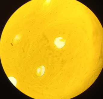
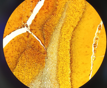
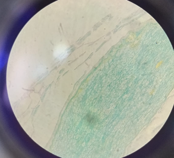
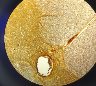
Omentum Majus Simple Squamous Epithelium - Silver Impregnation (AgNO3) (34a)
Part of the Peritoneum, which forms unique serous membrane (serosa)
built of simple squamous epithelium (mesothelium)
look from the top (surface) cells are polygonal
nuclei are not visible
Cells connect thanks to interdigitations
What is this?
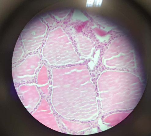
Glandula Thyroidea Simple Cuboidal Epithelium - HE (2)
Endocrine gland = hormones (iodine based)
built of follicles
on top of basal membrane = simple cuboidal epithelium (nucleus = circle)
looks like 2 layers of cells
colloid content - pre-hormones, iodine ions, and hormones

What is this?
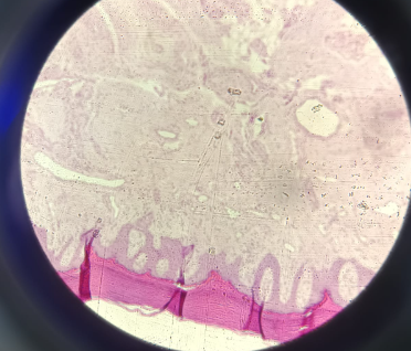
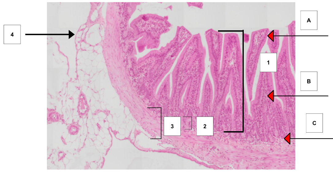
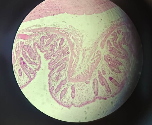
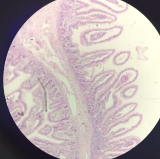
Small Intestine Simple Columnar Epithelium - HE (3, 44)
Tunica Mucosa - mucosa
A. Lamina Epithelialis - simple columnar resorptive epithelium with microvilli; columnar cells with microvilli (elongated, basal), goblet cells (triangular, basal) and stem cells (cylinder, middle)
B. Lamina Propria - Loose connective tissue containing blood vessels
C. Muscularis Mucosae - muscle layer, contains connective tissue and smooth musclesTela Submucosa - loose connective tissue, simple tubular glands, goblet cells
Tunica Muscularis - 2 types of smooth muscle cells
D. Stratum Circulare
E. Stratum longitudinaleTunica Serosa (mesothelium) or Tunica Adventitia (loose connective Tissue)
What is this?
Trachea pseudostratified epithelium - HE (22)
part of the respiratory tract
pseudostratified and columnar ciliated epithelium
3 types of cells
columnar ciliated cells
stem cells (basal cells)
goblet cells
Tunica Mucosa
Lamina Propria - LCT to glands
Tunica fibromusculocartilaginea - contains cartilage (hyaline cartilage), 2 ends of cartilage are linked together by muscle layer
Tunica Adventitia - loose connective tissue
What is this?
Ureter Transitional Epithelium - HE (59)
surrounded by a lot fat tissue (adipose tissue)
Tunica Mucosa
a. Lamina epithelialis
- (3 layers) (wrinkle) transitional epithelium of henle
- stratified epithelium, a lot of membrane (looks like stripes);
- very small cylindrical shaped cells (stratum basale)
- on top, polygonal cells (tennis rocket shaped)
- on top, only one cell (umbrella cells; may have 2 nucleus)
- outer membrane of the cells, invagination (wrinkling) of the membrane, linked together by uroplakins (proteins) (protects cells from toxicity)b. Lamina Propria
Tunica Muscularis - 2/3 layers of cells
Tunica adventitia - has a lot of big blood vessels
What is this?
Esophagus Stratified Squamous nonkeratinized - HE (18)
Membrane invaginations (wrinkling)
all of the cells are alive and has nuclei
stratified squamous nonkeratinized epithelium
Top of basal membrane - layer of small cells (stratum germinativum / stratum basale); small cylindrical shaped
On top, bigger cells - Polygonal shaped
On top, flat cells - squamous epithelium cells
Tunica Mucosa
a. Lamina Epitheliaris
b. Lamina Propria - may see some glandsTela submucosa - big glands (esophageal glands, glandula esophage)
Tunica Muscularis
Tunica Adventitia
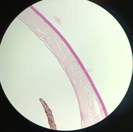
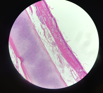
What is this?
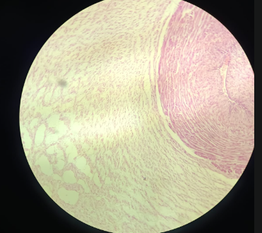
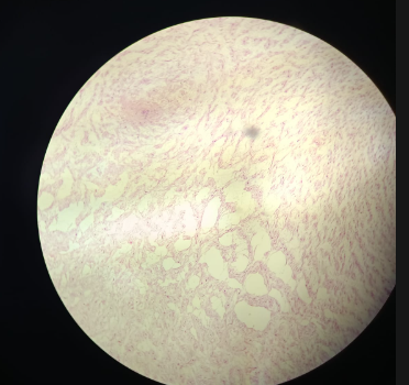
Cornea Stratified Squamous Nonkeratinized - HE (1)
Arch Structure
Cornea epithelium - Stratified squamous nonkeratinized
usually 6-7 layers of cells
Endothelium - origin Endothel
- epithelium anterius- stratified squamous non keratinized
- epithelium posterius- simple squamous epithelium
Basal Layer - Basal membrane is a straight line (has no blood vessels)
Stratum germinativum
Stratum basale
1-2 layers polygonal cells
1-2 layers of flat squamous cells
What is this?
Hairless Skin Stratified Squamous Keratinized - HE (19,25)
built of 3 layers
Epidermis - built in Tunica Mucosa
a. Epithelium - Stratified squamous keratinized epithelium
b. Stratum Basale - Keratin fiber, cuboidal cells
c. Stratum Spinosum - Desmosomes, polygonal cells
d. Stratum Granulosum - Keratohyalin, flat squamous epithelium, final layer with alive cells
e. stratum Lucidum - keratin
f. Stratum corneum - keratin, dead cells, cannot be seenDermis - LCT
hypodermis - LCT
Receptors
BOTH
Extra Cellular Matrix (ECM)
Fibers: Collagen type 1, elastic fibers, ground substance
Cells: Fibroblasts, fibrocytes
What is this?
Intestinum Tenue - Mucicarmine (3b)
Staining: Mucicarmine (not a lot of pink, but purples structures (vacuoles of goblet cells)
Microvilli also has staining, due to excretion produced by goblet cells and tubular glands and tubular glands are going to resurface, to scratch the surface of the cell
Tela Submucosa - big tubular glands (simple Tubular glands)
What is this?
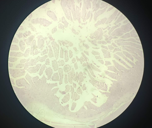
Intestinum Crassum (Large Intestine) (45) - HE
Epithelium: Same as the small intestine, Simple columnar resorptive epithelium with microvilli
Difference: Lamina Propria: tissue of glands (Simple Tubular Glands), Goblet cells usually dominate these glands
What is this?
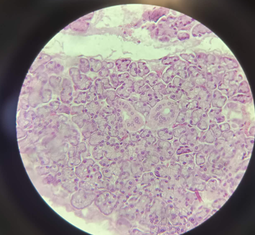
Glandula Parotis - HE (28)
Big saliva gland located behind the ears
Has fat tissue
contains only serosa acini (circular nucleus, with pyramidal outline, new epithelial cells, no lumen) (with ribosomes, produces protein rich solutions)
Epithelium: Cuboidal Epithelium
What is this
Glandula Submandibularis - HE (30)
under the mandibula bone (jaw)
2 different structures:
1. serosa tsinin (no lumen, circular nucleus, pyramidal outline, ribosomes, new epithelial cells) 70% (purple)
2. mucosa tsinin (big lumen, squamous nucleus, squamous like outline, new epithelial cells) 30%, but it looks big so 50-50% ratiov (pink)
What is this?
Glandula Sublingualis - HE (29)
located underneath the spine
more light structures
mucosa tsinin are dominating than serosa tsinin
also mixed atsinin (mucoctyes dominates the serocytes)
What is this?
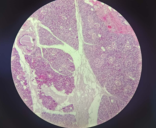
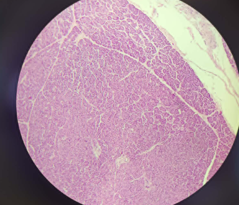
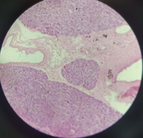
Pancreas - HE (47)
Exocrine
fully serosa tsini (they don’t have new epithelial cells), but will gain centroacinal cells inside the lumenEndocrine
- lining house ions - endocrine (hormones)islets of langerhans
What is this?
Placenta - HE
chorionic villi goes inside the endometrium
placenta is formed by the maternal and the fecal parts
placenta is normally formed in the endometrium of the mother
Structure of villi:
2 layers of cells surrounding the embryonic part
cytotrophoblast (inside)
syncytiotrophoblast (outside)
with time, cytotrophoblast will disappear and left with syncytiotrophoblast
Chorion villi has a lot of blood vessels, because placenta helps get nutrition and oxygen from mother to babies
these two tissues never mix
mucous embryonic connective tissue - built of collagen type 1 small fibers and round cells, very limited amount of ground substance
What is this?
Umbilical Cord - HE
structure which carries out blood vessels (looks like Loose connective tissue)
3 types of blood vessels:
1. venaumbilicaries
2. arterieumbilicaries
Epithelium: Amion, simple cuboidal epithelium
Wharton Jelly - very soft tissue, built of small non-refinigated microcollagen type 1 fibers
Stem cells: triangular shaped from the umbilical cord
Empty space is filled with ground substance
Umbilical cord is very
What is this?
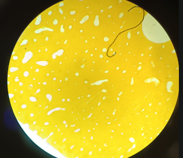
Hypoderma - HE
no blood vessels inside
example of loose connective tissue
loose connective tissues built of collagen and elastic fibers
Collagen fibers: big fibers and cells attach to them
elastic fibers: thin and smaller, cells don’t attach to them ( because they change composition, stretched and relaxed)
Cells that attach to collagen fibers are called Adherent oblique cells (fibroblast/fibrocytes)
fibroblasts - cells which produce the whole component of extracellular matrix (fibers, collagen, ground substances)
Defense cells - not attach, but moving, come from blood vessels (granulocytes, macrophages, lipocytes)
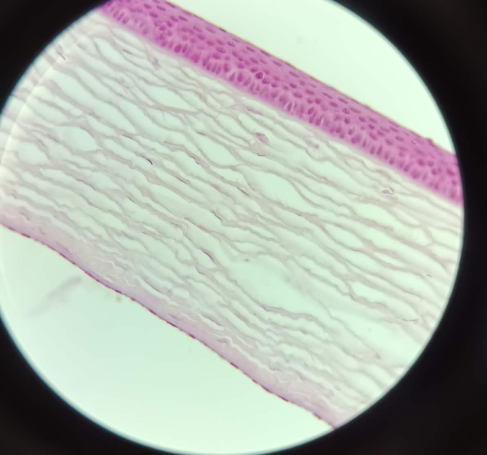
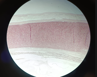
What is this?
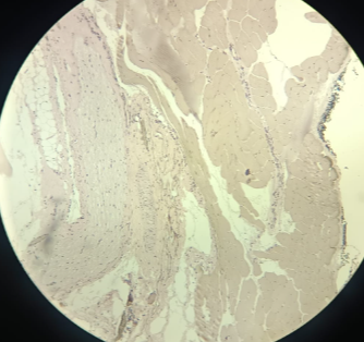
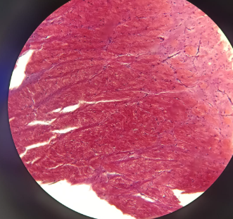
Tendon - Van-Gison
Dense irregular connective tissue
usually don’t have blood vessels
avascular aminavein structure (IDK)
collagen tendon organ
tenocytes - cells of tendons, and maintains collagen fibers inside
blood vessels in the sheaths
What is this?
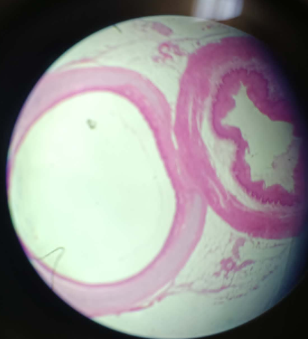
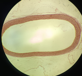
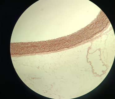
Aorta - Orcein
Orcein stains elastic structure only
its a blood vessel, with epithelium (simple squamous epithelium, endothlium)
Tunica etimum - contains endothelium
Suped utelium - loose connective tissue
2 layers:
top: membrana elastica externa
inside: tunica media which contains membrana elastica interna
Tunica adventitia
Smooth muscle cells
What is this?
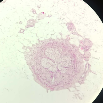
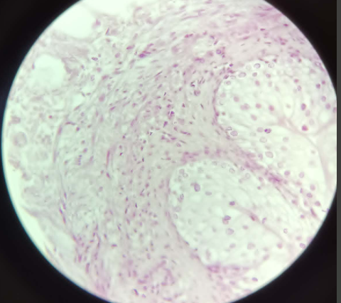
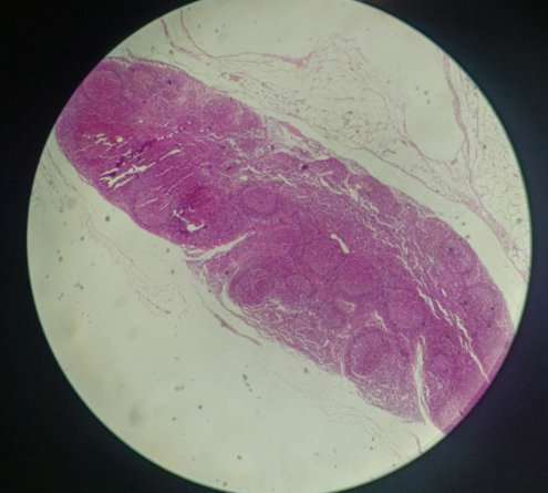
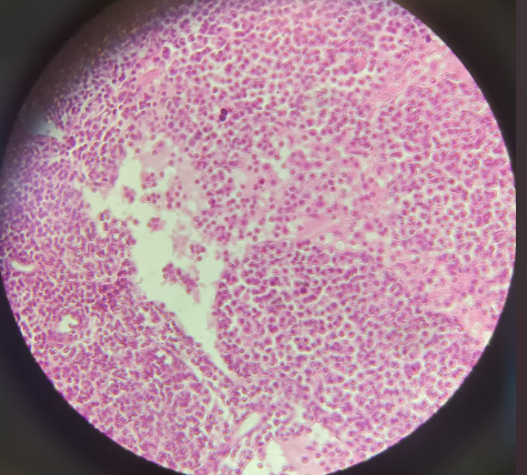
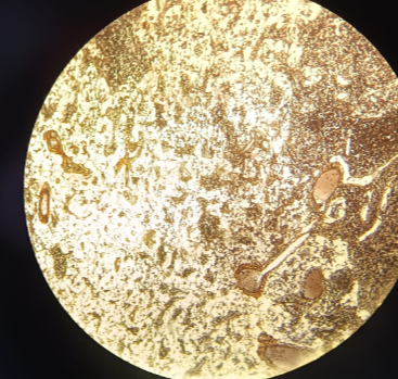
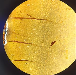
Lymph node - AgNO3
reticular connective tissue
has 2 surfaces
corticol: primary and secondary follicles
mendular part (inside)
reticular fibers (spider web) - collagen type 3 fibers has the affinity to silver ions
cells: reticular cytes
cells inside: lymphocytes
What is this?
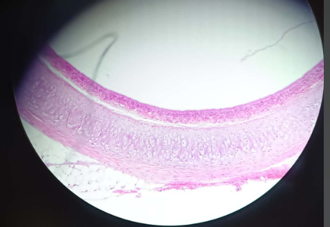
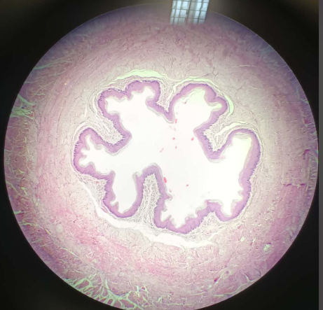
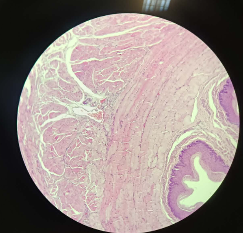
Trachea - HE
Epithelium - pseudo stratified and columnar ciliated epithelium
Loose connective tissue - contains the blood vessels
Hyaline cartilage - has 2 perichondrium (top and bottom)
perichondrium = chondroblasts
inside = chondrocytes in the hyaline cartilage forms huge isogenic groups. Halo around is called territorial matrix
Bright pink - interterritorial matrix, built of collagen type 2 and aggrecan, very robust?
tunica adventitia
Tunica fibromusculocartilaginea - it contains smooth muscle and fibers that connect two edges of the cartilage caalled “U-shaped” or “Horseshoe cartillage”
What is this
Epiglottis - Orcein
located at the end of mouth cavity
2 different types of epithelium:
Facing the mouth: stratified squamous non-keratinized epithelium
Facing the larynx: pseudo stratified columnar ciliated epithelium
Orcein - stains elastic structures of elastic cartilage
has 2 perichondrium
Deep zone of cartilage - middle has a lot of cells, very narrow space between cells stained with orcein
What is this?
Bone - Schmorl Staining
dense bone tissue contains osteons
osteons - center hole is called haversian channel that carries out blood vessels
concentric circle of collagen type 1 and hydroxyapatite
Lacunae (holes) - places where osteocytes reside
cracks of the bones (small channels “canaliculus”) - forms pathways towards center and also communicate between osteocytes
What is this?
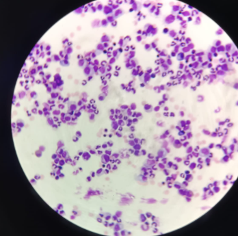
Blood Film - Wright-Giemsa
blood is a liquid connective tissue
extracellular matrix is liquid called blood plasma
blood plasma contains water and organic and non organic components
RBC - males (4-6×10^12 cells/L) Females (3.5-5.5 ×10^12 cells/L)
Erythrocytes are cells without nucleus, but have a lot of hemoglobin inside
on top of erythrocytes has a lot of factors (blood lines factors, A Group, B Group, AB Group, and O group)
has also rhesus factor
WBC - 4-10×10^9 Cells / L, all has nucleus, and all has lysosome (phagocytic cells)
granulocytes:
Neutrophils (55-60%) - contains segmented low nucleus (3-5), has granules but not stained
Eosinophils (2-4% or 1-3%) - specific cells, they will not divide in the blood stream
Basophils (0.1-1%) - biggest granulocytes, contains granules which are stained, contains inside hestamine and serotonin inflamatory cells
Agranulocytes:
Lymphocytes - built of B and T cells Ratio (20-80%), B cells don’t produce antibodies, they have the ability to turn inside the plasmacytes the one which are going to produce the antibody. They have the ability to differentiate
monocytes (4-5%) - the biggest white blood cells macrophages, kidney shaped nucleus, phagocytic cell has the ability to migrate closer to connective tissue
platelets - are not cells (200-400×10^9 Units/Liter)
What is this?
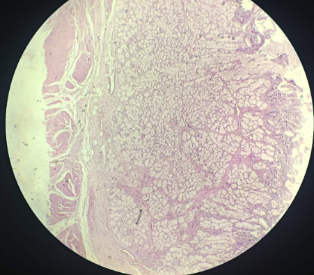
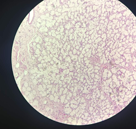
Bone Marrow - Wright-Giemsa
reticular connective tissue
built of collagen type 3 fibers
inside the bone marrow - formation of several blood lineages
most likely to observe the stages of erythropoiesis (basophilic, erythroblast, polychromotophic, etc)
observe stages of granulopoiesis
very big cells (megakaryocytes) which produce platelets
What is this?
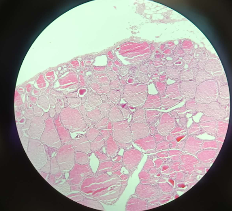
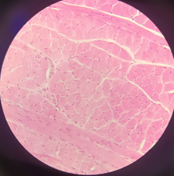
Tongue (lingua) - HE
Epithelium: Stratified squamous (non-keratinized).
Papillae: Highly deep structures containing taste buds.
Tunica Submucosa: Contains serous glands (circular nuclei) and mucous glands.
Muscle Cells: Rhabdomyocytes (striated voluntary muscle).
Nuclei: Up to 100 nuclei per cell, located underneath the sarcolemma (plasma membrane).
Connective Tissue Sheets: Endomysium (individual cells), perimysium (bundles), and epimysium (whole muscle)
What is this?
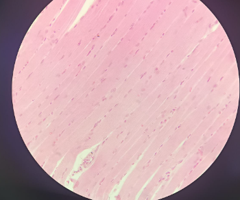
Tongue (Lingua) - FeH
Purpose: Specific stain to visualize striation.
Visuals: Consecutive dark and bright bands (isotropic and anisotropic).
Scale: One cell contains up to 10,000 sarcomeres
What is this?
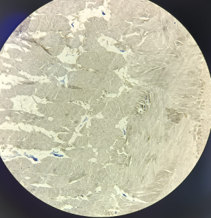
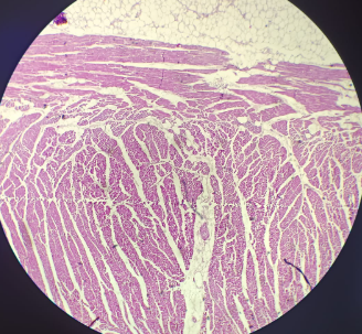
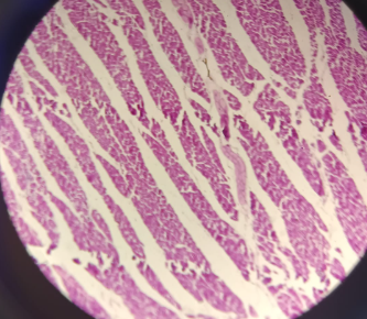
Heart (cor) - HE
Cells: Cardiomyocytes; typically a single-nucleus cell.
Organization: Each cell is wrapped in its own endomysium.
Connections: Linked by intercalated discs.
Conducting System: Large conducting cells (Purkinje fibers) that are not red because they lack myofibrils.
Endocrine Cells: Produce atrial natriuretic factor to control blood pressure/kidneys
What is this?
Small Intestine (intestinum tenue) - HE
Epithelium: Simple columnar resorptive epithelium with microvilli.
Cell Types: Columnar cells (elongated nuclei), goblet cells, and stem cells (circular nuclei).
Stem Cell Activity: Nuclei can be euchromatic (active/bright) or heterochromatic (resting/dark).
Lamina Propria: Loose vascular connective tissue with Type IV collagen.
Glands: Lieberkühn glands (simple tubular) dominated by goblet cells.
Muscle Layers: Two layers of smooth muscle (leiomyocytes): stratum circulare and longitudinale.
Outer Layer: Tunica serosa (mesothelium/simple squamous) or tunica adventitia (loose connective tissue)
What is this?
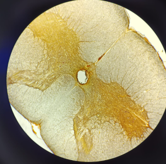
Cerebrum - AgNO3
Purpose: Used because astrocytes have an affinity for gold.
Visuals: Astrocytes look like "spiders or like stars".
Color: Appears as a "pink purple structure" even though gold is used.
What is this?
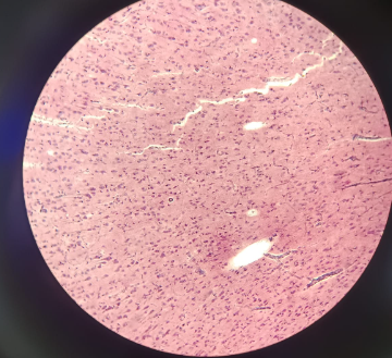
Cerebrum- HE
Layers: Contains six layers of cells in the gray matter.
Organization: Gray matter is on the outside (bodies and dendrites); white matter is on the inside (axons).
Key Cells: Features pyramidal neurons, including the "big pyramidal cells of Betz" in the fifth layer.
Glial Cells: Contains protoplasmic astrocytes that maintain the balance between neurons.
Staining: This is the slide referred to as the "pink purple staining
What is this?
Cerebrum - Fuschin
What is this?
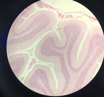
Cerebellum - HE
Organization: Gray matter outside, white matter inside.
Layers: Contains three layers of cells.
Key Cell: Features large Purkinje cells (referred to by the professor as "protein cells")
What is this?
Cerebellum - AgNO3
Organization: Gray matter outside, white matter inside.
Layers: Contains three layers of cells.
Key Cell: Features large Purkinje cells (referred to by the professor as "protein cells")
What is this?
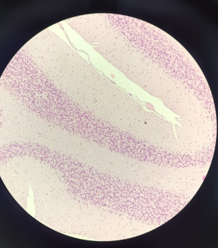
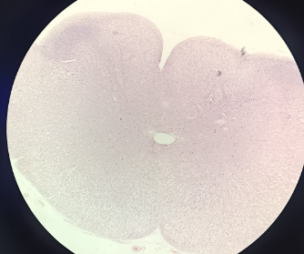
Medulla Spinalis - HE
Organization: White matter outside, gray matter inside (butterfly or 'H' shape).
Anterior Horn: Contains very large alpha motor neurons that innervate skeletal muscles.
Central Canal: Lined with ependymal cells (simple columnar with microvilli/cilia) which produce cerebrospinal fluid.
Note: The description is the same for HE or Silver staining, but silver clearly shows the cell shape
What is this?
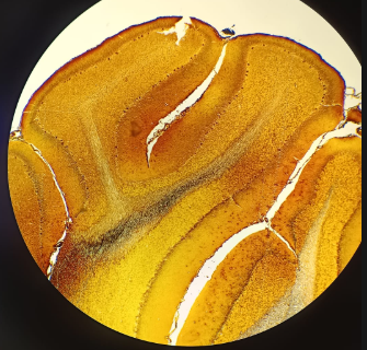
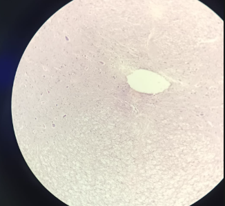
Medulla Spinalis - AgNO3
Organization: White matter outside, gray matter inside (butterfly or 'H' shape).
Anterior Horn: Contains very large alpha motor neurons that innervate skeletal muscles.
Central Canal: Lined with ependymal cells (simple columnar with microvilli/cilia) which produce cerebrospinal fluid.
Note: The description is the same for HE or Silver staining, but silver clearly shows the cell shape
What is this?
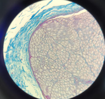
Peripheral Nerve - Azan
Focus: Used specifically to identify non-myelinated neurons.
Visuals: Axons appear as red dots with a blue outline called the endoneurium.
Layers: Includes perineurium (red line) and epineurium (outermost loose connective tissue)
What is this?
Peripheral Nerve - OsO4
Focus: Used specifically to visualize myelinated neurons.
Visuals: The axon is in the center, wrapped in a dark/black myelin sheath.
Connective Tissue: Also identifies the endoneurium, perineurium, and epinurium
What is this?
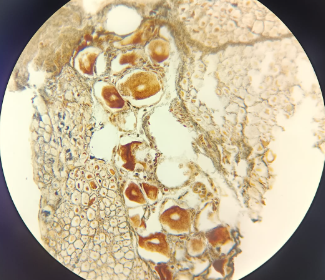
Ganglion Spinale - HE
Neurons: Contains pseudounipolar neurons (T-shaped neurons).
Satellite Cells: Neuron bodies are surrounded by a "mud-like" layer of satellite cells for protection/trophic function.
Note: There are no synapses in this structure; the description is identical for both HE and Silver slides.
What is this?
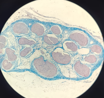
Ganglion Spinale - AgNO3
Neurons: Contains pseudounipolar neurons (T-shaped neurons).
Satellite Cells: Neuron bodies are surrounded by a "mud-like" layer of satellite cells for protection/trophic function.
Note: There are no synapses in this structure; the description is identical for both HE and Silver slides.
What is this?
Cutis (skin) - HE
Location: Found in the deep dermis.
Structure: Looks like a large capsule filled with Type I collagen and fibroblasts arranged in concentric circles.
Function: These are pressure and vibration receptors; when you squeeze or touch them, the internal structure indents to trigger the bare neuron inside.