sensation, ear, eye
1/102
There's no tags or description
Looks like no tags are added yet.
Name | Mastery | Learn | Test | Matching | Spaced | Call with Kai |
|---|
No analytics yet
Send a link to your students to track their progress
103 Terms
Sensation
Activation of sensory receptor cells by a stimulus
Perception
Central processing and interpretation of sensory stimuli into meaningful patterns
Difference between sensation and perception
Sensation is receptor activation while perception is CNS interpretation of stimuli
General senses
Senses distributed throughout the body with receptors in many organs and tissues
Special senses
Senses associated with specialised organs such as eye, ear, tongue and nose
Examples of general senses
Touch, pressure, pain, temperature, proprioception and visceral sensation
Examples of special senses
Vision, hearing, balance, taste and smell
Somatosensation
Group of sensory modalities associated with touch, proprioception and interoception
Proprioception is:
the sense of the position and movement of your body parts.
Interoception is:
sensing what is happening inside the body.
Somatosensory modalities
the different types of body sensations that the somatosensory system can detect.
Pressure, vibration, light touch, tickle, itch, temperature, pain, proprioception and kinesthesia
Location of somatosensory receptors
Skin, muscles, tendons, ligaments, joint capsules and walls of visceral organs
Proprioception
Sense of body position
Kinesthesia
Sense of body movement
Interoception
Sense of internal organ condition and movement
Sensory receptor
Cell or structure that detects stimuli and converts them into nervous system signals
Chemoreceptor
Receptor that detects chemical stimuli such as taste and smell
Thermoreceptor
Receptor sensitive to temperature changes
Photoreceptor
Receptor in the eye responding to light, colour and movement
Mechanoreceptor
Receptor responding to physical stimuli such as pressure, vibration and stretch
Baroreceptor
Receptor detecting pressure changes within vessels and organs
Nociceptor
Receptor stimulating pain responses from damaging stimuli
Nociception
Pain perception caused by potentially damaging mechanical, chemical or thermal stimuli
Cause of nociceptor activation
Stressed or damaged tissues release chemicals activating nociceptors
Temperature receptors
Receptors sensitive to heat or cold stimuli
Taste (gustation)
Special sense associated with chemical detection by the tongue
Smell (olfaction)
Special sense responsive to airborne chemical stimuli
Papillae
Raised bumps on tongue containing taste buds
Taste buds
Structures containing gustatory receptor cells
Gustatory receptor cells
Specialised chemoreceptors detecting chemicals in food
Function of gustatory receptor cells
Release neurotransmitters based on chemicals detected in food
Cranial nerves involved in taste
Facial nerve 7 and vagus nerve 10
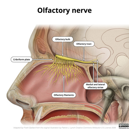
Olfactory epithelium
Specialised sensory epithelium in superior nasal cavity containing olfactory neurons
Location of olfactory epithelium
Superior nasal cavity
Olfactory receptor neurons
Bipolar sensory neurons detecting smell stimuli
Pathway of olfactory neurons
Axons pass through cranial cavity floor to reach brain
Brain regions receiving olfactory input
Primary olfactory cortex (Located in the inferior temporal lobe), limbic system and hypothalamus
Unique feature of smell pathway
Only sensory modality that does not synapse in thalamus before cerebral cortex
Most sensory pathways follow this pattern:
Receptor→Thalamus→Cerebral cortex
The Olfaction pathway is unique because:
smell information reaches the cerebral cortex BEFORE going to the thalamus.
Smell pathway Olfactory receptors→Olfactory bulb→Olfactory cortex
Reason smells trigger memories
Olfactory system has close connections with limbic system and hypothalamus
Audition
Hearing; transduction of sound waves into neural signals
Equilibrium
Sense of balance and body position
Major divisions of ear
External ear, middle ear and inner ear
Function of external ear
Funnels sound waves toward tympanic membrane
Auricle/Pinna
Large elastic cartilage structure directing sound waves
External auditory canal (external acoustic meatus)
Approximately 2.5 cm canal directing sound to tympanic membrane
Tympanic membrane
a thin membrane that vibrates when struck by sound waves.
the eardrum
Middle ear
Air-filled cavity containing auditory ossicles
Auditory ossicles
Malleus, incus and stapes
Malleus
Ossicle attached to tympanic membrane
Incus
Ossicle between malleus and stapes
Stapes
Ossicle attached to oval window of inner ear
Function of auditory ossicles
Amplify and transmit sound vibrations to inner ear
Eustachian tube (pharyngotympanic tube)/auditory tube
Tube connecting middle ear to pharynx to equalise air pressure across tympanic membrane
Function of Eustachian tube
Equalises pressure across tympanic membrane
Inner ear
Contains cochlea and vestibule for hearing and balance
Cochlea
Spiral-shaped structure containing receptors for hearing
Vestibule
Inner ear chamber containing receptors for balance
Oval window
Location where stapes attaches to inner ear
Hair cells with stereocilia
Mechanoreceptors in vestibule detecting head movement and balance
Vestibulocochlear nerve
Cranial nerve transmitting hearing and balance information to brain
Structures responsible for hearing
Cochlea and auditory pathway
Structures responsible for balance
Vestibule and vestibular apparatus
Vision
Special sense of sight based on photoreceptors responding to light
Location of eyes
Within the bony orbits of skull
Function of bony orbit
Protects eyeball and anchors soft tissues
Eyelids
Protect eye from abrasions and foreign particles
Conjunctiva
Membrane connecting eyelids to sclera
Lacrimal gland
Gland producing tears
located above eye
Lacrimal ducts
Drain tears across eye surface
in corner of eye
Function of tears
Wash away foreign particles and lubricate eye
Extraocular muscles
Six skeletal muscles controlling eye movement
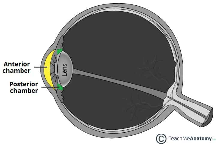
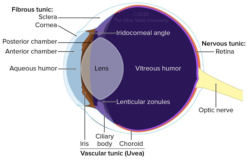
Anterior cavity of eye
Space between cornea and lens filled with aqueous humour
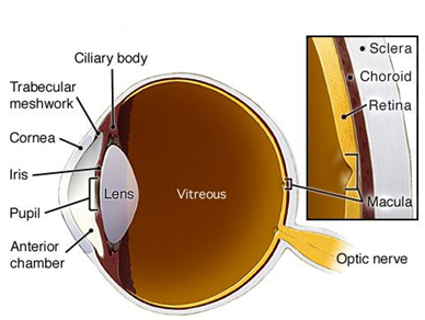
Posterior cavity of eye
Space posterior to lens filled with vitreous humour
Aqueous humour
Watery fluid filling anterior cavity of eye
Vitreous humour
Gel-like substance filling posterior cavity of eye
made by cells in retina
Three tunics of eye
Fibrous tunic, vascular tunic and neural tunic
Fibrous tunic
Outermost layer consisting of sclera and cornea
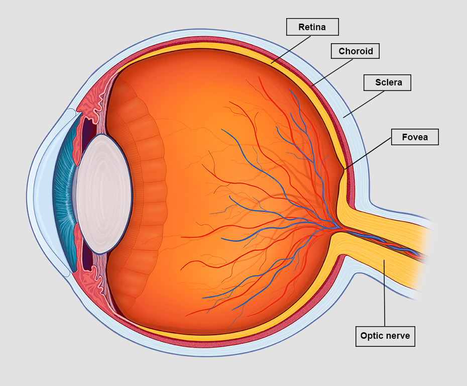
Sclera
White dense connective tissue forming most of eye surface
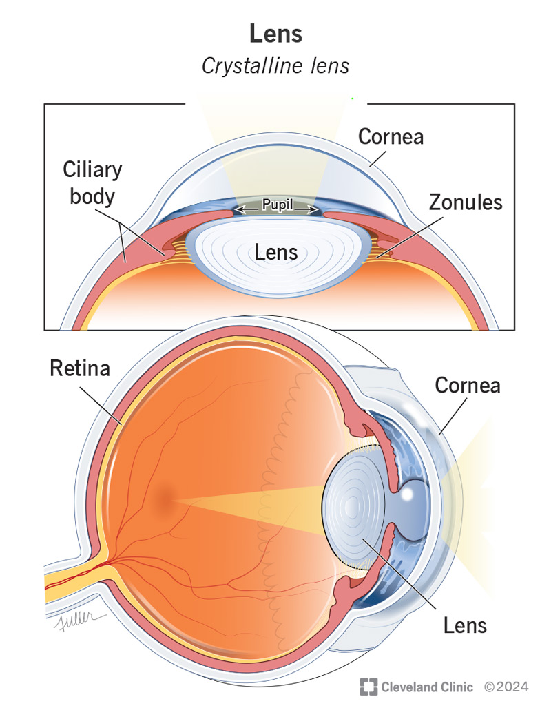
Cornea
Transparent fibrous coat allowing light into eye and helping focus light
Vascular tunic
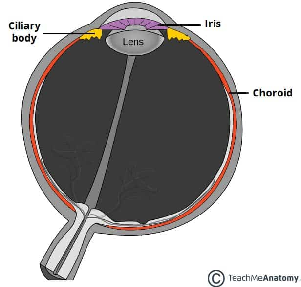
Middle layer consisting of choroid, ciliary body and iris, pupil, lens
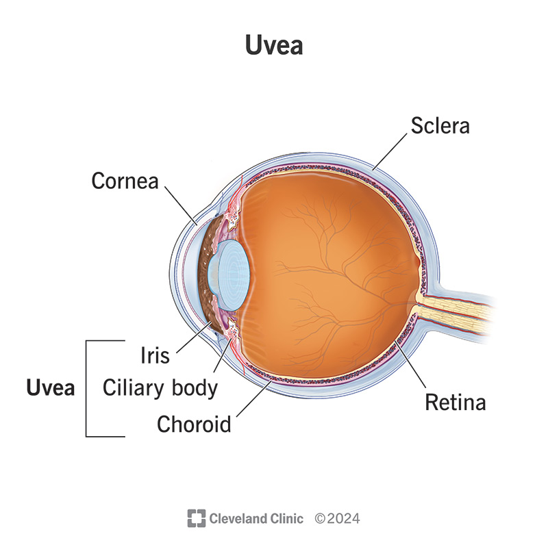
Choroid
Highly vascular connective tissue supplying blood to eye
deep to sclera
Ciliary body
Muscular structure attached to lens via suspensory ligaments
produces aquous humour. located anterior half of eye
Function of ciliary body
Changes lens shape for near and distant vision
Suspensory ligaments (zonule fibres)
Fibres attaching lens to ciliary body
Lens
Transparent elastic structure focusing light onto retina whcih are
posterior half of eye
Iris
Coloured smooth muscle regulating light entry
Pupil
Hole in centre of iris allowing light into eye
Response of iris to bright light
Constriction of pupil
Response of iris to dim light
Dilation of pupil
Neural tunic (retina)
Innermost layer containing photoreceptors and neurons
Function of retina
Receives and processes visual information
highly vascular
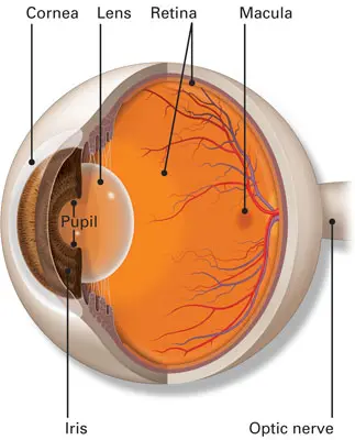
Optic disc
Point where optic nerve leaves eye
blind spot of eye
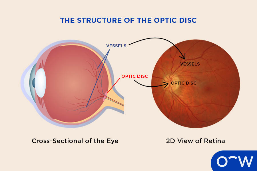
Blind spot
Area lacking photoreceptors at optic disc
Cause of blind spot
Optic nerve exits retina where no photoreceptors are present
Photoreceptors
Cells in retina responding to light stimuli
Optic nerve
Carries visual information from retina to brain
Fovea centralis
Region of retina associated with sharp vision
Structures responsible for vision
Eye, retina, optic nerve and visual pathways
Special sense organs
Eye, ear, tongue and nose
Main receptor type for vision
Photoreceptors
Main receptor type for hearing
Mechanoreceptors
Main receptor type for balance
Mechanoreceptors