other bone diseases
1/46
Earn XP
Description and Tags
lecture given 6/2/2026
Name | Mastery | Learn | Test | Matching | Spaced | Call with Kai |
|---|
No analytics yet
Send a link to your students to track their progress
47 Terms
bone dysplasias
fibrous dysplasia, cemento osseous dysplasia (COD)
other lesions of bone
central giant cell granuloma, aneurysmal bone cyst, cherubism, paget’s disease
fibrous dysplasia
normal bone metabolism replaced by fibrous tissue containing varying amounts of abnormal appearing bone
solitary/monostotic ~70%
most common sites are ribs, femur, tibia, maxilla, mandible
older age group individuals
multiple/polystotic more common in ~10 yo
what are radiographic features of fibrous dysplasia?
maxilla > mandible 2:1, posterior aspect and unilateral more often
ill defined, blending of the trabseculae into abnormal pattern, in young lesions it will appear corticated
density and trabecular pattern varies considerable, radiolucent, mixed and radiopaque, short/thin/irregularly shaped trabeculae
small lesions have no effects on surrounding structures, larger lesions can expand, thin the cortex, PDL space may be decreased/displace/or interfere with eruption, in rare cases root resorption, displace the canal in superior direction
DD: hyperparathyroidism, paget’s, PCD
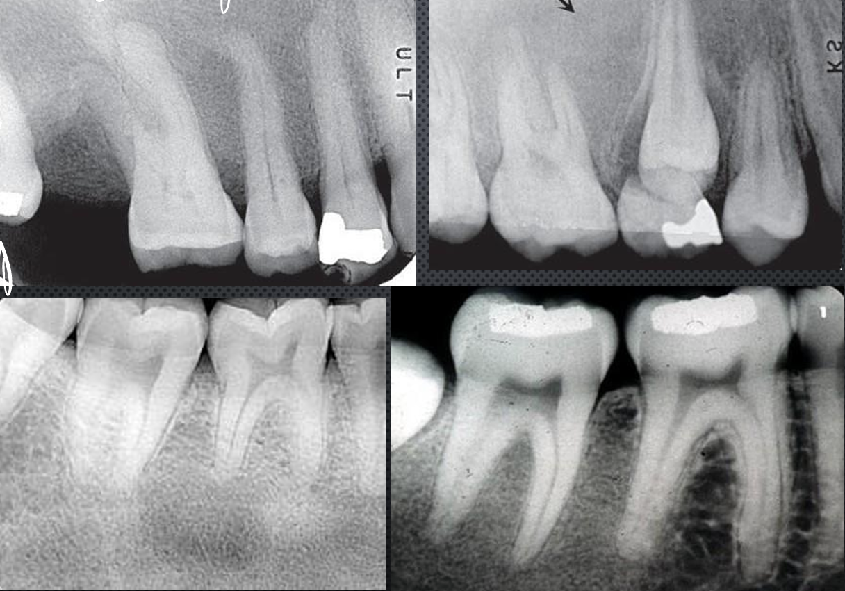
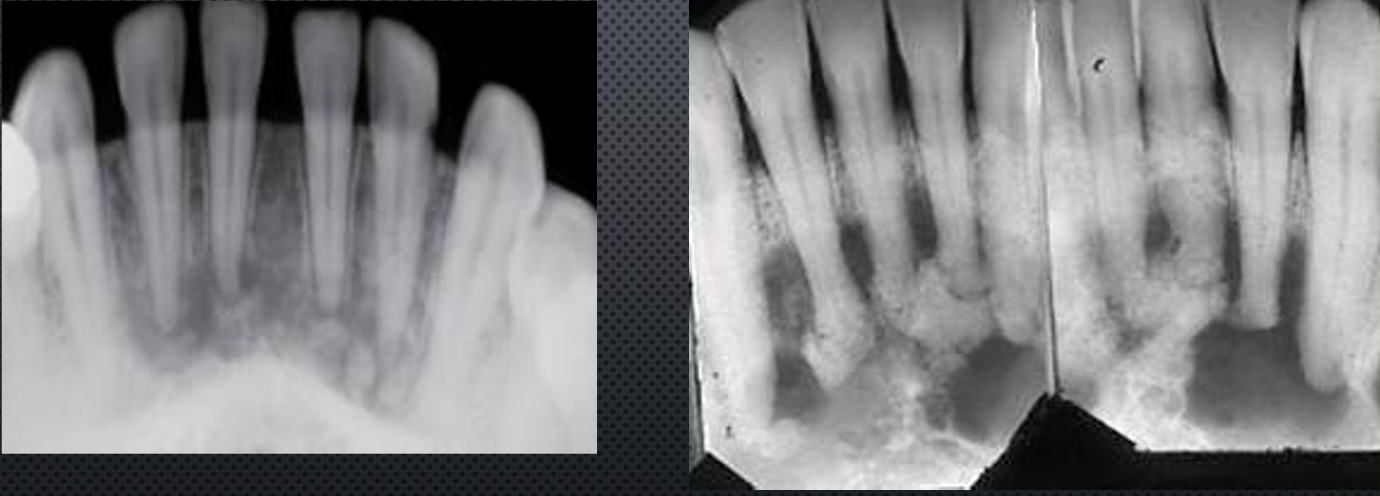
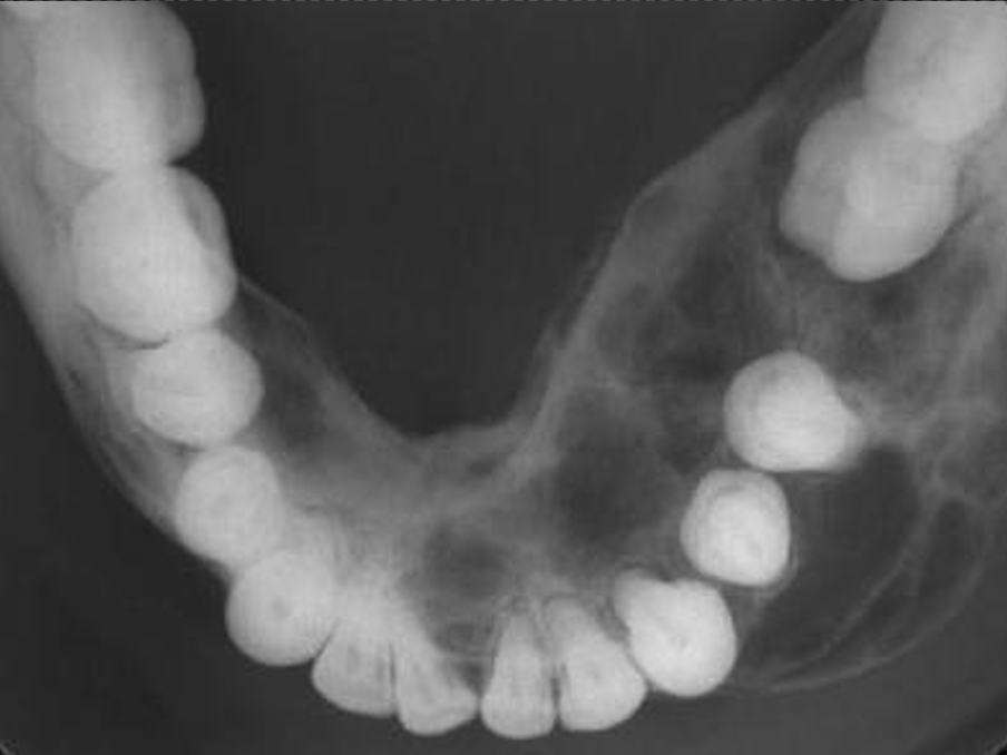
fibrous dysplasia
fibrous dysplasia
fibrous dysplasia
fibrous dysplasia
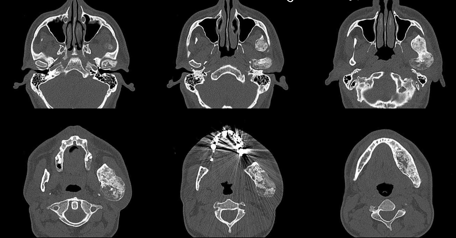
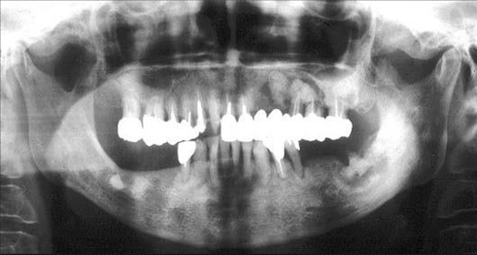
what is this, and how can you tell?
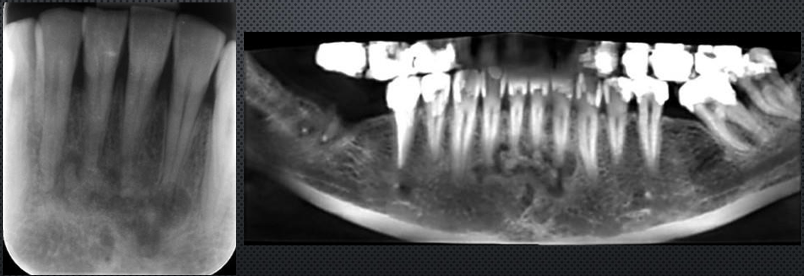
fibrous dysplasia
shape is maintained, it just grows bigger
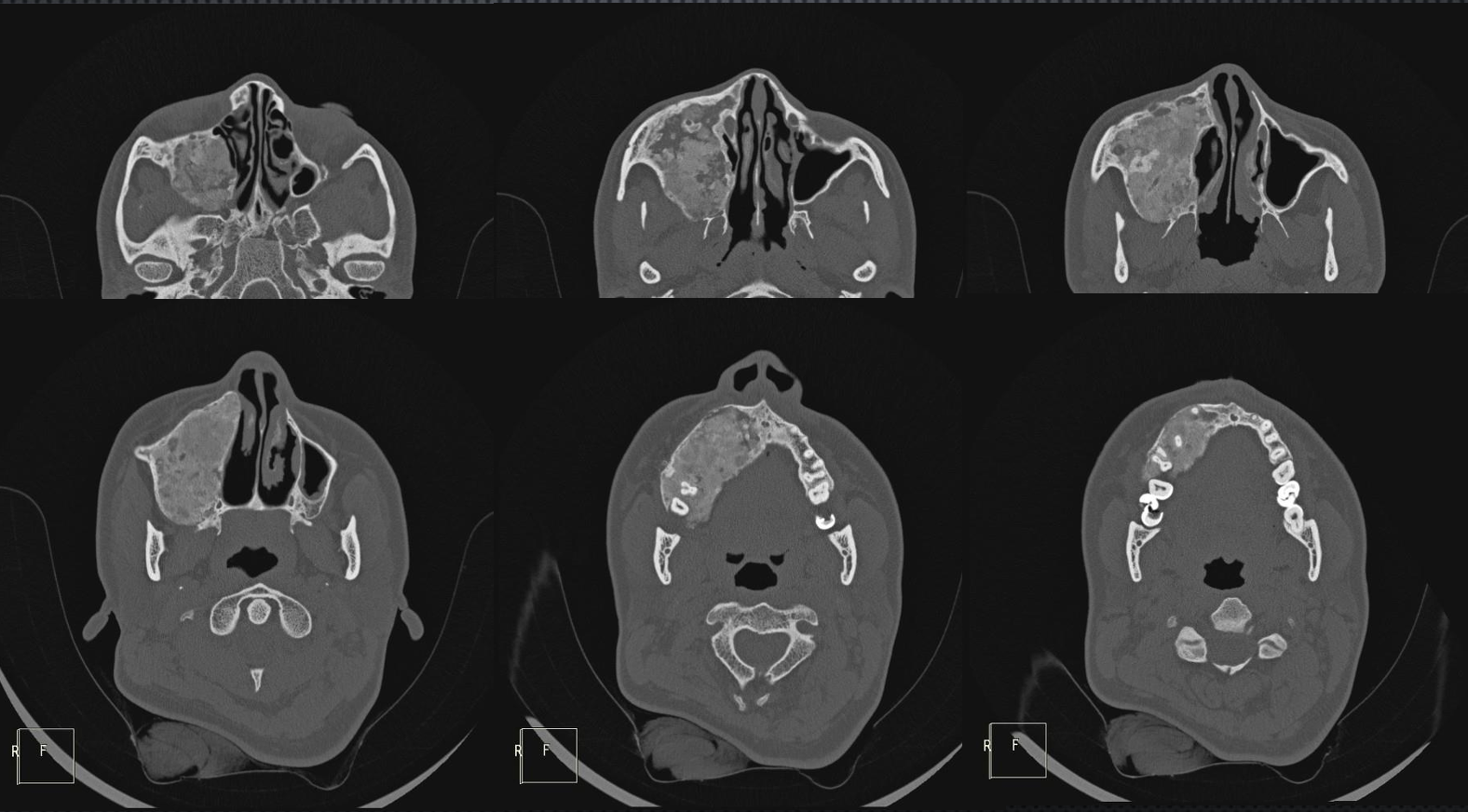
fibrous dysplasia
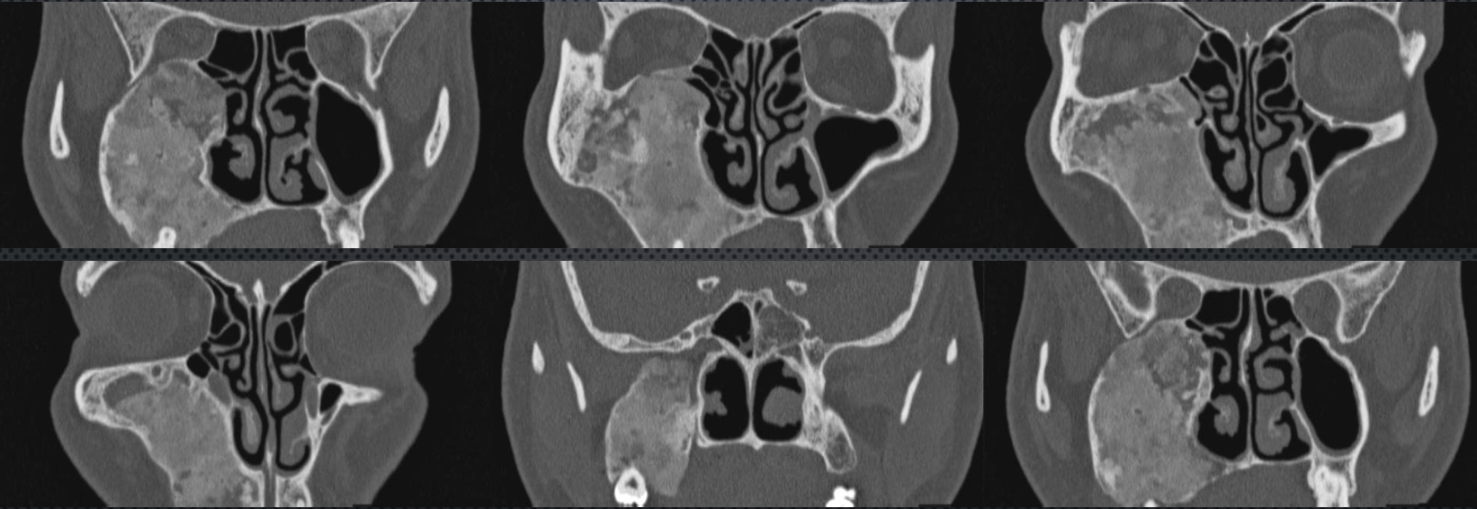
fibrous dysplasia
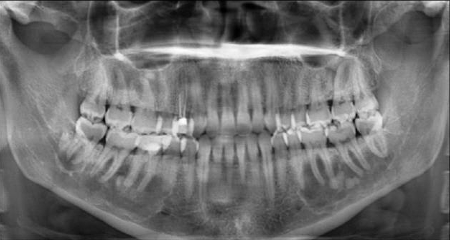
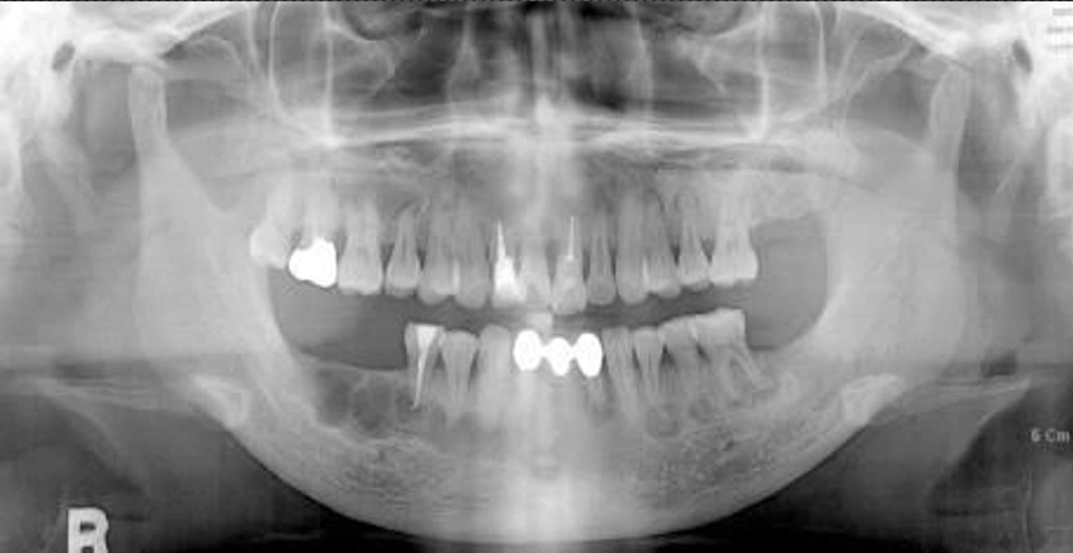
cemento-osseous dysplasias
periapical cemento-osseous dysplasia, florid osseous dysplasia
periapical cemento-osseous dysplasia
aka cementoma, fibrocementoma, sclerosing cementoma, periapical fibrocementoma, periapical osteofibrosis, periapical fibrous dysplasia
localized change in normal bone metabolism, resulting in cancellous bone replaced by fibrous tissue and cementum like material
middle aged women, females > males, more common in asians, blacks, and hispanics
radiographic features of periapical cemento-osseous dysplasia
apical to the root apex, mandibular anteriors more often but can occur at any tooth
well defined, corticated, round, or oval, sometimes irregular, secondary infection sclerotic
early stage radiolucent, intermediate mixed, mature stage radiopaque
loss of PDL space, widened PDL, expansion
DD: rarefying osteitis, inflammatory lesions, cementoblastoma
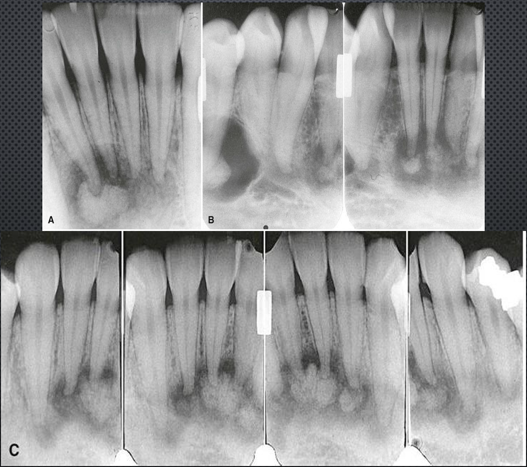
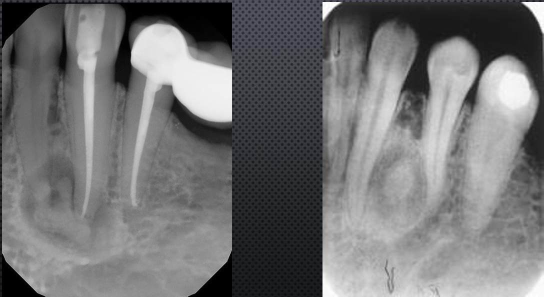
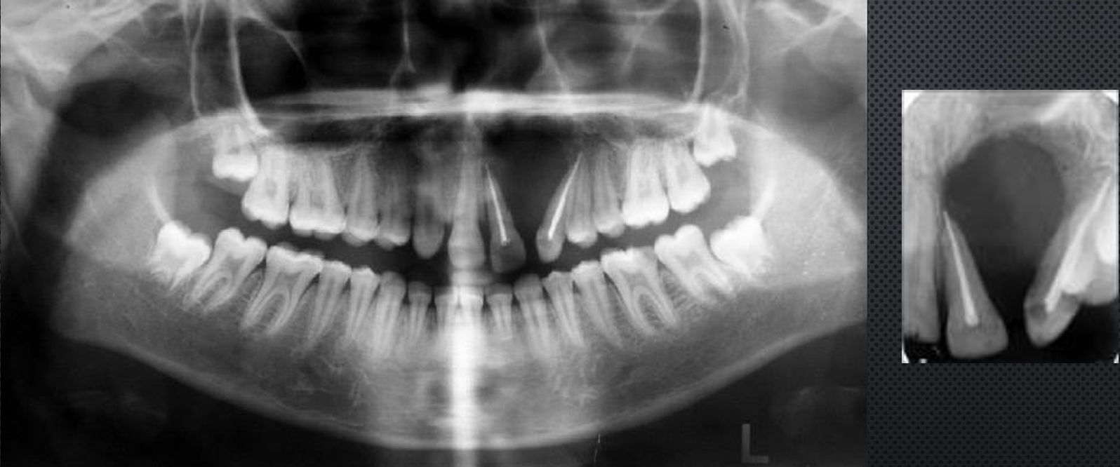
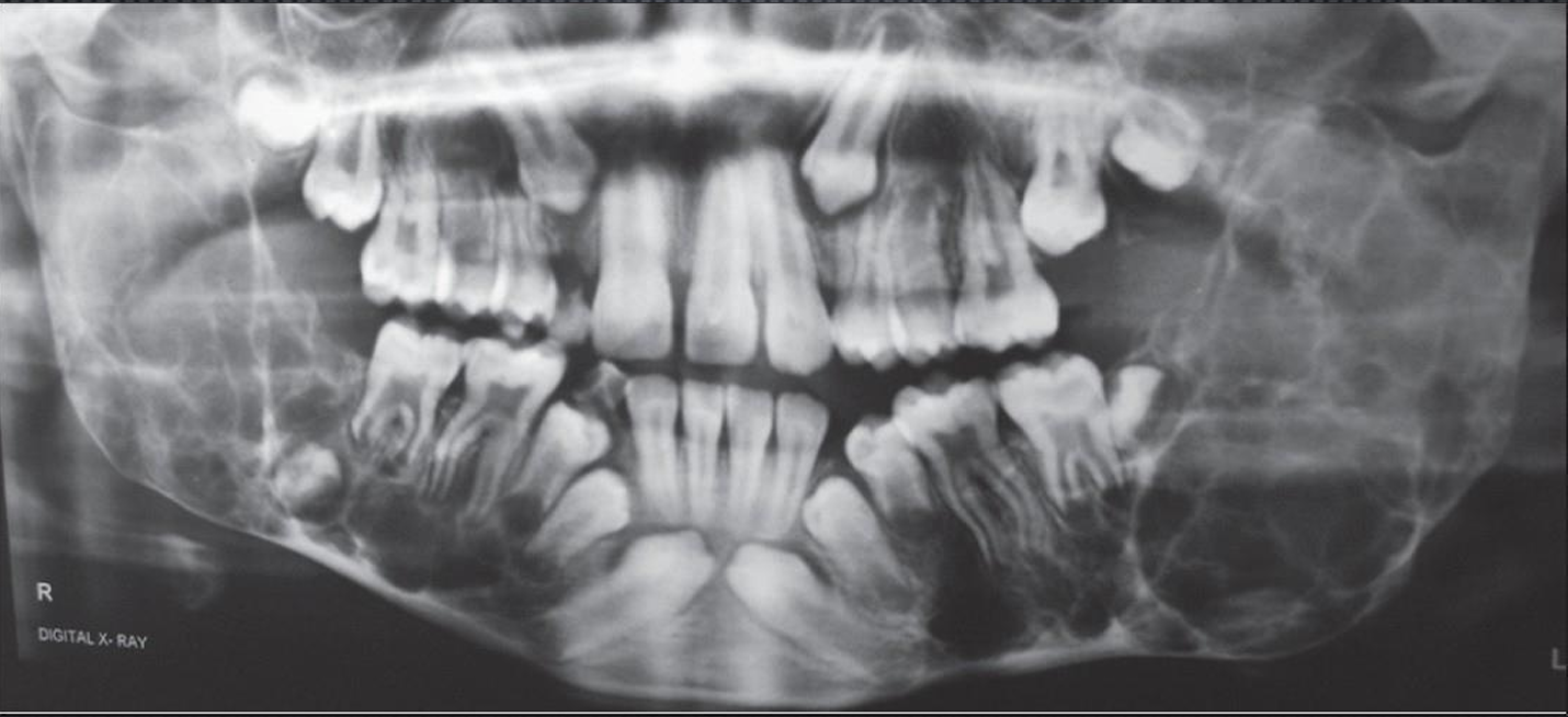
periapical cemento-osseous dysplasia
periapical cemento-osseous dysplasia
periapical cemento-osseous dysplasia
periapical cemento-osseous dysplasia
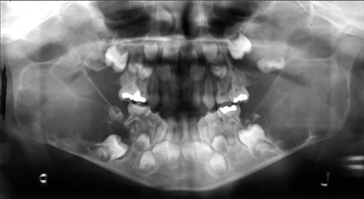
focal cemento osseous dysplasia
focal cemento osseous dysplasia
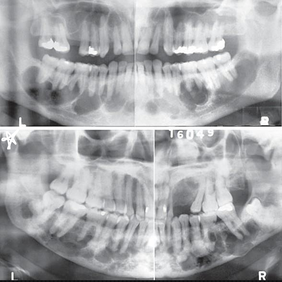
florid osseous dysplasia
florid cemento-osseous dysplasia, gigantiform cementoma, familial multiple cementoma
FCOD is wide spread PCD, normal cancellous bone replaced by fibrous tissue and cemento-osseous tissue, poorly vascularized
same clinical features as PCD, females > males, asians, blacks, hispanics, occasionally involved with SBC
what are radiographic features of florid osseous dysplasia?
bilateral and in both jaws or throughout in one jaw, apical to the teeth (>/= 2 quadrants)
well defined, corticated borders
radiolucent, radiopaque and mixed depending on the stage of the lesion, irregular amorphous calcifications
can displace IAN, expansile
DD: paget’s, sclerosing osteomyelitis
*
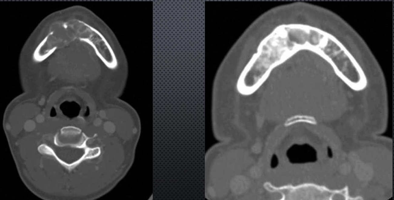
florid osseous dysplasia
*low vascularity
florid osseous dysplasia
florid osseous dysplasia
florid osseous dysplasia
central giant cell granuloma
aka giant cell reparative granuloma, giant cell lesion, giant cell tumor
thought to be a reactive lesion, w/ unknown stimulus
60% in young individuals ~20’s, slow growing with some rapid growth
what are the radiographic features of central giant cell granuloma?
mandible > maxilla 2:1, anterior to the 1st molar
periphery may not have cortication, the lesion is well defined in the mandible, maybe ill defined in the maxilla
radiolucent, wispy septa
displace and resorb teeth and roots, expansile, missing lamina dura, displaced IAC, may have an uneven undulating appearance
DD: ameloblastoma, odontogenic myxoma, aneurysmal bone cyst
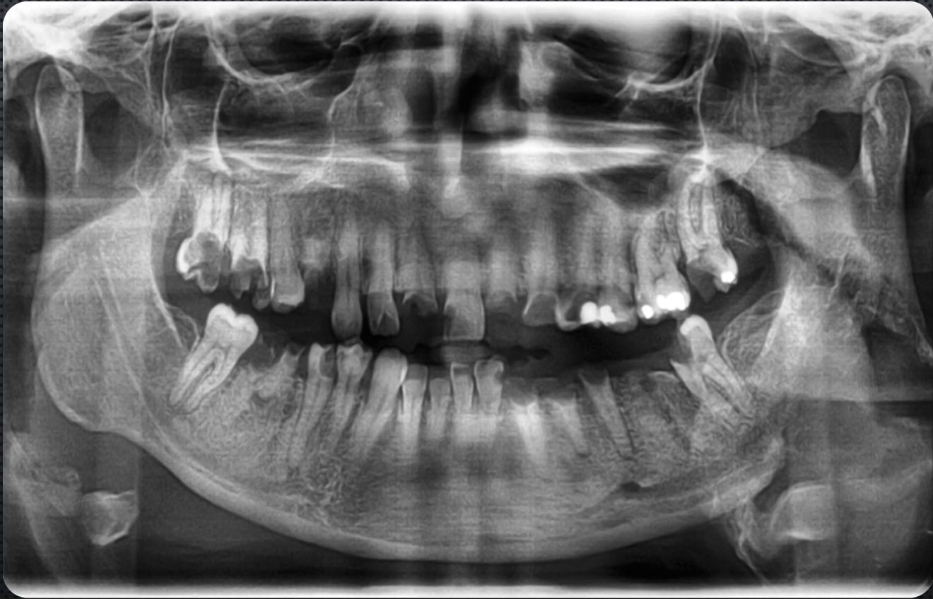
central giant cell granuloma
central giant cell granuloma
central giant cell granuloma
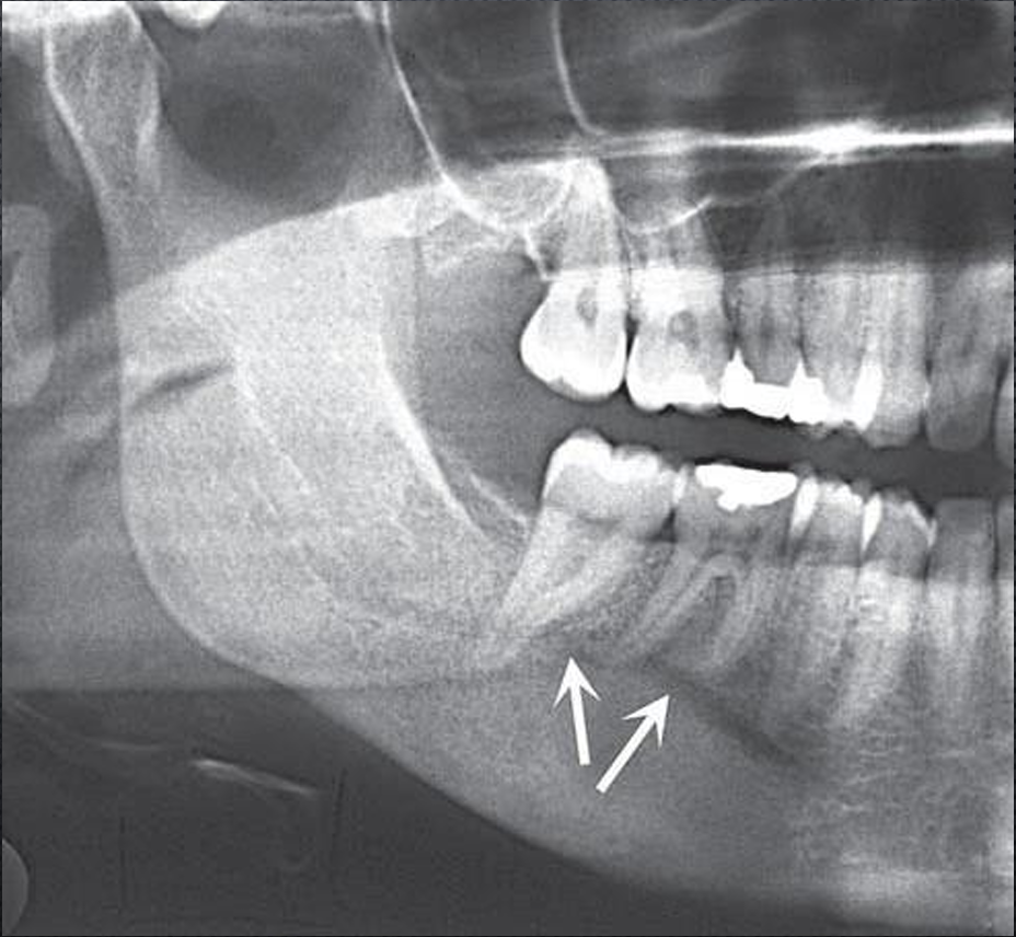
aneurysmal bone cyst
believed to represent an exaggerated localized proliferative response of vascular tissue in bone
rapid bony swelling, tenderness on palpation, pain occasionally
what are radiographic features of aneurysmal bone cyst?
mandible > maxilla, ramus and molar > anterior region
well defined and circular or hydraulic
small lesion, no internal structure evidence, larger lesions have mutlilocular appearance, septa are at right angle to outer cortex
as the lesion expands the cortical plates expand, can displace and resorb teeth
DD: giant cell granuloma
aneurysmal bone cyst
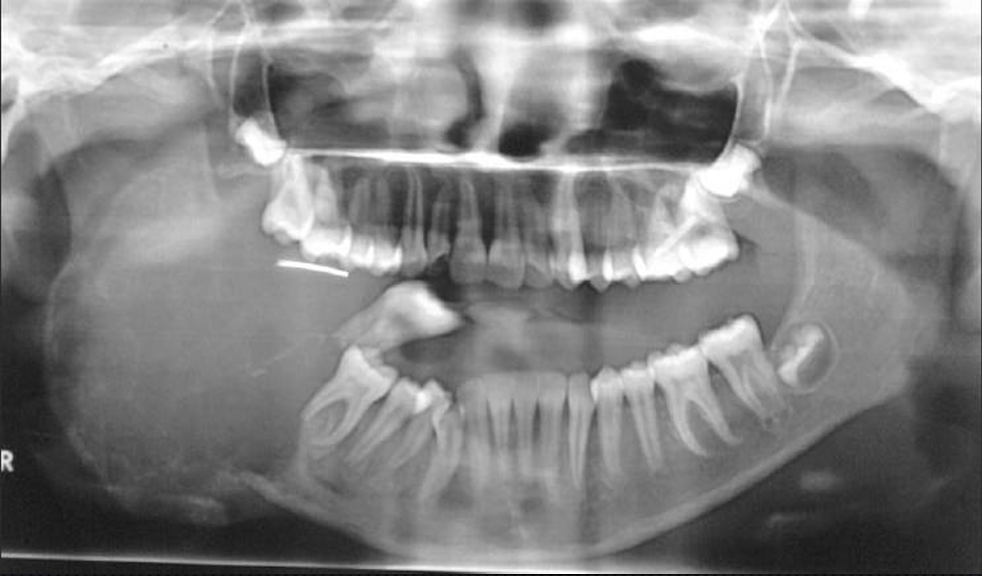
aneurysmal bone cyst
aneurysmal bone cyst
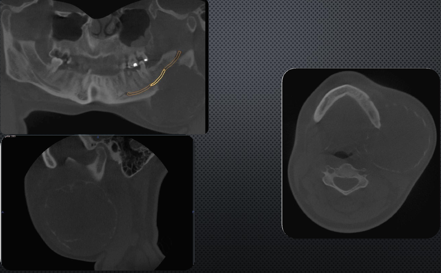
aneurysmal bone cyst
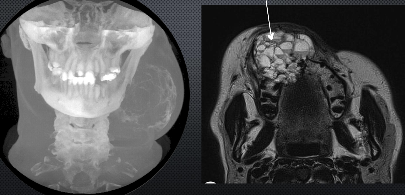
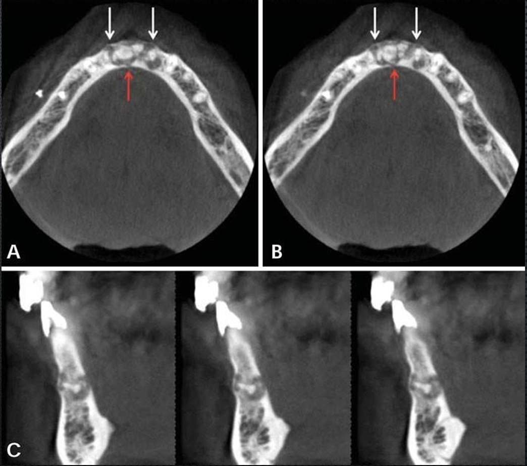
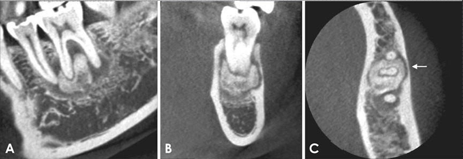
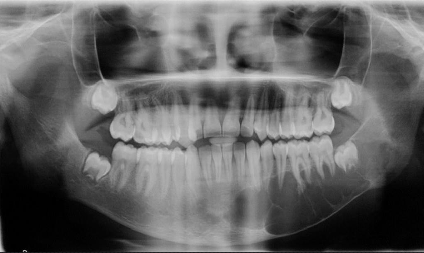
what is this and what is the white arrow pointing at?
aneurysmal bone cyst
blood
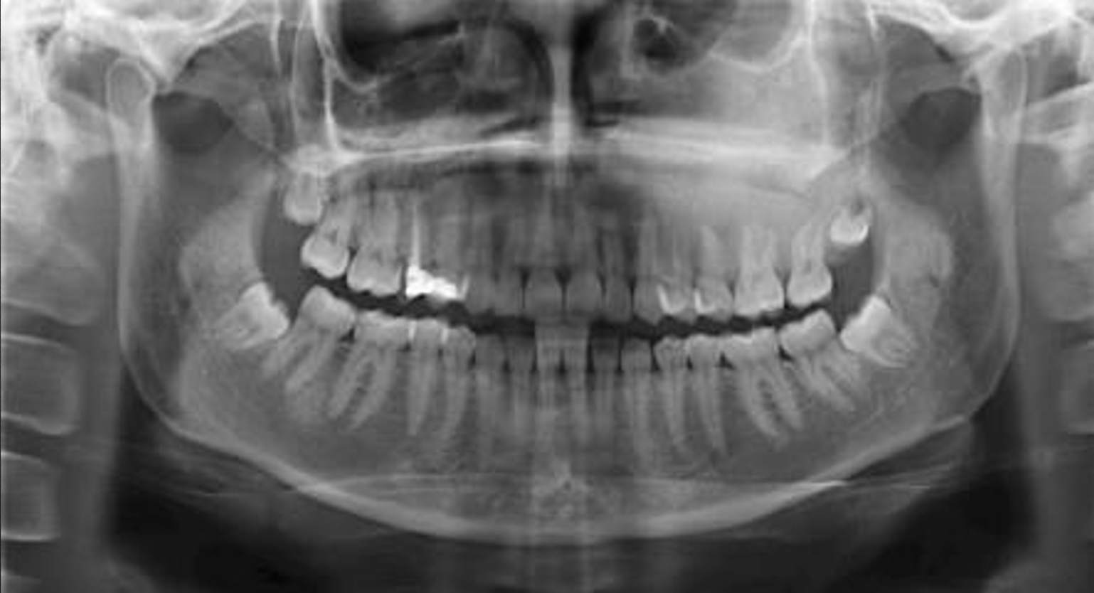
cherubism
*aka familial fibrous dysplasia
rare inherited autosomal dominant disease
age 2-6, bilateral enlargement of lower face
what are the radiographic features of cherubism?
bilateral affecting both jaws, when it is present in one jaw most often in mandible
well defined with some corticated borders
septated, wispy septa, multilocular appearance
expand the cortical plates, displace teeth in some instances tooth buds are destroyed
DD: fibrous dysplasia, BCNS
cherubism
cherubism
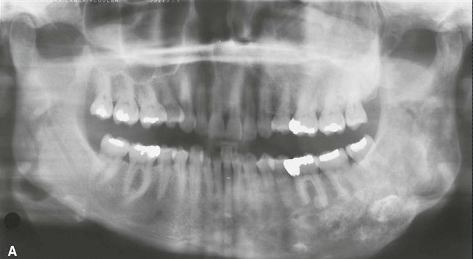
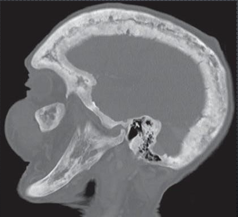
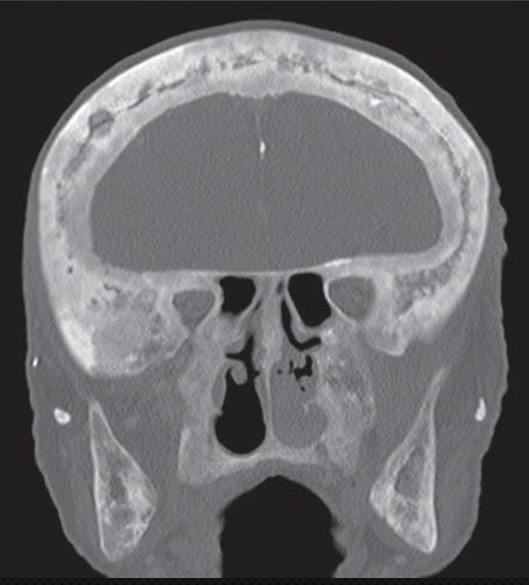
paget’s disease
aka osteitis deformans
abnormal resorption and apposition of osseous tissue in one or more bones
males > females 2:1, older than 40 yo, ill defined neurologic pain
what are the radiographic features of paget’s disease?
pelvis, femur, skull and vertebrae, infrequently jaws, maxilla > mandible 2:1
depending on the stage of development of the disease internal structure can vary from radiolucent to mixed to radiopaque, increased bone density
pagetoid skull 3 to 4x the normal skull thickness
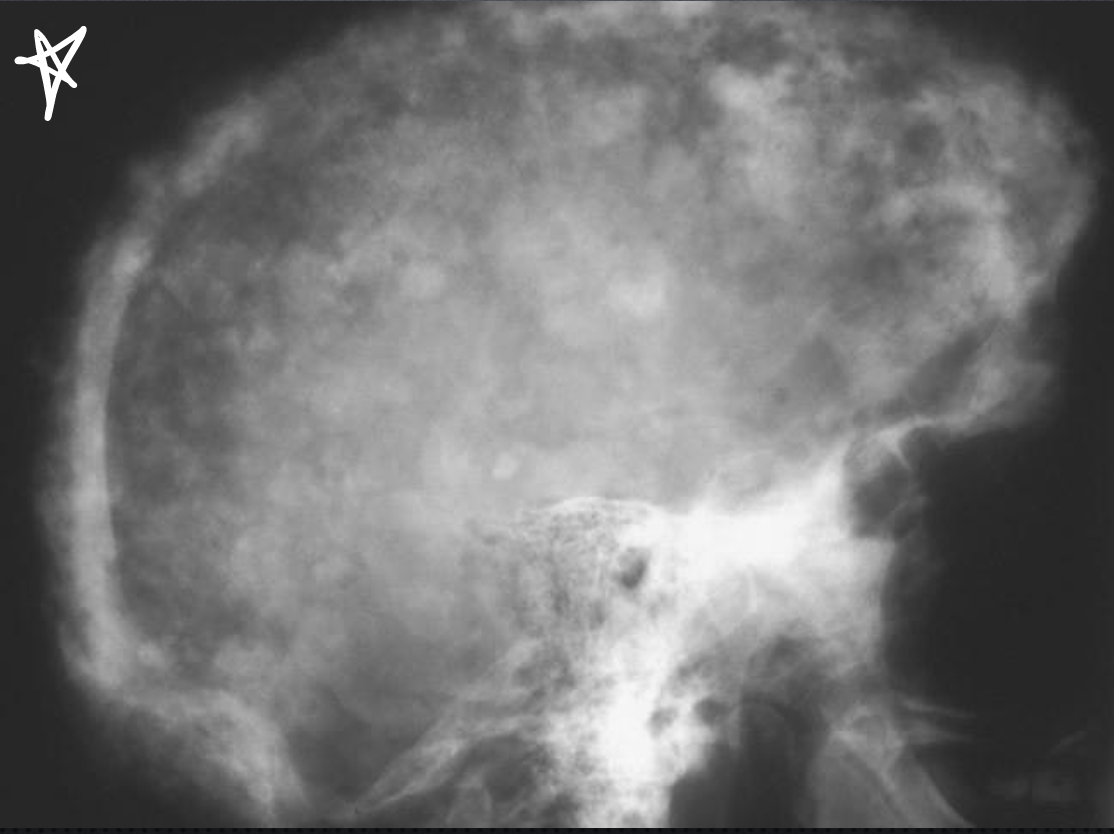
*what is this, and how do you know?
paget’s disease
cotton wool appearance of skull
paget’s disease
paget’s disease