Cellular Pathology: Applied Problem Solving Tasks
1/21
There's no tags or description
Looks like no tags are added yet.
Name | Mastery | Learn | Test | Matching | Spaced |
|---|
No study sessions yet.
22 Terms
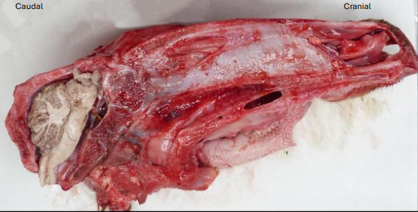
What does this image show?
hemi-sectioned head of a day-old calf
What is the abnormality affecting the brain?
cerebral agenesis

What does this image show?
right AV valve in a young dog

What is the abnormality affecting the AV valve?
vascular dysplasia
If you diagnosed AV dysplasia in a live puppy, is there a significant concern that the dysplastic tissue is at increased risk of transformation into neoplastic tissue?
NO

Which kidney is abnormal?
TOP
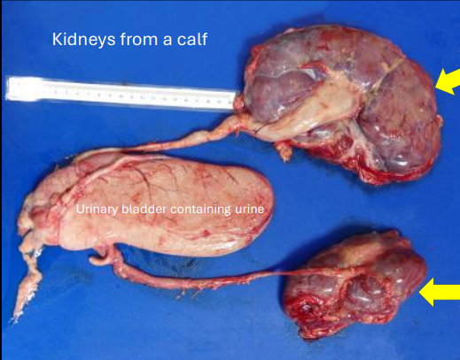
What term is best used to describe the top kidney?
enlarged
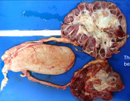
What has been done to these kidneys?
hemisectioned
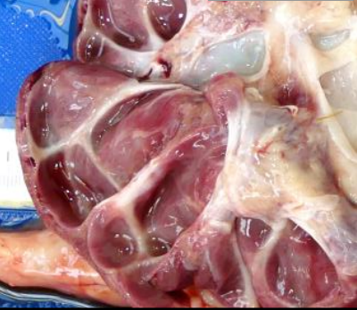
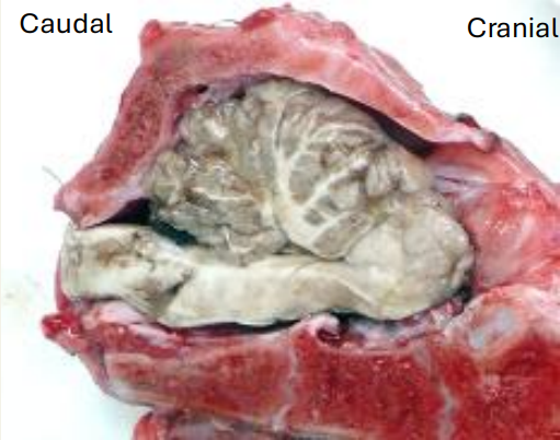
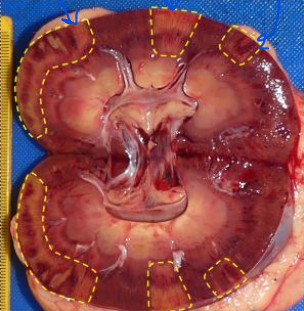
What does this image show?
cut surface of the abnormal kidney
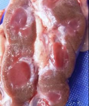
What does this image show?
cut surface of the normal kidney
What type of pathological change is most likely to have affected the renal parenchyma?
atrophy

What has caused the parenchymal atrophy?
pressure build up repressing blood supply to tissue

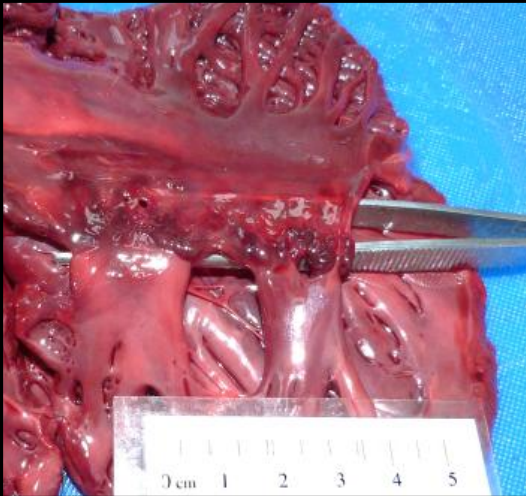
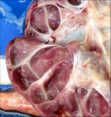
What does this image show?
abnormal kidney with the ureter opened
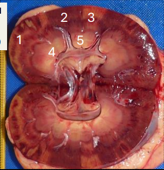
What areas or the kidney, labelled as 1-5 are abnormal?
1&3
What has occurred in the areas highlighted?
obstruction to blood supply that restricts blood supply to paler areas

What type of change has occurred in the renal parenchyma?
coagulative necrosis

What do you think about the colour of the liver?
abnormal, paler than normal
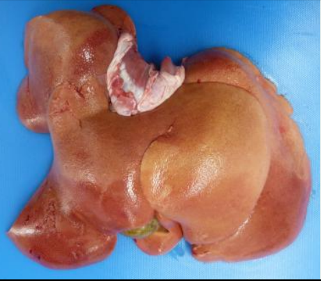
List 4 different cellular pathological changes affecting the hepatocytes that might account for the colour of the liver
hepatocellular vacuolar change with water accumulation (hydropic change)
hepatocellular vacuolar change with lipid accumulation (fatty change)
hepatocellular vacuolar change with glycogen accumulation
hepatocellular necrosis

What does this image show?
lungs and mediastinal tissues from a sheep with 2 masses incised

In what anatomical structure are these masses likely to have arisen?
lymph nodes

What term would best describe the appearance of the abnormal material?
caseous
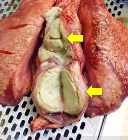
What is a likely cause of the pathology in this case?
bacterial infection